
Autophagy and Aging
Foundational science and methods. A research review published by South Beach Longevity.
Autophagy and Aging
A graded account of recycling pathways, flux measurement, and the claims that do not surviveAutophagy is a set of regulated recycling pathways, not a consumer cleanse that switches on at a clock hour. The cell biology is deep. The human aging and fasting claims are not. This article grades that gap. It is not a fasting protocol.
Compiled by South Beach Longevity · 20 August 2026 Copyright 2026 Series SBL-41 / SP-AUTOPHAGY-AND-AGING · Register A scientific article Sources peer-reviewed cell, animal, and human studies · assay guidelines · verified NCBI records Constraint This document describes published research. It is not medical advice. No human use, dose, route or schedule is recommended anywhere in this document.
How to read this document Every finding is labelled, in the sentence that reports it, by the kind of study that produced it. In vitro means a cell or a reconstituted system. Animal names the species. Human means people. A static LC3 or p62 measurement is not autophagic flux. Ketosis is not an autophagy assay. Rodent or invertebrate lifespan is not a human outcome. A named fasting hour is an operational choice, not a receptor timeout. Amounts and durations appear only as reported experimental parameters, always with the system attached. Nothing here is a recommendation. Findings are graded in place as established, strongly supported, emerging, plausible, or speculative. Two further labels mark careful absences rather than verdicts: not established, where the evidence is too thin to place a claim on the ladder at all — untested or insufficient, an absence of proof rather than disproof; and not supported, where the weight of evidence leans against a claim but stops short of a formal refutation.
01 The controlling question
The market sentence is simple: autophagy is cellular cleansing; a sixteen-hour fast turns it on; more of it is how animals, and therefore people, age more slowly. The laboratory sentence is longer. Autophagy is a family of lysosome-dependent routes that capture and degrade cytoplasmic material (Mizushima and Komatsu, 2011; Galluzzi et al., 2017; Dikic and Elazar, 2018). Those routes are regulated, cargo-selective or bulk, and competent both to protect a cell and to damage it. They are measured properly only as flux — the rate of delivery to, and degradation in, the lysosome — not as the height of one Western-blot band (Mizushima and Yoshimori, 2007; Klionsky et al., 2016; Klionsky et al., 2021; Yoshii and Mizushima, 2017; Loos, du Toit, and Hofmeyr, 2014).
This article asks two questions and refuses to let them collapse. First: what is established about the mammalian machinery — macroautophagy, microautophagy, chaperone-mediated autophagy, the ULK and Beclin complexes, LC3, the lysosome, mTORC1, AMPK, mitophagy, lipophagy, and proteostasis? Second: what, if anything, does that machinery license about human aging, named fasting protocols, exercise, drugs, or dietary compounds?
The sibling title on fasting already ruled that a consumer hour-count is not a human autophagy-longevity switch. This title is the mechanism cut of the same problem. It keeps the cell biology. It drops the slogan.
02 Three pathways that must not collapse
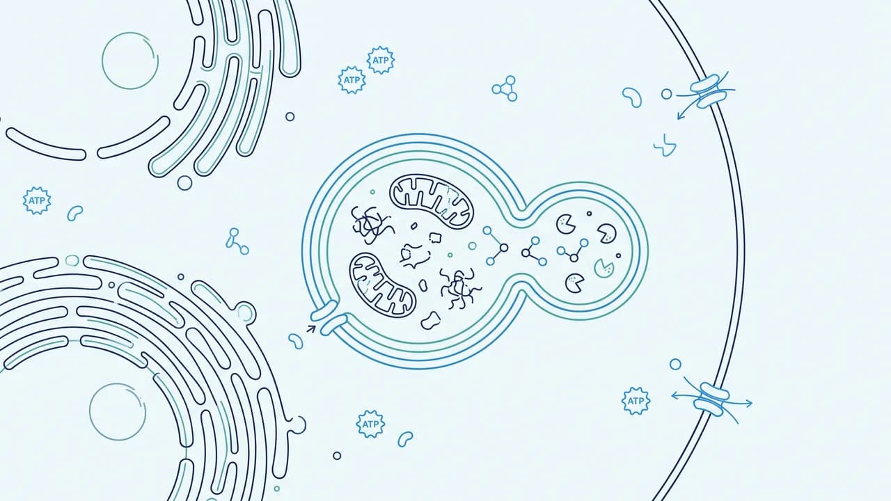
Macroautophagy sequesters cytoplasm inside a double-membrane autophagosome that later fuses with a lysosome (Mizushima and Komatsu, 2011; Galluzzi et al., 2017). It can be relatively non-selective, or it can use cargo receptors to choose mitochondria, protein aggregates, pathogens, or lipid droplets (Kirkin, McEwan, Novak, and Dikic, 2009; Kirkin and Rogov, 2019). This is the pathway almost every consumer sentence means, and the only one most LC3 blots can even pretend to see.
Microautophagy is direct lysosomal or endosomal engulfment of cytoplasm, without a separate sequestering vesicle as the defining intermediate. Oku and Sakai distinguished at least three membrane-dynamic classes in a 2018 synthesis (Oku and Sakai, 2018). Mammalian microautophagy remains thinner than the yeast literature. Treating it as “the other autophagy” without naming the membrane event is how the word becomes a synonym for any lysosomal eating.
Chaperone-mediated autophagy (CMA) is not vesicular at all. Cytosolic proteins bearing a KFERQ-like motif are delivered one-by-one to the lysosomal membrane by HSPA8/HSC70 and translocated through LAMP2A (Dice, 2007; Kaushik and Cuervo, 2018). Cuervo and Dice reported an age-related decline of CMA in rodent liver, measured as reduced lysosomal uptake of a CMA substrate and reduced LAMP2A (Cuervo and Dice, 2000). That is an animal finding, and it is strongly supported as a description of that assay in that tissue. It is not a proof that a human sixteen-hour fast restores neuronal CMA.
The three routes share a lysosome and a nutrient logic. They do not share a single switch, a single marker, or a single disease map (Galluzzi et al., 2017; Dikic and Elazar, 2018). Collapsing them into “autophagy” is the first commercial simplification this document refuses.
03 Autophagosome formation: ULK, Beclin, LC3
Macroautophagy is a construction project. The ULK1 (or ULK2) complex with ATG13 and FIP200 is a proximal kinase module. Hosokawa and colleagues showed, in cells, that mTORC1 associates with that complex when nutrients are present and that the association is nutrient-dependent (Hosokawa et al., 2009). Kim and colleagues showed that AMPK and mTOR regulate autophagy through direct and opposing phosphorylation of ULK1 (Kim, Kundu, Viollet, and Guan, 2011). Egan and colleagues connected AMPK phosphorylation of ULK1 to mitophagy under energy stress (Egan et al., 2011). Those papers are established as signalling biochemistry. They are not a human meal-timing protocol.
Downstream, the Beclin 1–VPS34 lipid-kinase complex produces phosphatidylinositol 3-phosphate on the forming membrane. Liang and colleagues identified Beclin 1 as a mammalian autophagy gene whose overexpression inhibited tumorigenesis in mice (Liang et al., 1999). He and Levine reviewed the Beclin interactome: activating and inhibitory binding partners, not a binary on-switch (He and Levine, 2010). Russell and colleagues showed that ULK1 phosphorylates Beclin 1 and thereby activates VPS34 (Russell et al., 2013). Established in cells. Not a supplement claim.
LC3 is the mammalian ATG8 that made the pathway visible. Kabeya and colleagues showed that LC3 is conjugated to phosphatidylethanolamine and localizes to autophagosome membranes after processing — the LC3-I to LC3-II conversion that every later blot copies (Kabeya et al., 2000). Mizushima, Yamamoto, Matsui, Yoshimori, and Ohsumi then placed a fluorescent LC3 reporter in mice and watched autophagosomes accumulate in tissues during nutrient starvation (Mizushima et al., 2004). That mouse is the ancestor of almost every “fasting turns on autophagy” slide. It is a mouse reporter. It is not a human brain biopsy.
Two interpretive rules follow immediately and will be enforced for the rest of the document. LC3-II can rise because more autophagosomes are being made, or because fewer are being cleared (Mizushima and Yoshimori, 2007; Klionsky et al., 2021). p62/SQSTM1 is both a selective-autophagy receptor and a substrate; its accumulation can mean failed clearance, and its disappearance can mean either successful flux or simple transcriptional change (Kirkin et al., 2009; Klionsky et al., 2021). A single snapshot decides neither.
04 The lysosome
Flux ends in the lysosome. Ballabio and Bonifacino reviewed lysosomes as dynamic regulators of cell and organismal homeostasis, not as static waste bags (Ballabio and Bonifacino, 2020). Acid hydrolases, membrane traffic, calcium, and the transcription factor TFEB sit in that review. An intervention that “induces autophagy” while impairing lysosomal function can raise LC3-II and still reduce net degradation. Chloroquine, hydroxychloroquine, and bafilomycin A1 are used in the laboratory precisely because they block the late step and thereby reveal the difference between formation and clearance (Klionsky et al., 2021; Yoshii and Mizushima, 2017; Amaravadi, Lippincott-Schwartz, Yin, et al., 2011).
This is why consumer language about “cleansing” is mechanistically backwards as often as it is merely vague. The clean-up, if the word must be used, is enzymatic digestion inside an acid organelle. The autophagosome is a delivery vehicle. Counting vehicles in a traffic jam is not proof that more cargo arrived.
05 Nutrient sensing: mTORC1 and AMPK
mTORC1 integrates amino acids, growth-factor input, and energy status and, when active, promotes anabolic work and restrains ULK-dependent autophagy (Saxton and Sabatini, 2017; Hosokawa et al., 2009; Kim et al., 2011). Wolfson and colleagues identified Sestrin2 as a leucine sensor for the mTORC1 pathway in cells (Wolfson et al., 2016). That paper is established as a leucine-sensing mechanism. It is not evidence that a human high-protein meal “shuts autophagy off for sixteen hours,” and it is not evidence that avoiding leucine at breakfast is a longevity intervention.
AMPK is the complementary energy charge sensor. Hardie’s 2005 review of the LKB1–AMPK pathway remains a compact statement of that logic (Hardie, 2005). AMPK phosphorylation of ULK1 is one of the cleanest biochemical bridges from energy stress to the autophagy machinery (Kim et al., 2011; Egan et al., 2011). Again: cells and animals. A gym session or a skipped lunch changes many hormones at once. Naming AMPK in a caption does not isolate autophagy as the cause of any human outcome.
Johnson, Rabinovitch, and Kaeberlein reviewed mTOR as a modulator of ageing and age-related disease, with rapamycin as the pharmacologic probe (Johnson, Rabinovitch, and Kaeberlein, 2013). Harrison and colleagues showed that rapamycin fed late in life extended lifespan in genetically heterogeneous mice (Harrison et al., 2009). That is an animal longevity result. It is strongly supported for those mice on that regimen. It is not a human autophagy-hour finding, and it is not a licence to treat rapamycin as a consumer autophagy activator.
06 Selective autophagy, mitophagy, lipophagy
Once receptors are admitted, “autophagy” stops meaning bulk recycling and starts meaning a set of named jobs.
Mitophagy removes mitochondria. Narendra, Tanaka, Suen, and Youle showed that Parkin is recruited selectively to impaired mitochondria and promotes their autophagy in cells (Narendra et al., 2008). Youle and Narendra reviewed the mechanisms (Youle and Narendra, 2011). Lazarou and colleagues showed that the ubiquitin kinase PINK1 recruits autophagy receptors to induce mitophagy (Lazarou et al., 2015). Pickrell and Youle placed PINK1, Parkin, and mitochondrial fidelity in the Parkinson disease argument (Pickrell and Youle, 2015). Palikaras, Lionaki, and Tavernarakis reviewed mitophagy in homeostasis and pathology (Palikaras, Lionaki, and Tavernarakis, 2018). Established as cell biology. Emerging as a quantitative explanation of any human neurodegenerative trial. Egan’s AMPK–ULK1 paper already warned that energy-stress mitophagy is a specific phospho-switch, not a general cleanse (Egan et al., 2011).
Lipophagy is autophagic consumption of lipid droplets. Singh and colleagues showed, in mice and cells, that autophagy regulates lipid metabolism and that inhibiting the pathway increased hepatic lipid content (Singh et al., 2009). Singh and Cuervo later named the connection explicitly (Singh and Cuervo, 2012). Ueno and Komatsu reviewed hepatic autophagy in health and disease (Ueno and Komatsu, 2017). Those papers make lipophagy strongly supported as a hepatic mechanism in rodents. They do not make a human fasting app a treatment for steatosis.
Selective autophagy receptors — p62/SQSTM1, NBR1, OPTN, NDP52, and others — bind cargo and LC3-family proteins (Kirkin et al., 2009; Kirkin and Rogov, 2019). The diversity of receptors is the mechanistic reason “more autophagy” is not a single dial. A cell can increase one selective route and leave another untouched.
07 Proteostasis
Autophagy is one limb of proteostasis, not the whole tree. Klaips, Jayaraj, and Hartl reviewed the pathways of cellular proteostasis in aging and disease: chaperones, the ubiquitin–proteasome system, and autophagy as interacting, not interchangeable, routes (Klaips, Jayaraj, and Hartl, 2018). Hipp, Kasturi, and Hartl reviewed the decline of that network in ageing (Hipp, Kasturi, and Hartl, 2019). Rubinsztein, Mariño, and Kroemer placed autophagy inside the aging argument without making it the only hallmarks chapter (Rubinsztein, Mariño, and Kroemer, 2011). Aman and colleagues restated the aging-and-disease map a decade later (Aman et al., 2021).
The load-bearing implication is negative as well as positive. A practice that changes one Western blot in circulating cells has not been shown to restore the proteostasis network of a seventy-year-old human neuron. Proteasome and chaperone capacity can fail while LC3-II looks busy. Busy is not cleared.
08 Age-related autophagic flux
The aging claim has a clean animal core and a muddy human edge.
Kuma and colleagues showed that autophagy is required during the early neonatal starvation period in mice: Atg5-deficient neonates die despite apparent nursing, with a metabolic failure that the paper attributes to missing autophagic amino-acid supply (Kuma et al., 2004). That is established as a developmental-starvation result in that genotype. It is not an adult intermittent-fasting result.
Hara and colleagues suppressed basal autophagy in neural cells and produced neurodegenerative disease in mice (Hara et al., 2006). Komatsu and colleagues deleted Atg7 in the central nervous system and likewise caused neurodegeneration, with ubiquitin-positive inclusions (Komatsu et al., 2006). Those two Nature papers are the strongest genetic argument that basal autophagy is required to keep a mammalian brain from accumulating junk. They are loss-of-function papers. They do not show that inducing extra autophagy in an already intact adult human extends life.
Cuervo and Dice’s CMA decline with age remains the clearest named-route aging measurement in rodent liver (Cuervo and Dice, 2000). Rubinsztein, Mariño, and Kroemer synthesized autophagy and aging across models (Rubinsztein, Mariño, and Kroemer, 2011). Madeo, Zimmermann, Maiuri, and Kroemer argued that autophagy is essential for several lifespan-extension paradigms in model organisms (Madeo, Zimmermann, Maiuri, and Kroemer, 2015). Hansen, Rubinsztein, and Walker reviewed autophagy as a promoter of longevity in those organisms; the record attached to that review in this bibliography is the 2018 publisher correction, which identifies the review and does not replace it (Hansen, Rubinsztein, and Walker, 2018). Aman and colleagues reviewed autophagy in healthy aging and disease (Aman et al., 2021).
What is strongly supported: some autophagic routes decline with age in rodent tissues; genetic loss of basal macroautophagy is catastrophic in mouse brain and muscle; several longevity interventions in invertebrates and mice require an intact autophagy machinery.
What is not established: a quantitative, tissue-resolved map of autophagic flux across human decades, measured with lysosomal blockade, that predicts a clinical aging outcome.
09 Tissue differences
Mizushima’s GFP-LC3 mouse already showed that starvation-induced autophagosome appearance is tissue-selective, not uniform (Mizushima et al., 2004). Liver, muscle, heart, and pancreas do not move together. CMA decline was measured in liver (Cuervo and Dice, 2000). Neural knockout phenotypes are brain phenotypes (Hara et al., 2006; Komatsu et al., 2006). Hepatic lipophagy is a liver argument (Singh et al., 2009; Ueno and Komatsu, 2017). Muscle autophagy is a myofibre argument (Masiero et al., 2009; Vainshtein, Grumati, Sandri, and Bonaldo, 2014).
A blood-cell LC3 change, even when real, is therefore not “body autophagy.” Jamshed and colleagues measured circulating markers during early time-restricted feeding and discussed autophagy-related transcripts among other clock and aging markers (Jamshed et al., 2019). That is a human feeding-window study. It is not a tissue-flux study. Treating a leukocyte snapshot as hepatic or neuronal autophagy is the tissue error this section exists to block.
10 Neurodegeneration
The mouse genetics are harsh and clear. Loss of basal autophagy in neurons causes degeneration and inclusion pathology (Hara et al., 2006; Komatsu et al., 2006). Nixon reviewed autophagy in neurodegenerative disease as both a clearance failure and, in some settings, a swollen, stalled pathway rather than a simple shortage (Nixon, 2013). Sarkar, Davies, Huang, Tunnacliffe, and Rubinsztein showed that trehalose enhanced autophagy independently of mTOR in cells and accelerated clearance of mutant huntingtin and α-synuclein (Sarkar et al., 2007). That is a cell enhancer paper. It is not a human trehalose-for-Huntington protocol.
PINK1/Parkin mitophagy supplies a Parkinson-facing mechanism (Narendra et al., 2008; Lazarou et al., 2015; Pickrell and Youle, 2015). The mechanism is established in cells. The step from that mechanism to a disease-modifying human intervention remains emerging at best. An intervention that raises LC3-II in a mouse cortex has not been shown, in this bibliography, to slow a human neurodegenerative endpoint.
Levine and Kroemer’s broader pathogenesis review already warned that autophagy’s role in disease is context-dependent: protective in some models, contributory in others (Levine and Kroemer, 2008). Neurodegeneration is not an exception to that warning. Stalled autophagosomes in an Alzheimer neuron can look like “more autophagy” on a snapshot and still be a failure of flux (Nixon, 2013; Klionsky et al., 2021).
11 Skeletal muscle
Masiero and colleagues showed that autophagy is required to maintain muscle mass in mice: muscle-specific Atg7 deletion produced atrophy, weakness, and accumulation of abnormal mitochondria and inclusions (Masiero et al., 2009). That is established as a loss-of-function result. It cuts in both directions. Basal autophagy protects muscle. Unrestrained autophagy can also eat muscle. “More” is not the clinical sentence.
Vainshtein, Grumati, Sandri, and Bonaldo reviewed the three-way relationship among skeletal muscle, autophagy, and physical activity (Vainshtein et al., 2014). He and colleagues showed that exercise-induced, BCL2-regulated autophagy was required for muscle glucose homeostasis in mice (He, Bassik, Moresi, et al., 2012). Lira and colleagues showed that autophagy was required for exercise-training adaptations and performance improvements in mice (Lira et al., 2013). Those are animal training papers. They are strongly supported for those genotypes and protocols. They are not a human hypertrophy programme.
Human muscle work exists and is smaller. Møller and colleagues reported that physical exercise increased autophagic signalling through ULK1 in human skeletal muscle (Møller et al., 2015). Schwalm and colleagues reported that activation of autophagy markers in human skeletal muscle depended on exercise intensity and AMPK activation (Schwalm et al., 2015). Fritzen and colleagues examined regulation of autophagy in human skeletal muscle under exercise, training, and insulin stimulation (Fritzen et al., 2016). Brandt and colleagues reported increases in autophagy markers after exercise and exercise training in human skeletal muscle (Brandt et al., 2018). Vendelbo and colleagues showed that fasting increased net phenylalanine release from human skeletal muscle and decreased mTOR signalling — a proteolysis and signalling paper, not a completed flux paper (Vendelbo et al., 2014).
Grade those human studies as emerging for signalling and marker movement, and not established for organ-level autophagic flux tied to a clinical aging endpoint. Several used static LC3/p62/ULK1 readouts. That is the measurement problem of Part Three, not a detail.
12 Liver
Singh’s lipophagy papers and Ueno and Komatsu’s hepatic review are the centre of the liver map (Singh et al., 2009; Singh and Cuervo, 2012; Ueno and Komatsu, 2017). Rodent liver is also where CMA aging was measured (Cuervo and Dice, 2000) and where GFP-LC3 starvation signals are often obvious (Mizushima et al., 2004). Human liver flux under a named fasting protocol is essentially absent from this bibliography. Ketone production during starvation is a hepatic metabolic fact (Cahill, 2006; Rothman, Magnusson, Katz, Shulman, and Shulman, 1991). It is established as fuel biochemistry. It is not an autophagy assay.
13 Immune system
Deretic, Saitoh, and Akira reviewed autophagy in infection, inflammation, and immunity (Deretic, Saitoh, and Akira, 2013). The pathway captures pathogens, presents antigens, and modulates inflammasomes. That is strongly supported as immunology. It is also the first place “more autophagy is always better” dies on contact with infection biology: a cell that cannot restrain the pathway can die, and a pathogen that hijacks the pathway can eat the host’s logistics. Aging of the immune system is not graded here as an autophagy trial outcome. No human immunosenescence endpoint in this bibliography is carried by a validated flux measurement.
14 Metabolic health
He and colleagues’ mouse exercise-autophagy paper tied the pathway to muscle glucose homeostasis (He et al., 2012). Singh’s lipophagy paper tied it to hepatic lipid (Singh et al., 2009). Saxton and Sabatini’s mTOR review and Wolfson’s leucine sensor sit under every fed-state metabolic cartoon (Saxton and Sabatini, 2017; Wolfson et al., 2016). Human metabolic outcomes of fasting and caloric restriction — weight, energy expenditure, oxidative-damage markers — have their own trial literature (Heilbronn et al., 2005; Redman et al., 2018; Most, Gilmore, Smith, Han, Ravussin, and Redman, 2018; de Cabo and Mattson, 2019). Those outcomes are not autophagy measurements.
The CALERIE-adjacent metabolic-slowing paper of Redman and colleagues is a human caloric-restriction physiology result (Redman et al., 2018). It is strongly supported for the endpoints it measured. It does not report tissue autophagic flux. Using it as proof that restriction “turned on autophagy” is citation laundering.
15 Fasting, intermittent fasting, caloric restriction
Starvation physiology in humans is real and old. Rothman and colleagues quantified hepatic glycogenolysis and gluconeogenesis in fasting humans with 13C NMR (Rothman et al., 1991). Cahill reviewed fuel metabolism in starvation (Cahill, 2006). Those papers are established as human metabolic description. Neither is an LC3-flux paper.
In mice, short-term fasting induces profound neuronal autophagy on a GFP-LC3 and electron-microscopy readout (Alirezaei et al., 2010). In the same species, whole-body starvation induces tissue-selective autophagosome appearance (Mizushima et al., 2004). Bagherniya, Butler, Barreto, and Sahebkar reviewed fasting or calorie restriction and autophagy induction and, honestly, found a literature dominated by cells and animals (Bagherniya et al., 2018).
Human intermittent fasting has a clinical-outcome literature that this series already graded in the sibling Fasting article. de Cabo and Mattson reviewed effects of intermittent fasting on health, aging, and disease for a general medical audience (de Cabo and Mattson, 2019). Heilbronn, Smith, Martin, Anton, and Ravussin tested alternate-day fasting in nonobese subjects on weight and energy metabolism (Heilbronn et al., 2005). Jamshed and colleagues tested early time-restricted feeding on 24-hour glucose and on circulating markers they related to clock, aging, and autophagy (Jamshed et al., 2019). Vendelbo and colleagues fasted humans and measured muscle amino-acid balance and mTOR signalling (Vendelbo et al., 2014).
None of those human papers reports organ autophagic flux with a lysosomal block in brain, liver, or muscle as a primary endpoint tied to a clinical event. Jamshed’s autophagy-related blood markers are the closest approach in this set. They are emerging as circulating molecular responses to an early eating window. They are speculative as proof that a named hour “switched on” tissue autophagy.
Caloric restriction without a named window has a cleaner model-organism longevity record and a thinner human flux record. Redman and Most supply human CR physiology without autophagy-flux endpoints (Redman et al., 2018; Most et al., 2018). Madeo’s claim that autophagy is essential for lifespan extension is a model-organism synthesis (Madeo et al., 2015). Crossing that synthesis with a human 16:8 brochure is the species error of §24.
Established: nutrient withdrawal induces autophagy in yeast, cells, and mice, including mouse neurons. Not established: a specific human fasting duration that turns autophagy on in a named human organ as a proven aging mechanism.
16 Exercise
The mouse requirement data are stronger than the human flux data. Exercise-induced BCL2-regulated autophagy was required for muscle glucose homeostasis in mice (He et al., 2012). Training-induced adaptation required autophagy in mice (Lira et al., 2013). Human studies show ULK1 signalling and autophagy-marker changes after acute exercise and some training, with intensity dependence in Schwalm’s hands (Møller et al., 2015; Schwalm et al., 2015; Fritzen et al., 2016; Brandt et al., 2018). Vainshtein and colleagues reviewed the triangle (Vainshtein et al., 2014).
Grade: strongly supported that exercise moves autophagy-related signalling and some static markers in human muscle; established that the pathway is required for selected metabolic and training phenotypes in mice; not established that exercise “turns on autophagy” in humans as a longevity mechanism, or that any marker change is the cause of the human outcomes exercise already owns in the sibling Exercise Intervention article.
17 Pharmacologic manipulation
Rapamycin extends lifespan in heterogeneous mice when fed late in life (Harrison et al., 2009) and sits inside the mTOR-ageing review (Johnson et al., 2013). That is strongly supported mouse longevity. Human rapamycin use is a transplant and, increasingly, a geroscience question that this title does not try. It is not graded here as a proven human autophagy-flux therapy.
Shoji-Kawata and colleagues identified a Tat-Beclin 1 peptide that induced autophagy in cells and mice (Shoji-Kawata et al., 2013). That is a research tool and a proof of pharmacologic inducibility. It is not a consumer product, and no dose is a recommendation.
Amaravadi and colleagues reviewed targeting autophagy for cancer treatment, including lysosomal-blocking antimalarials as inhibitors, not activators (Amaravadi et al., 2011). White reviewed the context-dependent role of autophagy in cancer: tumour-suppressive in some initiation settings, tumour-promoting in some established cancers (White, 2012). Galluzzi, Pietrocola, Bravo-San Pedro, et al. mapped autophagy in malignant transformation and progression (Galluzzi et al., 2015). Pharmacologic “activation” is therefore not a virtue word. In oncology it is often the opposite word.
18 Dietary compounds
Eisenberg and colleagues showed that spermidine induced autophagy and promoted longevity in model organisms (Eisenberg et al., 2009). Madeo, Eisenberg, Pietrocola, and Kroemer reviewed spermidine in health and disease (Madeo, Eisenberg, Pietrocola, and Kroemer, 2018). Morselli and colleagues linked caloric restriction and resveratrol to Sirtuin-1-dependent autophagy induction in cells and model organisms (Morselli et al., 2010). Sarkar’s trehalose paper is a cell-clearance enhancer (Sarkar et al., 2007).
Ryu and colleagues showed that urolithin A induced mitophagy and prolonged lifespan in C. elegans and increased muscle function in rodents (Ryu et al., 2016). Andreux and colleagues then reported that urolithin A was safe in humans and induced a molecular signature of improved mitochondrial and cellular health — a human molecular-signature trial, not a flux-in-muscle-under-blockade trial and not a mortality trial (Andreux et al., 2019).
Grade: established that several compounds move autophagy or mitophagy readouts in cells and invertebrates; emerging that urolithin A changes human molecular signatures; speculative that any of these is a human aging therapy because it “activates autophagy.” Bagherniya’s review of fasting and restriction already showed how easily a cell result is written as if it were a diet (Bagherniya et al., 2018). The same laundering applies to powders.
19 Why flux is hard to measure in living humans
This section is the methodological spine. The commercial literature treats it as a footnote. It is not.
Klionsky’s consensus guidelines — third and fourth editions — are the field’s refusal letter to naive blots (Klionsky et al., 2016; Klionsky et al., 2021). Mizushima and Yoshimori explained how to interpret LC3 immunoblotting: LC3-II must be read with a lysosomal inhibitor if flux is the claim (Mizushima and Yoshimori, 2007). Yoshii and Mizushima reviewed monitoring methods, including reporters that distinguish cytosolic LC3 from autophagosome-bound LC3 and assays that require a late-stage block (Yoshii and Mizushima, 2017). Loos, du Toit, and Hofmeyr defined autophagosome flux as a rate, not a level, and named the gap between the concept and what most papers plot (Loos, du Toit, and Hofmeyr, 2014).
A living human does not tolerate the standard block. Bafilomycin, high-dose chloroquine, and genetic reporter alleles are not routine clinical tools. Muscle biopsy is possible and has been used for signalling studies (Møller et al., 2015; Schwalm et al., 2015; Fritzen et al., 2016; Brandt et al., 2018; Vendelbo et al., 2014). Brain biopsy for an autophagy-hour study is not an ethics committee’s idea of a weekend protocol. Plasma ketones, leukocyte transcripts, and LC3 in circulating cells are accessible. They are also the wrong compartment for the claims people buy.
Even in muscle, most human papers report static markers or phosphorylation, not a paired-with-blockade flux. Without that pair, LC3-II up after exercise is compatible with more formation, less fusion, or both (Mizushima and Yoshimori, 2007; Klionsky et al., 2021). That is not pedantry. It is the difference between a cleared mitochondrion and a parking lot of undegraded autophagosomes — the difference Nixon already flagged in neurodegenerative tissue (Nixon, 2013).
| Assay | What it can show | What it cannot show | Typical setting |
|---|---|---|---|
| LC3-I/II blot, no inhibitor | A change in lipidated LC3 | Flux; formation versus stalled clearance | Cells, tissues, some human biopsies |
| LC3-II ± lysosomal block | Autophagic flux as the inhibitor-sensitive increment | Organ-wide human flux; a consumer hour-count | Cells; rare human designs |
| p62/SQSTM1 level | Compatible with failed clearance if it accumulates | Transcriptional confounds; flux alone | Cells and tissues |
| GFP-LC3 / reporter mice | Autophagosome appearance in named tissues | Human organs; net degradation without further tools | Mice (Mizushima et al., 2004; Alirezaei et al., 2010) |
| Electron microscopy | Double-membrane vesicles | Rate; identity of every cargo | Cells, animals, rare human tissue |
| Blood-cell LC3 or transcripts | A circulating molecular change | Brain, liver, or muscle flux | Human feeding trials (Jamshed et al., 2019) |
| Ketones / NMR glycogen | Starvation fuel use | Autophagy | Human fasting physiology (Rothman et al., 1991; Cahill, 2006) |
| ULK1 / AMPK phosphosites | Energy-stress signalling | Completed lysosomal degradation | Human muscle after exercise (Møller et al., 2015; Schwalm et al., 2015) |
20 “Autophagy equals cellular cleansing”
Cleansing is a household metaphor. Macroautophagy is vesicle construction, trafficking, and enzymatic digestion. Microautophagy is membrane engulfment. CMA is motif-guided translocation (Galluzzi et al., 2017; Dice, 2007; Oku and Sakai, 2018). Selective receptors choose cargo (Kirkin and Rogov, 2019). The lysosome can fail while vesicles pile up (Ballabio and Bonifacino, 2020; Nixon, 2013). Cancer cells can use the same pathway as a survival kit (White, 2012; Galluzzi et al., 2015). Immune cells use it against microbes and can be harmed by its excess (Deretic, Saitoh, and Akira, 2013).
The metaphor erases selectivity, context, and failure modes. It also implies a dirty cell that becomes clean, which is not a measured variable in any paper in this bibliography. Speculative as science; useful only as a warning label on marketing.
21 Fixed fasting-hour switches
No paper in this set calibrates a human clock hour at which autophagy in a named organ switches from off to on. Alirezaei’s neuronal induction is a mouse fast (Alirezaei et al., 2010). Mizushima’s reporter is a mouse (Mizushima et al., 2004). Rothman’s glycogen map is human fuel, not LC3 (Rothman et al., 1991). Jamshed’s early window is a human glycaemic and circulating-marker study (Jamshed et al., 2019). Wolfson’s leucine sensor has no hour constant (Wolfson et al., 2016). Hardie’s AMPK review has no sixteen (Hardie, 2005).
Hours in human protocols are operational: sleep, work, adherence, clinic visits. They are not receptor timeouts. The sibling Fasting red-team already dropped the 16-hour switch. This title restates the drop with the assay literature attached.
22 LC3 and p62 misread
Kabeya made LC3-II famous (Kabeya et al., 2000). Mizushima and Yoshimori then spent a methods paper telling the field not to treat the band as a scoreboard (Mizushima and Yoshimori, 2007). The guidelines repeated the warning for a decade (Klionsky et al., 2016; Klionsky et al., 2021). p62 is a receptor and a substrate (Kirkin et al., 2009). Rising p62 can mean jammed flux. Falling p62 can mean flux or less transcription. Publishing one without the inhibitor pair, then captioning “autophagy activated,” is the error the guidelines exist to prevent.
23 Static markers versus flux
Loos named the concept–reality gap (Loos, du Toit, and Hofmeyr, 2014). Yoshii and Mizushima catalogued the tools that close it (Yoshii and Mizushima, 2017). Human exercise papers in this set mostly report markers and signalling (Møller et al., 2015; Schwalm et al., 2015; Fritzen et al., 2016; Brandt et al., 2018). Those data are real. They are not flux. A commercial sentence that upgrades a marker to a clearance event is a category error, not a simplification.
24 Model-organism longevity extrapolated
Spermidine extends life in model systems via autophagy (Eisenberg et al., 2009). Urolithin A extends C. elegans life and helps rodent muscle (Ryu et al., 2016). Rapamycin extends heterogeneous-mouse life (Harrison et al., 2009). Autophagy is required for several longevity paradigms (Madeo et al., 2015). Hansen, Rubinsztein, and Walker reviewed the model-organism case (Hansen, Rubinsztein, and Walker, 2018). None of that is a human mortality trial of “autophagy activation.” de Cabo and Mattson’s intermittent-fasting review is explicit that human aging and disease claims outrun the human outcome data (de Cabo and Mattson, 2019). Species translation is a table, not a slogan.
| Object | Where it is established | What does not follow |
|---|---|---|
| Starvation-induced autophagosomes | Yeast, cells, GFP-LC3 mice, mouse neurons | A human 16:8 neuronal cleanse |
| Basal autophagy required in brain and muscle | Mouse Atg5/Atg7 knockouts | That inducing extra autophagy helps intact humans |
| CMA decline with age | Rodent liver assays | That human TRE restores CMA |
| Rapamycin longevity | Heterogeneous mice | A human autophagy-hour drug |
| Spermidine longevity | Model organisms | A human supplement outcome |
| Urolithin A mitophagy | Worms, rodents; human molecular signature | Human lifespan or disease-event benefit |
| Exercise autophagy markers | Human muscle signalling papers | Longevity causation via autophagy |
| Human CR metabolic slowing | CALERIE-adjacent physiology | Measured tissue flux |
25 The assumption that more is always better
Masiero’s muscle knockout shows that too little autophagy atrophies muscle (Masiero et al., 2009). The same pathway, unrestrained, is a wasting mechanism the muscle field has never treated as an unalloyed good (Vainshtein et al., 2014). White and Galluzzi show that established tumours can require autophagy (White, 2012; Galluzzi et al., 2015). Amaravadi’s oncology review treats inhibition as the therapeutic direction in several settings (Amaravadi et al., 2011). Deretic’s immunity review is full of contexts in which the pathway must be timed, not maximised (Deretic, Saitoh, and Akira, 2013). Nixon’s degenerating neuron can be full of autophagosomes that are not finishing the job (Nixon, 2013). Levine and Kroemer said “pathogenesis,” not “panacea,” in the title (Levine and Kroemer, 2008).
More autophagy is not a monotonic health function. The honest sentence is that the right amount, in the right cargo route, in the right cell, at the right time, is the variable — and that variable is not what a brochure sells.
26 The strongest case against commercial fasting/autophagy rhetoric
The strongest case is not that autophagy is fake. The strongest case is that the rhetoric commits six substitutions, each of which is individually forbidden by the papers it pretends to cite.
It substitutes one word for three pathways (Galluzzi et al., 2017; Dice, 2007; Oku and Sakai, 2018). It substitutes a snapshot for flux (Mizushima and Yoshimori, 2007; Klionsky et al., 2021; Loos, du Toit, and Hofmeyr, 2014). It substitutes a mouse neuron for a human organ (Alirezaei et al., 2010; Mizushima et al., 2004). It substitutes ketones or a clock hour for a lysosomal-block assay (Cahill, 2006; Rothman et al., 1991; Jamshed et al., 2019). It substitutes invertebrate or mouse lifespan for a human outcome (Eisenberg et al., 2009; Harrison et al., 2009; Madeo et al., 2015; Ryu et al., 2016). It substitutes more for appropriate, against the cancer, immunity, and muscle-wasting records (White, 2012; Deretic, Saitoh, and Akira, 2013; Masiero et al., 2009).
After those substitutions are refused, what remains of the commercial sentence is a nutrient-sensing pathway that is real in cells and animals, a human fasting and exercise literature that can stand on metabolic and functional endpoints without autophagy as a mascot, and a measurement problem that living humans have not solved. That is not a small remainder. It is also not what was advertised.
| Human evidence | Design | Autophagy readout | Outcome readout | Grade for “autophagy as the mechanism” |
|---|---|---|---|---|
| Rothman et al., 1991 | Fasting NMR | None | Hepatic glycogenolysis / gluconeogenesis | Not an autophagy paper |
| Heilbronn et al., 2005 | Alternate-day fasting | None | Weight, energy metabolism | Not an autophagy paper |
| Vendelbo et al., 2014 | Human fast, muscle A-V balance | mTOR signalling, not flux | Phenylalanine release | Signalling only |
| Jamshed et al., 2019 | Early TRE | Circulating autophagy-related markers | 24-hour glucose | Emerging markers; not organ flux |
| Møller et al., 2015 | Acute exercise, muscle biopsy | ULK1 signalling | None clinical | Emerging signalling |
| Schwalm et al., 2015 | Exercise intensity, muscle | Autophagy markers, AMPK | None clinical | Emerging markers |
| Fritzen et al., 2016 | Exercise / insulin, muscle | Autophagy regulation | None clinical | Emerging regulation |
| Brandt et al., 2018 | Exercise and training, muscle | Autophagy markers | None clinical | Emerging markers |
| Redman et al., 2018 | Human CR | None | Metabolic slowing, oxidative-damage markers | Not an autophagy paper |
| Andreux et al., 2019 | Urolithin A, humans | Mitochondrial molecular signature | Safety, signatures | Emerging mitophagy-adjacent signature |
| Fasting or exercise study | Species | Intervention as reported | Autophagy claim the paper can carry | Claim it cannot carry |
|---|---|---|---|---|
| Mizushima et al., 2004 | Mouse | Nutrient starvation | Tissue-selective reporter autophagosomes | Human hour-switch |
| Alirezaei et al., 2010 | Mouse | Short-term fast | Neuronal autophagy on reporter/EM | Human 16:8 neuron cleanse |
| Kuma et al., 2004 | Mouse | Neonatal starvation, Atg5 null | Autophagy required neonatally | Adult TRE |
| He et al., 2012 | Mouse | Exercise | BCL2-regulated autophagy needed for glucose homeostasis | Human longevity |
| Lira et al., 2013 | Mouse | Exercise training | Autophagy required for adaptation | Human marker equals adaptation |
| Møller / Schwalm / Fritzen / Brandt | Human | Acute or training exercise | Signalling and static markers in muscle | Flux; aging events |
| Jamshed et al., 2019 | Human | Early TRE | Glucose window; circulating markers | Organ flux |
| Heilbronn / Redman / Most | Human | ADF or CR | Weight and metabolic physiology | Autophagy mechanism |
| de Cabo and Mattson, 2019 | Review | IF family | Narrative of health claims | A calibrated human autophagy dose |
27 The honest remainder
Autophagy is one of the better-understood recycling systems in cell biology. The ULK, Beclin, LC3, mTORC1, and AMPK logic is established. Selective routes, including mitophagy and lipophagy, are established as mechanisms and emerging as quantitative human explanations. Age-related decline of some routes is strongly supported in rodents. Basal autophagy is established as necessary for mouse brain and muscle integrity.
Human fasting and exercise do not need a fake autophagy switch to be interesting. They have fuel maps, glycaemic trials, training adaptations, and safety problems of their own. Those objects should be argued in their own units. Autophagy may participate. Participation is a hypothesis. It is not a caption on a sixteen-hour graphic.
28 Contexts in which autophagy is not a virtue
Do not induce for its own sake in a document that cannot recommend induction. The scientific point, not a prescription, is that context can invert the sign. Established tumours may depend on the pathway (White, 2012; Galluzzi et al., 2015; Amaravadi et al., 2011). Infection and inflammation require timing (Deretic, Saitoh, and Akira, 2013). Muscle needs basal autophagy and can be damaged by excess (Masiero et al., 2009). Degenerating neurons may already be full of unfinished autophagosomes (Nixon, 2013). A peptide that forces the pathway exists as a research tool (Shoji-Kawata et al., 2013). Tools are not virtues.
Standing constraint This document describes published research. It is not medical advice. No human use, dose, route or schedule of fasting, exercise, rapamycin, spermidine, urolithin A, trehalose, chloroquine, or any other intervention is recommended anywhere in this document.
References
Verified NCBI records, sorted by first author, replace this stub at build.
Evidence handling
Study type is labelled in the reporting sentence. Animal and in-vitro results are never phrased as human outcomes. Static LC3, p62, ULK1 phosphosites, circulating transcripts, and ketone concentrations are treated as different objects from autophagic flux. When a mouse reporter and a human feeding trial are both cited, the species and the assay stay in the sentence. A newer methods guideline supersedes a naive blot reading; it does not delete the blot’s existence. Project 06 was queried read-only for orientation and is not a human flux corpus. Project 05 remains not imported and is not this title. Local Firecrawl was used only to retrieve open full text already identified by PMID.
Limitations and sibling titles
This title does not re-try the matched-calorie intermittent-fasting outcome record; that lives in Fasting and Intermittent Fasting (SBL-41/SP-FASTING). It does not re-try exercise as a clinical intervention; that lives in Exercise Intervention (SBL-41/SP-EXERCISE-INTERVENTION). It is a South Beach Longevity science article, not a Radix peptide title. Its two figures are original schematics, captioned as projected; no commissioned or third-party plate is used.
Coverage is limited to the 79 MEDLINE records that survived title-checked NCBI resolution. Recalled PMIDs that mapped to unrelated papers were discarded before drafting. The Hansen, Rubinsztein, and Walker 2018 review is cited from the publisher-correction record that identifies it. Human flux-with-blockade studies, if they exist outside this set, are a coverage gap and are not invented.
Glossary
Autophagic flux. The rate of autophagosome (or CMA substrate) delivery to, and degradation in, the lysosome; measured as the increment caused by a late-stage block.
CMA. Chaperone-mediated autophagy; KFERQ-bearing proteins translocated via LAMP2A.
LC3-II. Lipidated LC3 on autophagosome membranes; a marker that rises with formation or with failed clearance.
Macroautophagy. Vesicular sequestration of cytoplasm for lysosomal digestion.
Microautophagy. Direct lysosomal or endosomal engulfment without a separate autophagosome as the defining step.
Mitophagy. Selective autophagy of mitochondria.
p62/SQSTM1. Selective-autophagy receptor and substrate; not a stand-alone flux meter.
Static marker. A level at one time point, without a blockade pair.
Continue reading