
What Is Cellular Senescence?
A cell that stops dividing but refuses to die — protective early in life, and a driver of ageing when these cells accumulate and their inflammatory signals turn chronic.
Abstract
Cellular senescence is a stress-induced, essentially stable cell-cycle arrest — a distinct cell state rather than simply an old cell — in which the cell stops dividing, resists apoptosis, and adopts a pro-inflammatory secretory program (the SASP). It is triggered by telomere attrition, oncogene activation, or DNA-damaging stress, and identified by a panel of markers, no one of which is definitive. Senescence is beneficial in the short term — it suppresses tumours, aids wound healing, and shapes embryonic development, after which senescent cells are normally cleared — but harmful when clearance fails and the cells accumulate with age, sustaining chronic inflammation. In mice, genetically or pharmacologically clearing senescent cells delays age-related disease and extends median lifespan; in humans, senolytic testing so far is limited to small, uncontrolled, open-label pilots that show target engagement and a preliminary functional signal, not proven clinical benefit. No senolytic is approved for ageing or any senescence-driven disease.
Key findings
- Cellular senescence is a stress-induced, essentially stable cell-cycle arrest — a distinct cell state, not merely an old cell — in which the cell stops dividing yet actively resists programmed death (Gorgoulis et al., 2019; Zhu et al., 2015).
- It is triggered by telomere shortening, an activated oncogene, or DNA-damaging stress, and is recognised by a panel of markers (SA-β-galactosidase, p16, p21, DNA-damage foci) — no single one of which is definitive (Serrano et al., 1997; Gorgoulis et al., 2019).
- Senescent cells secrete a pro-inflammatory mixture, the SASP, that can push neighbouring cells into senescence — shown in cell and animal models (Coppé et al., 2008; Acosta et al., 2013).
- Senescence has two faces: it suppresses tumours, aids wound healing, and shapes the embryo early in life, but drives inflammation and dysfunction when senescent cells accumulate with age (Muñoz-Espín & Serrano, 2014).
- In mice, genetically clearing senescent cells delayed age-related disease and extended median lifespan; the same causal chain is not yet demonstrated in humans (Baker et al., 2011; Baker et al., 2016).
- Senolytic drugs clear senescent cells in mice, but human testing remains limited to small, uncontrolled, open-label pilots, and no senolytic is approved for ageing or any senescence-driven disease (Justice et al., 2019; Hickson et al., 2019).
A senescent cell is one that has stopped dividing for good but has not died. Something has damaged it or pushed it to a limit — worn-down telomeres, an activated cancer gene, a dose of DNA damage — and instead of repairing itself or self-destructing, it settles into a distinct, lasting state: it withdraws from the cell cycle, resists the usual signals for programmed death, and stays alive and metabolically active. Biologists call this state cellular senescence, and the operative word is state, not age. A senescent cell is not simply an old cell; it is a cell that has thrown a switch (Gorgoulis et al., 2019).
That switch is one of biology's genuinely double-edged tools. In the short term, and early in life, senescence protects: it stops damaged cells from turning cancerous, helps wounds close, and even helps shape the developing embryo, after which the immune system clears the senescent cells away. The trouble comes later, when clearance falters and senescent cells accumulate — because a senescent cell is not quiet. It secretes inflammatory molecules that can inflame the surrounding tissue and, in animal models, drive the dysfunction we recognise as ageing.
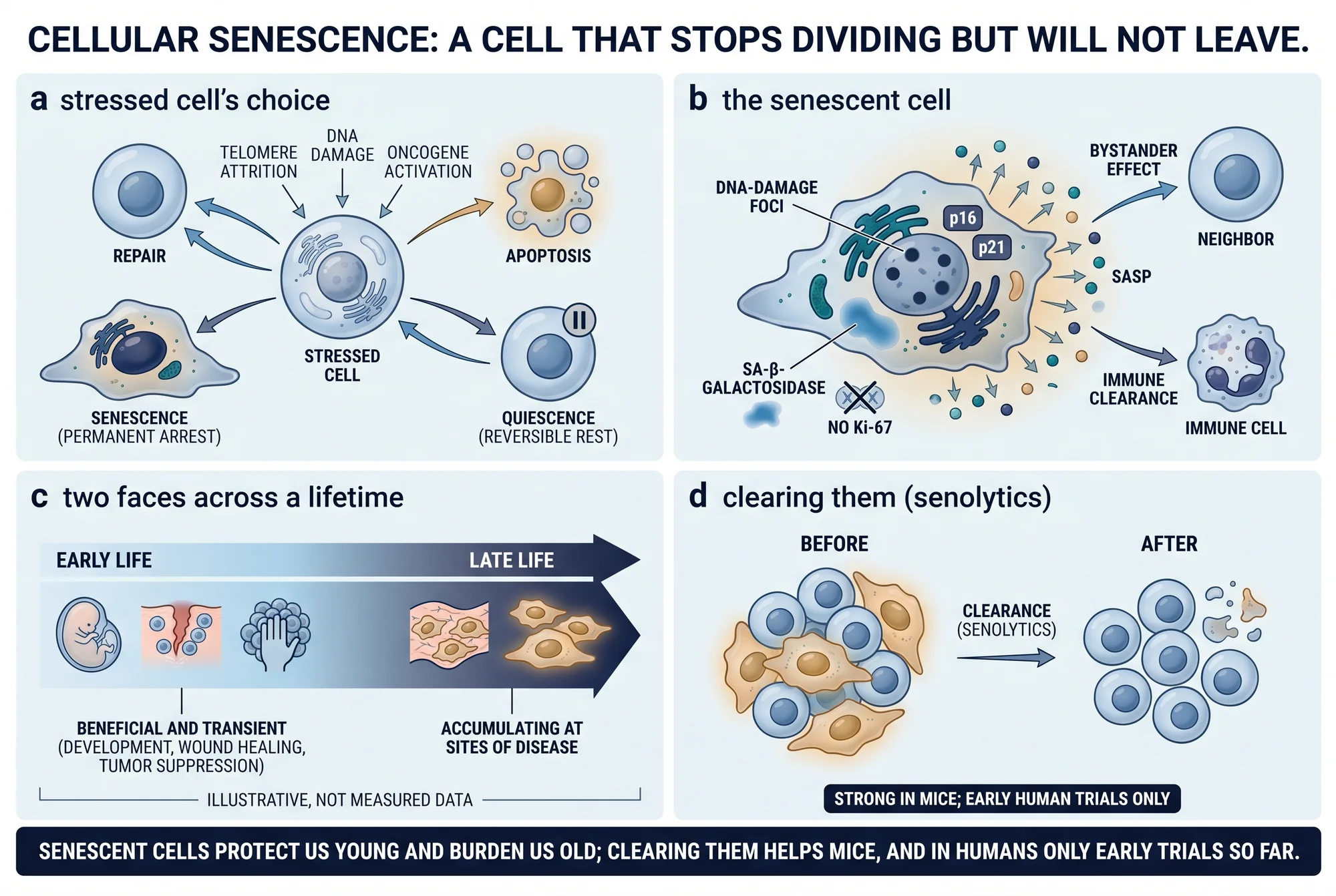
What a senescent cell actually is
The defining feature of senescence is a cell-cycle arrest that does not reverse under ordinary conditions. When a healthy cell is merely resting — paused for lack of growth signals — it can re-enter the cycle and divide again; that reversible pause is called quiescence. A senescent cell is different: given the same growth signals, it stays put. The classic senescence stain, SA-β-galactosidase, marks senescent human fibroblasts but is absent from quiescent ones and from fully differentiated cells (Dimri et al., 1995), which is one way researchers separate a genuinely senescent cell from a merely idle one.
Two further properties complete the picture. First, the arrest is durable — reviews describe it as essentially stable and self-sustaining rather than a temporary stall (Gorgoulis et al., 2019; Hernández-Segura et al., 2018) — though it is better called very stable than absolutely irreversible, because some experimental settings can coax cells back out of it. Second, and central to everything that follows, senescent cells actively resist apoptosis, the body's program for orderly cell death. They keep pro-survival machinery switched on (Zhu et al., 2015, working in human cells and mice), which is exactly why they persist instead of being quietly disposed of — and, later, why a class of drugs tries to force them to die.
What tips a cell into senescence
Senescence is a response to stress, and biologists have mapped three main roads into it.
The first was found in a culture dish more than sixty years ago. Normal human cells do not divide indefinitely; they multiply a finite number of times — a few dozen divisions for many cell types — and then stop (Hayflick & Moorhead, 1961; Hayflick, 1965). The explanation arrived decades later. Each division shortens the telomeres, the protective caps on the ends of chromosomes; telomeres were shown to shorten as human fibroblasts aged in culture (Harley et al., 1990), though at the time it was unclear whether the shortening caused the arrest or merely accompanied it. The causal link came when researchers supplied telomerase, the enzyme that rebuilds telomeres, to normal human cells: the cells lengthened their telomeres and kept dividing well past their usual limit — by at least twenty divisions — with normal chromosomes (Bodnar et al., 1998). Mechanistically, a critically short telomere is read by the cell as a broken chromosome, so telomere-triggered senescence sets off the same DNA-damage alarm a double-strand break would (d'Adda di Fagagna et al., 2003, in human cells). This is replicative senescence.
The second road is oncogene-induced senescence, and it is a piece of built-in cancer defence. When an activated cancer gene — the founding study used oncogenic ras — is switched on in a normal cell, the cell does not race into malignant growth; it slams into a permanent arrest instead, accumulating the tumour-suppressor proteins p53 and p16 and becoming, in the researchers' description, indistinguishable from a senescent cell (Serrano et al., 1997, in human and rodent cells). Senescence here is not a tally of past divisions but an emergency response to a dangerous signal.
The third road is straightforward damage. Enough genotoxic or oxidative stress — radiation, reactive oxygen species, the byproducts of failing mitochondria, cancer therapies — will drive a cell into senescence regardless of telomere length (Coppé et al., 2008; reviewed by Hernández-Segura et al., 2018 and Di Micco et al., 2021).
Recognising one: markers, and why no single one is enough
There is no single test that says "this cell is senescent," and stating that plainly is part of the science. Researchers instead look for a combination of signs. SA-β-galactosidase, detectable as a blue stain, was the first widely used marker and remains the most common; it also rises in human skin as donors age — the earliest direct evidence that senescent cells accumulate in people over a lifetime (Dimri et al., 1995). Two cell-cycle-inhibitor proteins, p16 and p21, enforce and mark the arrest; p16 in particular is the flag researchers would later use to tag and remove senescent cells in mice (Serrano et al., 1997; Baker et al., 2011). Persistent DNA-damage foci signal ongoing alarm, and the absence of proliferation markers such as Ki-67 confirms the cell truly is not dividing (d'Adda di Fagagna et al., 2003; Hernández-Segura et al., 2018).
The catch is that each of these signs turns up in some non-senescent cells too, so no marker on its own is sufficient. The international consensus on senescence is explicit that identification requires several signals used together, and that detecting senescent cells reliably inside living tissue is still an unsolved methods problem (Gorgoulis et al., 2019; Sharpless & Sherr, 2015).
The senescent cell is not silent: the SASP
If senescence were only a silent brake on one cell, it would matter far less. What makes it consequential for a whole tissue is that senescent cells secrete. They release a complex mixture of inflammatory signalling proteins — cytokines such as interleukin-6 and interleukin-8, chemokines, growth factors, and tissue-remodelling enzymes — known collectively as the senescence-associated secretory phenotype, or SASP (Coppé et al., 2008, in human cells and tumour tissue). The SASP is not an instant switch; it builds over several days after damage severe enough to trigger senescence, and its recipe varies with the trigger and the cell type rather than being one fixed formula.
The SASP is also why senescence can spread. The same secretions that summon immune cells can push healthy neighbours into senescence — so-called bystander, or paracrine, senescence — an effect shown both in culture and in animal and human tissue models (Acosta et al., 2013). Transplant senescent cells into a young mouse and senescence spreads into the animal's own tissues (Xu et al., 2018, in mice). This dual character — helpful when brief and local, harmful when chronic and widespread — is what one review called the dark side of tumour suppression (Coppé et al., 2010).
The useful face: cancer defence, wound healing, development
The reason evolution kept a program this costly is that, used briefly, it is valuable. Its oldest job is tumour suppression: by arresting a cell on the verge of malignant transformation, senescence removes it from the pool that could become cancer (Serrano et al., 1997). In mice, restoring the tumour-suppressor p53 to liver cancers pushed the cancer cells into senescence and, together with an immune response, cleared the tumours (Xue et al., 2007, in mice).
Senescence also helps repair tissue. In a healing skin wound, senescent cells appear early and speed the wound's closure by secreting a growth factor, PDGF-AA; remove them and closure is delayed, an effect reversed by supplying the growth factor back (Demaria et al., 2014, in mice).
Most striking, senescence is part of normal development. During mammalian embryonic development, cells become senescent on a set schedule as a sculpting mechanism — a form that depends on p21 but, unlike the stress-induced kind, needs neither p53 nor DNA damage — and are then cleared by immune cells as the tissue is remodelled; the same pattern appears in human embryonic tissue (Muñoz-Espín et al., 2013; Storer et al., 2013, in mouse and chick embryos). The unifying theme across all three roles is timing: senescence, prompt immune clearance, and regeneration form a normal, transient sequence (Muñoz-Espín & Serrano, 2014). The damage begins when that sequence breaks down.
The harmful face: when senescent cells accumulate
When immune clearance wanes with age, senescent cells stop being a passing phase and start to accumulate in tissues and organs (van Deursen, 2014). They build up slowly — in mice, lineage tracing shows this creeping in around ten to twelve months of age, concentrated in the endothelial cells lining the liver's small vessels (Grosse et al., 2020) — and they collect at sites of disease. In mice, senescent foam cells inside arterial plaques are harmful at every stage of atherosclerosis (Childs et al., 2016).
The pivotal question is whether accumulated senescent cells cause age-related decline or merely mark it, and the strongest answers come from mice. Using a genetic switch that selectively kills p16-positive senescent cells, researchers showed that clearing them throughout life delayed the onset of age-related deterioration in fat, muscle, and eye in a fast-ageing (progeroid) mouse strain, and that clearing them late in life slowed problems already underway — evidence that senescence is causally involved in producing age-related changes, not just associated with them (Baker et al., 2011, in progeroid mice). The same tool was then applied to ordinary, naturally ageing mice: clearing senescent cells from one year of age extended their median lifespan and eased the deterioration of the kidney, heart, and fat (Baker et al., 2016, in mice). A complementary experiment ran the logic in reverse — transplanting senescent cells into young mice caused lasting physical dysfunction and shortened survival, while an oral senolytic drug combination improved function and increased post-treatment survival by 36% in naturally aged mice (Xu et al., 2018, in mice).
Every one of those lifespan and healthspan results is a mouse result. Together they show that senescent cells can cause age-related dysfunction in animals, and make it plausible the same holds in humans — but plausibility is not proof, and the human evidence is thinner.
Clearing them out: senolytics
The discovery that senescent cells survive by keeping their self-destruct programs switched off suggested an obvious strategy: switch those programs back on. Drugs that do this — selectively killing senescent cells while sparing healthy ones — are called senolytics. The first were identified by mapping the survival networks senescent cells depend on: the cancer drug dasatinib killed senescent human fat-cell progenitors, the plant flavonoid quercetin was more effective against senescent human endothelial cells, and the two together reduced the senescent-cell burden in aged, irradiated, and progeroid mice, improving heart and blood-vessel function in old mice within five days of a single dose (Zhu et al., 2015, in cells and mice). A second flavonoid, fisetin, emerged from a screen as the most potent of ten tested and extended both median and maximum lifespan when given late in life (Yousefzadeh et al., 2018, in mice).
Then came the first human tests — and their limits need to be stated as plainly as their promise. Two small pilot trials have been reported. In the first, fourteen people with idiopathic pulmonary fibrosis, a progressive scarring lung disease, took intermittent dasatinib and quercetin (the trial administered 100 mg and 1,250 mg respectively, three days a week for three weeks). It was an open-label study with no control group, and its primary aim was simply to show the approach was feasible; measures of physical function such as walking distance and gait speed improved, but lung function and self-reported health did not, one serious adverse event occurred, and the effect on the SASP was inconclusive (Justice et al., 2019). In the second, nine people with diabetic kidney disease took a three-day course of the same two drugs; this was the first study to show directly, in humans, that senolytics reduce senescent-cell burden — fewer senescent cells and lower inflammatory markers in fat and blood, measured eleven days later — but again with no control group and only surrogate, non-clinical endpoints (Hickson et al., 2019).
The status of these compounds is easy to misread. Dasatinib is an FDA-approved drug — for chronic myeloid leukaemia, not for senescence or ageing, in which it is only investigational. Quercetin and fisetin are dietary flavonoids sold as supplements, not approved senolytic medicines. No senolytic is approved to treat ageing or any senescence-driven disease. The two human trials tested feasibility and whether the drugs engaged their target, in a handful of patients, without controls; they did not test whether removing senescent cells prevents disease or extends life.
What remains uncertain
The honest edges of this field matter as much as its headline.
The human causal chain is still inferential. That senescent cells drive ageing and that removing them helps is established in mice; in humans there is, so far, evidence that senolytics engage their target (Hickson et al., 2019) and a preliminary signal that physical function can improve (Justice et al., 2019) — but a fall in a biomarker is not the same as preventing a disease or extending a life, and no trial has shown the latter.
Detection itself is imperfect. Because no marker is definitive and identifying senescent cells inside living human tissue is still an active methods problem (Gorgoulis et al., 2019; Sharpless & Sherr, 2015), any claim about how many senescent cells a person carries, or how far a treatment lowered them, carries real uncertainty.
Not every senescent cell is disposable. In mice, some p16-positive senescent cells — largely the endothelial cells lining liver vessels — turn out to be indispensable, and removing them damages the barriers between blood and tissue and causes fibrosis and declining health (Grosse et al., 2020). Because the SASP also does useful work in repair and development, a senolytic that is too broad could cost the body functions it needs.
Finally, the gap between mouse biology and human marketing is wide. Robust animal results and two tiny, uncontrolled human pilots do not justify anti-ageing claims, yet quercetin and fisetin senolytic protocols are already sold well ahead of any human outcome data. The accurate framing is the one the trialists themselves use: senolytics are promising and investigational, and the larger randomised controlled trials that would show whether they help people have not yet been done (Justice et al., 2019).
The most common misunderstanding
The most common mistake is to treat "senescent" as a synonym for "old" — to picture a senescent cell as simply a worn-out one running down. It is closer to the opposite. A senescent cell has made an active commitment to stop dividing and to stay alive, and it works to keep itself from dying while broadcasting signals to the tissue around it. That is why the interesting biology is not the cell's age but its behaviour, and why the therapeutic idea is not to rejuvenate these cells but to remove them. A second, subtler error follows from the first: assuming that clearing senescent cells is a proven route to a longer human life. In mice, the evidence for that is genuinely strong. In people, it remains a well-motivated hypothesis under early test — and keeping those two apart is the whole discipline of reading this field well.
This review covers what cellular senescence is, how it is triggered and identified, the SASP it secretes, its beneficial and harmful roles, the evidence that clearing senescent cells helps in animals, and the early human testing of senolytics — drawn from primary studies and consensus statements retrieved from PubMed and PubMed Central. Evidence cutoff: sources as retrieved 23 August 2026; the human senolytic literature in particular is early and moving quickly. It is educational and is not medical advice, a diagnosis, or a treatment recommendation, and it names no dose or protocol for human use; the trial parameters cited are reported with the population, design, and duration that produced them. For the broader framework, see The Hallmarks of Aging, and browse the Aging biology hub.
References
- 1.Acosta JC, Banito A, Wuestefeld T, et al. A complex secretory program orchestrated by the inflammasome controls paracrine senescence. Nat Cell Biol. 2013;15(8):978-990. doi:10.1038/ncb2784
- 2.Baker DJ, Wijshake T, Tchkonia T, et al. Clearance of p16Ink4a-positive senescent cells delays ageing-associated disorders. Nature. 2011;479(7372):232-236. doi:10.1038/nature10600
- 3.Baker DJ, Childs BG, Durik M, et al. Naturally occurring p16Ink4a-positive cells shorten healthy lifespan. Nature. 2016;530(7589):184-189. doi:10.1038/nature16932
- 4.Bodnar AG, Ouellette M, Frolkis M, et al. Extension of life-span by introduction of telomerase into normal human cells. Science. 1998;279(5349):349-352. doi:10.1126/science.279.5349.349
- 5.Childs BG, Baker DJ, Wijshake T, et al. Senescent intimal foam cells are deleterious at all stages of atherosclerosis. Science. 2016;354(6311):472-477. doi:10.1126/science.aaf6659
- 6.Coppe JP, Patil CK, Rodier F, et al. Senescence-associated secretory phenotypes reveal cell-nonautonomous functions of oncogenic RAS and the p53 tumor suppressor. PLoS Biol. 2008;6(12):2853-2868. doi:10.1371/journal.pbio.0060301
- 7.Coppe JP, Desprez PY, Krtolica A, Campisi J. The senescence-associated secretory phenotype: the dark side of tumor suppression. Annu Rev Pathol. 2010;5:99-118. doi:10.1146/annurev-pathol-121808-102144
- 8.d'Adda di Fagagna F, Reaper PM, Clay-Farrace L, et al. A DNA damage checkpoint response in telomere-initiated senescence. Nature. 2003;426(6963):194-198. doi:10.1038/nature02118
- 9.Demaria M, Ohtani N, Youssef SA, et al. An essential role for senescent cells in optimal wound healing through secretion of PDGF-AA. Dev Cell. 2014;31(6):722-733. doi:10.1016/j.devcel.2014.11.012
- 10.Di Micco R, Krizhanovsky V, Baker D, d'Adda di Fagagna F. Cellular senescence in ageing: from mechanisms to therapeutic opportunities. Nat Rev Mol Cell Biol. 2021;22(2):75-95. doi:10.1038/s41580-020-00314-w
- 11.Dimri GP, Lee X, Basile G, et al. A biomarker that identifies senescent human cells in culture and in aging skin in vivo. Proc Natl Acad Sci USA. 1995;92(20):9363-9367. doi:10.1073/pnas.92.20.9363
- 12.Gorgoulis V, Adams PD, Alimonti A, et al. Cellular Senescence: Defining a Path Forward. Cell. 2019;179(4):813-827. doi:10.1016/j.cell.2019.10.005
- 13.Grosse L, Wagner N, Emelyanov A, et al. Defined p16High Senescent Cell Types Are Indispensable for Mouse Healthspan. Cell Metab. 2020;32(1):87-99.e6. doi:10.1016/j.cmet.2020.05.002
- 14.Harley CB, Futcher AB, Greider CW. Telomeres shorten during ageing of human fibroblasts. Nature. 1990;345(6274):458-460. doi:10.1038/345458a0
- 15.Hayflick L, Moorhead PS. The serial cultivation of human diploid cell strains. Exp Cell Res. 1961;25:585-621. doi:10.1016/0014-4827(61)90192-6
- 16.Hayflick L. The limited in vitro lifetime of human diploid cell strains. Exp Cell Res. 1965;37:614-636. doi:10.1016/0014-4827(65)90211-9
- 17.Hernandez-Segura A, Nehme J, Demaria M. Hallmarks of Cellular Senescence. Trends Cell Biol. 2018;28(6):436-453. doi:10.1016/j.tcb.2018.02.001
- 18.Hickson LJ, Langhi Prata LGP, Bobart SA, et al. Senolytics decrease senescent cells in humans: Preliminary report from a clinical trial of Dasatinib plus Quercetin in individuals with diabetic kidney disease. EBioMedicine. 2019;47:446-456. doi:10.1016/j.ebiom.2019.08.069
- 19.Justice JN, Nambiar AM, Tchkonia T, et al. Senolytics in idiopathic pulmonary fibrosis: Results from a first-in-human, open-label, pilot study. EBioMedicine. 2019;40:554-563. doi:10.1016/j.ebiom.2018.12.052
- 20.Munoz-Espin D, Canamero M, Maraver A, et al. Programmed cell senescence during mammalian embryonic development. Cell. 2013;155(5):1104-1118. doi:10.1016/j.cell.2013.10.019
- 21.Munoz-Espin D, Serrano M. Cellular senescence: from physiology to pathology. Nat Rev Mol Cell Biol. 2014;15(7):482-496. doi:10.1038/nrm3823
- 22.Serrano M, Lin AW, McCurrach ME, Beach D, Lowe SW. Oncogenic ras provokes premature cell senescence associated with accumulation of p53 and p16INK4a. Cell. 1997;88(5):593-602. doi:10.1016/s0092-8674(00)81902-9
- 23.Sharpless NE, Sherr CJ. Forging a signature of in vivo senescence. Nat Rev Cancer. 2015;15(7):397-408. doi:10.1038/nrc3960
- 24.Storer M, Mas A, Robert-Moreno A, et al. Senescence is a developmental mechanism that contributes to embryonic growth and patterning. Cell. 2013;155(5):1119-1130. doi:10.1016/j.cell.2013.10.041
- 25.van Deursen JM. The role of senescent cells in ageing. Nature. 2014;509(7501):439-446. doi:10.1038/nature13193
- 26.Xu M, Pirtskhalava T, Farr JN, et al. Senolytics improve physical function and increase lifespan in old age. Nat Med. 2018;24(8):1246-1256. doi:10.1038/s41591-018-0092-9
- 27.Xue W, Zender L, Miething C, et al. Senescence and tumour clearance is triggered by p53 restoration in murine liver carcinomas. Nature. 2007;445(7128):656-660. doi:10.1038/nature05529
- 28.Yousefzadeh MJ, Zhu Y, McGowan SJ, et al. Fisetin is a senotherapeutic that extends health and lifespan. EBioMedicine. 2018;36:18-28. doi:10.1016/j.ebiom.2018.09.015
- 29.Zhu Y, Tchkonia T, Pirtskhalava T, et al. The Achilles' heel of senescent cells: from transcriptome to senolytic drugs. Aging Cell. 2015;14(4):644-658. doi:10.1111/acel.12344
Disclosures
Educational review of published evidence. Not medical advice, diagnosis, or a treatment recommendation. No senolytic is approved to treat ageing or any senescence-driven disease; study parameters are reported with the population, design, and duration that produced them.