
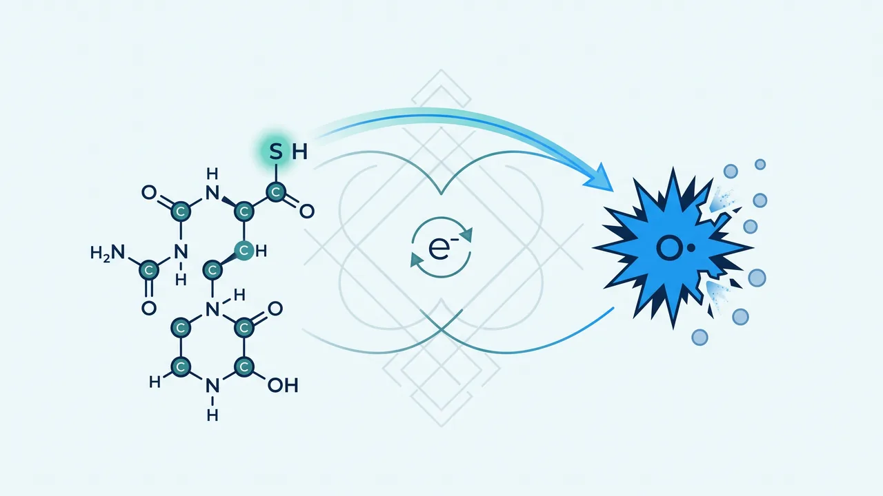
Glutathione
Amino acids and derivatives. A research review published by South Beach Longevity.
Evidence is labelled by study type in the sentence that reports it. In vitro and animal findings are never phrased as human outcomes. Human randomized means people under allocation. A meta-analysis is named as such. An agency review is an agency review. A blood glutathione increment is a biomarker. A clinical event is a clinical outcome. The two are not interchangeable here.
Findings are graded in place as established, strongly supported, emerging, plausible, or speculative. Industry affiliation is named where it bears on weight. GlyNAC, N-acetylcysteine, L-glutamine, glutathione S-transferase polymorphisms, and glutathione-peroxidase activity are different interventions or different molecules. They are not rewritten as glutathione-supplementation evidence. No product, dose, or regimen is recommended.
Abstract
Glutathione (GSH) is the most abundant low-molecular-weight thiol in mammalian cells. It is assembled in the cytosol from glutamate, cysteine and glycine by glutamate–cysteine ligase and glutathione synthetase; cysteine availability and GCL activity are the usual rate controls (Wu et al., 2004; Lu, 2013). The GSSG/2GSH couple is the dominant intracellular redox pair; its half-cell potential has been used as a quantitative index of the cellular redox environment (Schafer and Buettner, 2001). That couple is not in equilibrium with thioredoxin or with extracellular cysteine/cystine, and the potentials of mitochondria, nuclei, secretory pathway and plasma are maintained as distinct, non-equilibrium states (Go and Jones, 2008; Jones and Go, 2010). Liver is the principal biosynthetic organ and a major exporter of plasma GSH. Mitochondria import GSH and keep a more reducing potential than cytosol. None of this chemistry, by itself, answers a delivery question.
Oral glutathione meets γ-glutamyltransferase in the gut. Witschi and colleagues gave about 3 g as a single dose to seven volunteers and recorded no significant rise in plasma GSH, cysteine or glutamate over 270 minutes (Witschi et al., 1992). Park later confirmed that free plasma and blood-cell GSH do not rise after a large oral dose, and located a transient signal only in the protein-bound plasma fraction (Park et al., 2014). The independently funded four-week NIH RCT at 1,000 mg/day found no change in erythrocyte GSH or in urinary F2-isoprostanes and 8-OHdG (Allen and Bradley, 2011). The six-month placebo-controlled trial that did report 20–31 percent rises in whole-blood GSH used Setria product supplied and funded by Kyowa Hakko Bio; cysteine and GCL activity did not move, and results were never posted to NCT01044277 (Richie et al., 2015). The liposomal study most often cited as superiority evidence is an uncontrolled twelve-person pilot with a manufacturer-supplied product (Sinha et al., 2018). A 2026 single-dose crossover of a branded micellar formulation versus standard glutathione reported higher whole-blood incremental AUC (Solnier et al., 2026). That is a formulation-PK finding in fourteen people, not tissue repletion and not a clinical outcome.
Human outcome evidence for glutathione itself is thin and often commercially aligned. The largest intravenous RCT — Alliance N08CA, n = 185 — did not prevent paclitaxel/carboplatin neuropathy and favoured placebo on two secondary endpoints (Leal et al., 2014). Blinded Parkinson trials did not reproduce the open-label “42 percent” claim (Sechi et al., 1996; Hauser et al., 2009; Mischley et al., 2017). Inhaled GSH in cystic fibrosis produced a 3.5 percent FEV1 gain at three months that was no longer significant at six, with no effect on quality of life, BMI or exacerbations (Ciofu et al., 2019). Oral GSH for NAFLD is a single-arm pilot (Honda et al., 2017). Skin trials are small, short, confined largely to sun-exposed sites in Fitzpatrick IV–V Asian cohorts, and the longest oral RCT missed its primary melanin endpoint (Weschawalit et al., 2017; Dilokthornsakul et al., 2019). FDA’s 2022 compounding review judged glutathione insufficiently effective across 24 proposed uses. GlyNAC trials are precursor trials (Kumar et al., 2021, 2023). In glutathione-synthetase deficiency, the documented management is not GSH supplementation (Ristoff et al., 2001; Ristoff and Larsson, 2007). The chemistry is real. The delivery claim is not settled. A blood increment is not a health proof.
Contents
- The molecule
- The GSH/GSSG couple
- Synthesis from cysteine, glycine and glutamate
- Glutathione reductase and the peroxidases
- Compartments: extracellular versus intracellular pools
- Liver and mitochondria
- Redox signaling is not a supplement claim
- What oral glutathione encounters
- The oral-store contest
- Liposomal, sublingual and other delivery claims
- Precursor strategies are different interventions
- Stability and formulation quality
- Biomarkers are not health
- Metabolic, immune and exercise outcomes
- Skin
- Clinical disease
- Aging
- Safety and regulators
- Five questions the literature does not close
- References and evidence handling
01 The molecule
Glutathione is γ-L-glutamyl-L-cysteinyl-glycine. The peptide bond between glutamate and cysteine is formed at the γ-carboxyl of glutamate, not at the α-carboxyl used by ribosomal protein synthesis. That unusual linkage is the reason ordinary peptidases do not digest GSH as they digest most peptides, and the reason γ-glutamyltransferase is the enzyme that does (Meister and Anderson, 1983). The reactive function that dominates its biology is the cysteine thiol. Two GSH molecules can be oxidised to glutathione disulfide (GSSG) by formation of a disulfide between those cysteines. Reduced GSH is the species cells keep in large excess; GSSG is the minority partner under ordinary conditions (Wu et al., 2004; Forman et al., 2009).
Meister and Anderson’s 1983 review remains the load-bearing chemical statement: GSH is present in millimolar concentrations in most mammalian cells; it participates in peroxide disposal, electrophile conjugation, amino-acid transport through the γ-glutamyl cycle, and maintenance of protein thiols (Meister and Anderson, 1983). Wu and colleagues later restated the same architecture in nutritional language: synthesis from glutamate, cysteine and glycine; regulation by GCL activity, cysteine availability and GSH feedback inhibition; and a list of physiological roles that runs from antioxidant defence to glutathionylation (Wu et al., 2004). Forman, Zhang and Rinna reviewed measurement and biosynthesis and warned, correctly, that “GSH status” is not a single number (Forman et al., 2009). Those reviews describe intracellular chemistry. They are not pharmacokinetic papers.
02 The GSH/GSSG couple
Schafer and Buettner defined the cellular redox environment through the Nernst equation applied to GSSG/2GSH. Because two GSH molecules form one GSSG, the potential depends on [GSH]2/[GSSG], not merely on the ratio. They estimated half-cell potentials near −240 mV in proliferating cells, −200 mV in differentiating cells, and −170 mV in apoptosis (Schafer and Buettner, 2001). Those values are quantitative biology of intracellular couples. They are not plasma-GSH cut-offs, and they are not targets for a capsule.
Go and Jones, and later Jones and Go, established that the major thiol/disulfide couples — thioredoxin-1, thioredoxin-2, GSH/GSSG, and extracellular cysteine/cystine — are not in equilibrium with each other and are held at distinct potentials in mitochondria, nuclei, the secretory pathway and the extracellular space (Go and Jones, 2008; Jones and Go, 2010). Mitochondria are the most reducing and the most oxidation-sensitive. Nuclei are reducing and relatively oxidation-resistant. Extracellular compartments are stably oxidising. The practical consequence for this article is severe: a change in plasma GSH, whole-blood GSH, or even erythrocyte GSH does not report the mitochondrial matrix, the nucleus, or a hepatocyte’s cytosolic potential. Established as compartmental chemistry. Not transferable as a supplement-efficacy metric.
03 Synthesis from cysteine, glycine and glutamate
Biosynthesis is cytosolic and two-step. Glutamate–cysteine ligase (GCL; also called γ-glutamylcysteine synthetase) condenses glutamate and cysteine to γ-glutamylcysteine. Glutathione synthetase adds glycine. GCL is a heterodimer of a catalytic subunit (GCLC) and a modifier subunit (GCLM). The usual rate controls are cysteine availability, GCL activity, and feedback inhibition by GSH (Wu et al., 2004; Lu, 2013). Lu’s hepatic reviews add the transport and trans-sulfuration detail that matters in liver: cysteine, cystine and methionine uptake, and methionine-to-cysteine conversion, determine precursor supply (Lu, 1998, 2013). Anderson showed that γ-glutamylcyst(e)ine can be used directly for GSH synthesis, which is a biochemical route, not a retail product class (Anderson and Meister, 1983).
Transcriptional control of GCL and GS includes Nrf2 acting at antioxidant-response elements, with AP-1 and NF-κB also implicated (Lu, 2013). That regulation explains why cells can raise GSH synthesis under electrophile or oxidative stress. It does not explain how an intact oral tripeptide would appear inside a lymphocyte or a hepatocyte after intestinal GGT has had its chance. Richie and colleagues later reported that six months of oral GSH did not change cysteine concentrations or GCL activity even in the arm that reported higher blood GSH (Richie et al., 2015). If the oral molecule were being hydrolysed and rebuilt, those two measurements were the ones that should have moved. They did not. The chemistry of synthesis and the pharmacology of a capsule are different problems.
Glycine is the third residue and is usually abundant relative to cysteine. That is why cysteine, not glycine, is classically described as limiting (Wu et al., 2004). GlyNAC trials later treat glycine as co-limiting in older adults (Kumar et al., 2021, 2023). Those trials are discussed as precursor interventions in Part Three. They are not evidence that oral GSH supplies glycine in a physiologically decisive way.
04 Glutathione reductase and the peroxidases
Glutathione reductase (GR) reduces GSSG to GSH using NADPH. The glutathione peroxidases (GPx1 and related selenoproteins; phospholipid-hydroperoxide GPx4) oxidise GSH to GSSG while reducing hydrogen peroxide or lipid hydroperoxides (Forman et al., 2009). Glutathione S-transferases conjugate GSH to electrophiles. Those enzymes are not the tripeptide. Papers that report “low GPx activity,” a GST polymorphism, or a GPx4 cell-cycle effect are enzyme or genotype papers (Wang et al., 2003, is an example of the last). This article does not treat them as supplementation outcomes.
The cycle is conceptually simple: GPx (or a related peroxidase) consumes GSH; GR restores it if NADPH is available. Cells that cannot keep the couple reduced do not fail for lack of a capsule in the gut; they fail because synthesis, NADPH supply, export, or consumption has changed. Inborn errors of the γ-glutamyl cycle make that point without metaphor. Glutathione-synthetase deficiency produces haemolytic anaemia, metabolic acidosis, 5-oxoprolinuria, and, in the severe form, central-nervous-system injury and recurrent infection. Residual enzyme activity and erythrocyte GSH do not predict neurologic phenotype. Documented long-term management in the largest cohort was acidosis correction plus vitamins C and/or E, not GSH administration (Ristoff et al., 2001; Ristoff and Larsson, 2007). Established as rare autosomal-recessive disease. Not a model that licenses over-the-counter GSH for ordinary aging.
05 Compartments: extracellular versus intracellular pools
Plasma GSH is a small pool. Richie and colleagues stated that absolute plasma concentrations in their trial were of the order of 0.2–8 nmol/mL and “<1% of levels in whole blood” (Richie et al., 2015). Whole-blood GSH is dominated by erythrocytes. Lymphocyte and buccal-cell GSH are different compartments again. Mitochondrial GSH is still another. Extracellular cysteine/cystine, not GSH/GSSG, is the principal plasma thiol couple (Go and Jones, 2008). Mixing these pools in a marketing sentence is the most common error in the consumer literature.
Park’s LC-MS/MS work is the usual missing clause in oral-GSH arguments. After 50 mg/kg oral GSH, deproteinised plasma GSH and the blood-cell fraction did not change significantly. The protein-bound plasma fraction rose between 60 and 120 minutes (Park et al., 2014). That reconciles Witschi (no free-plasma rise) with the possibility of a small, transient bound signal. It is a two-hour binding observation. It is not tissue repletion.
06 Liver and mitochondria
Liver is the principal site of GSH synthesis and a major contributor to plasma GSH (Lu, 1998, 2013). Hepatic GCL is transcriptionally and post-transcriptionally regulated; cholestasis and endotoxin models in mice can suppress synthesis (Yang et al., 2009; Tomasi et al., 2014). Those are animal mechanism papers. They support the statement that hepatic GSH synthesis can be dysregulated in liver injury. They do not support oral GSH as a treatment for MASLD. The only direct oral-GSH trial in NAFLD located is Honda et al. (2017): open-label, single-arm, 300 mg/day for four months after a lifestyle run-in, ALT as primary surrogate. That design cannot separate regression to the mean from a drug effect. It is discussed as such in Part Four.
Mitochondria do not synthesise GSH. They import it and maintain a more reducing potential than cytosol, with high electron-transfer flux and high sensitivity to oxidation (Go and Jones, 2008; Jones and Go, 2010). Mitochondrial GSH is therefore a compartment of genuine biological interest and of almost no human-trial measurement in the oral-supplement literature. Claims that a retail liposome “repletes mitochondrial glutathione” are, in the records retrieved for this title, speculative. GlyNAC papers report mitochondrial fuel-oxidation measurements after precursor supplementation (Kumar et al., 2021, 2023). Those are precursor papers.
07 Redox signaling is not a supplement claim
GSH participates in glutathionylation, peroxide tone, and the kinetic poise of sulfur switches (Forman et al., 2009; Jones and Go, 2010). That is signaling chemistry. It is not a warrant that raising whole-blood GSH by 20 percent will change a clinical trajectory. Wu et al. (2004) listed diseases in which GSH deficiency has been invoked. Association is not depletion, and depletion is not a licence to administer the tripeptide. The operational rule for this title: do not treat “GSH is low in disease X” as a warrant for glutathione supplementation.
γ-Glutamyltransferase is the enzyme that defeats supplemental GSH in more than one organ. In the gut it hydrolyses oral GSH (Witschi et al., 1992). In the cystic-fibrosis airway, rising sputum GGT can degrade inhaled GSH and generate pro-oxidant products; Corti and colleagues suggested that patients with rising sputum GGT may be harmed rather than helped (Corti et al., 2017). Any delivery-route argument in this article has to contend with GGT. Liposomes and micelles are attempts to evade it. Whether they do so in a way that matters to tissue, as opposed to whole-blood incremental AUC, is the question Part Three takes up.
08 What oral glutathione encounters
Witschi, Reddy, Stofer and Lauterburg gave 0.15 mmol/kg oral GSH — about 3 g — to seven healthy volunteers and sampled for 270 minutes. Baseline plasma GSH was 6.2 µmol/L. Plasma glutathione, cysteine and glutamate did not increase significantly. The authors concluded that systemic availability is negligible in man and that intestinal and hepatic γ-glutamyltransferase hydrolyse the dose (Witschi et al., 1992). The paper is an open-label single-dose study. It is high confidence for acute free-plasma GSH. It is not a chronic-dosing study and it did not measure erythrocytes, lymphocytes or tissue.
Park and colleagues later administered 50 mg/kg oral GSH and split plasma into deproteinised and protein-bound fractions by LC-MS/MS. Free plasma GSH and the blood-cell fraction did not rise. The protein-bound plasma fraction rose from 60 to 120 minutes (Park et al., 2014). That is the reconciliation, not the refutation, of Witschi. Sharma and colleagues, advocating an orobuccal film, still wrote that absorption from the gastrointestinal tract is poor and that general consensus remains against oral glutathione for hyperpigmentation (Sharma and Sharma, 2022). An advocacy review that concedes the oral route is useful as an admission against interest. It is not independent pharmacokinetics.
09 The oral-store contest
Two placebo-controlled oral trials sit at the same 1,000 mg/day dose and disagree.
Allen and Bradley randomised adults without acute or chronic disease to 500 mg twice daily or placebo for four weeks (n = 40 randomised, 39 completed). Urinary F2-isoprostanes and 8-OHdG did not change. “Total reduced, oxidized, and ratio measures of GSH status were also unchanged.” Funding was NIH extramural (Allen and Bradley, 2011). Design quality is that of a short, independently funded RCT. Duration is the legitimate defence against it.
Richie and colleagues randomised 54 non-smoking adults to placebo, 250 mg/day or 1,000 mg/day Setria GSH for six months plus a one-month washout (NCT01044277). Whole-blood GSH rose 31 percent at 1,000 mg and 20 percent at 250 mg at six months versus baseline. Erythrocytes rose about 35 percent at the high dose. Plasma rose only in the high-dose arm at three and six months. Lymphocytes rose about 30 percent, high-dose only. Buccal cells rose only in the high-dose arm at six months — the source of the “260 percent” figure, with high variation. The GSSG/GSH ratio fell at six months. NK-cell cytotoxicity rose at three months in a within-group paired comparison in the high-dose arm (P_paired = 0.01), not as a stated between-arm contrast. Neutrophil phagocytosis and respiratory burst did not change consistently. After washout, high-dose blood GSH remained above baseline. Cysteine and GCL activity did not change in any arm. Changes at six months were not correlated with baseline GSH. Kyowa Hakko Bio funded the work, supplied product and placebo, and provided travel funds to the corresponding author. The paper states the company had no role in conduct, analysis or writing. Results were never posted to the registry (Richie et al., 2015). Guilford’s published commentary came from a liposomal-glutathione vendor (Guilford, 2015).
On design duration, Richie is stronger. On independence, Allen and Bradley is stronger. The only quantitative pooled estimate located in the sibling dossier (Mangkalopakorn 2024, three studies, not PubMed-indexed) was statistically null in both erythrocytes and plasma; it is not a numbered NCBI authority in this session and is mentioned only as a discovery note. Defensible position: oral glutathione at 1,000 mg/day for three to six months may produce a modest rise in blood-compartment GSH, but that finding rests on one manufacturer-funded trial, is contradicted at the same dose by the only independently funded RCT, has no demonstrated mechanism in that trial’s own precursor and enzyme data, and has not been shown to change oxidative-stress biomarkers in the independent RCT. Oral bioavailability is not presented as settled in either direction.
| Study | Design | Dose / route | n | What moved | Independence |
|---|---|---|---|---|---|
| Witschi 1992 | Open-label single dose | ~3 g oral | 7 | No free-plasma GSH rise at 270 min | No industry sponsor stated |
| Allen & Bradley 2011 | RCT, placebo | 1,000 mg/d × 4 wk | 39 | GSH status and F2-isoP / 8-OHdG unchanged | NIH |
| Park 2014 | Open-label single dose | 50 mg/kg oral | small | Free plasma and cells unchanged; bound plasma fraction rose 60–120 min | Not a tissue study |
| Richie 2015 | RCT, placebo, 6 mo | 250 / 1,000 mg/d oral Setria | 54 | Blood +20–31%; RBC ~35%; plasma high-dose only; Cys and GCL unchanged | Kyowa Hakko funded and supplied |
| Schmitt 2015 | Crossover, no placebo | Sublingual vs oral vs NAC, 3 wk | 20 | Sublingual > oral on plasma GSH/GSSG | Two authors at Laboratoires Le Stum |
| Sinha 2018 | Open-label, no placebo | 500 / 1,000 mg/d liposomal | 12 | Peak blood >40% vs baseline | Product supplied by Researched Nutritionals |
| Solnier 2026 | Crossover single dose | Micellar 300 mg vs standard 500 mg vs liposomal | 14 | Higher whole-blood iAUC for branded micellar vs standard | Branded LipoMicel formulation; 30-day open-label safety arm |
Table. Formulation pharmacokinetic and store-raising human studies of glutathione itself. Every quantitative cell was checked against the NCBI record or the published results text cited in the research dossier. A blood increment is not a clinical outcome.
10 Liposomal, sublingual and other delivery claims
Sinha and colleagues gave phosphatidylcholine liposomal GSH (Tri-Fortify, Researched Nutritionals) at 500 or 1,000 mg/day for one month to twelve healthy non-smokers aged 50–80, six per arm, without placebo. Peak whole-blood GSH exceeded 40 percent versus baseline in the 500 mg arm at two weeks; erythrocytes about 28 percent; plasma about 25 percent; PBMCs nearly two-fold. The abstract says “100 percent” in PBMCs; the discussion says “200 percent.” A two-fold rise is a 100 percent rise. Plasma 8-isoprostane fell 35 percent at two weeks in the 500 mg arm. NK cytotoxicity was reported up to +400 percent versus baseline. Effects tended to recede at four weeks. The authors reported inverse correlations with baseline GSH (r = 0.6–0.8), which directly contradicts Richie 2015 (same senior author), where baseline correlations were non-significant. The authors stated that the design did not use a placebo control (Sinha et al., 2018). This study is not a replication of liposomal superiority. It is a twelve-person uncontrolled pilot. Confidence for a clinical or comparative-delivery claim is low.
Ly and colleagues reported that liposomal GSH in HIV-infected individuals restored TH1 cytokine responses to Mycobacterium tuberculosis in a 13-week placebo-compared cohort; outcomes were immunological and in-vitro PBMC assays, not clinical tuberculosis endpoints (Ly et al., 2015). Confidence for a clinical claim is low.
Schmitt and colleagues compared sublingual GSH, oral GSH and NAC in a three-week randomised crossover in twenty volunteers with metabolic syndrome. Sublingual GSH raised total and reduced plasma GSH and the GSH/GSSG ratio versus oral GSH (p = 0.003). Plasma vitamin E rose only in the sublingual arm. Two of four authors were affiliated with Laboratoires Le Stum, whose sublingual product was the winner (Schmitt et al., 2015). Treat “sublingual is superior” as a manufacturer-supported crossover without a placebo arm. Confidence low to moderate.
Solnier and colleagues (2026) ran a randomised double-blind crossover in fourteen healthy adults of micellar glutathione (LipoMicel, 300 mg) versus standard GSH (500 mg) and a Setria liposomal arm, then a 30-day open-label safety follow-up of the micellar product at 600 mg/day. Baseline-adjusted whole-blood iAUC0–24 was 1287.5 ± 179.0 versus 517.8 ± 180.0 µg·mL·h for micellar versus standard (p = 0.0064); ΔCmax 103.9 versus 42.8 µg/mL (p = 0.0003). GSSG exposure did not differ; the GSH/GSSG ratio was higher after the micellar product. Clinical chemistry (ALT, AST, ALP, creatinine) did not change over 30 days. Registered as NCT06345950 (Solnier et al., 2026). This is the most recent oral-PK trial in the reviewed record. It is a branded-formulation comparison of single-dose whole-blood kinetics in fourteen people. It does not measure tissue GSH, does not report clinical outcomes, and does not independently replicate Sinha’s uncontrolled liposomal pilot. Emerging as formulation PK. Not a justification for expensive delivery as a health intervention.
Handog and colleagues used a buccal glutathione lozenge, 500 mg daily for eight weeks, in a single-arm study of thirty Filipino women. Melanin indices fell from baseline. There was no control group. The paper itself concedes that oral administration has been less successful in elevating plasma GSH (Handog et al., 2016). Confidence for efficacy is low.
11 Precursor strategies are different interventions
N-acetylcysteine supplies cysteine. GlyNAC supplies glycine plus NAC. Neither is glutathione. Wu et al. (2004) already listed NAC, cystine, methionine and L-2-oxothiazolidine-4-carboxylate as cysteine precursors that can support tissue GSH synthesis in nutritional studies. Transferring a precursor result to a GSH capsule is a category error.
Kumar, Sekhar and colleagues reported an open-label 24-week GlyNAC series in eight older adults, with withdrawal for twelve weeks: RBC GSH, oxidative-stress markers, mitochondrial fuel oxidation, inflammation, endothelial markers, insulin resistance, cognition and strength improved on supplement and receded off it (Kumar et al., 2021). The 2023 RCT randomised 24 older adults 1:1 to GlyNAC or isonitrogenous alanine for 16 weeks (NCT01870193). The abstract reports correction of GSH deficiency and of a broad aging-phenotype panel on GlyNAC and not on placebo, without effect sizes or confidence intervals in the abstract record (Kumar et al., 2023). An earlier open-label HIV series used the same product concept in eight people (Kumar et al., 2020). Babu Balagopal et al. (2024) tested NAC, not GSH, in thirteen children with MASLD. Schmitt used NAC as an active comparator, not as GSH (Schmitt et al., 2015). GlyNAC evidence cannot be scored as glutathione-supplementation evidence. The sibling articles on NAC and GlyNAC own those interventions.
| Agent | What it is | Best human design in the reviewed record | May be cited as GSH evidence? |
|---|---|---|---|
| Oral GSH | The tripeptide | Allen 2011 RCT; Richie 2015 RCT | Yes — with the funding conflict named |
| Liposomal / micellar GSH | Encapsulated tripeptide | Sinha 2018 n=12 uncontrolled; Solnier 2026 n=14 PK | Yes — as formulation, not as proven superiority |
| Sublingual GSH | Mucosal tripeptide | Schmitt 2015 crossover | Yes — manufacturer-affiliated |
| NAC | Cysteine precursor | Schmitt 2015; Babu Balagopal 2024; Ciofu oral NAC subgroup | No |
| GlyNAC | Glycine + NAC | Kumar 2021 open-label; Kumar 2023 n=12/arm RCT | No |
| L-glutamine (SCD) | Amino acid, NADH argument | Not GSH; excluded from scoring | No |
Table. Precursor comparison. Scoring a precursor trial as a glutathione trial is a labelling error, not a conservative reading.
12 Stability and formulation quality
GSH oxidises to GSSG in solution. FDA’s 2022 Pharmacy Compounding Advisory Committee materials treated chemical stability as one reason the committee later voted to keep glutathione available for compounding after FDA staff had recommended exclusion for insufficient effectiveness. That vote was about access and stability, not about a new efficacy finding. A 2019 compounding risk alert cited potentially high endotoxin in bulk glutathione powder. Those are quality and compounding facts. They are not bioavailability proofs.
Sinha’s viscous liposomal product was spoon-dosed; the authors themselves suggested possible self-dosing drift as an explanation for late-week fade (Sinha et al., 2018). Retail liposomes vary in phospholipid, payload, particle-size claim and oxidation control. This article did not assay commercial products. Formulation quality is a plausible modifier of a blood-PK signal. It has not been shown, independently and repeatedly, to convert oral GSH into a tissue-relevant or clinically justified intervention.
13 Biomarkers are not health
The standing instruction for this title is not decorative. A rise in blood glutathione is a measurement. It is not proof of improved health. Allen and Bradley measured the oxidative-stress markers that the oral-GSH story most often invokes — urinary F2-isoprostanes and 8-OHdG — and they did not move (Allen and Bradley, 2011). Richie reported blood-compartment GSH changes and a within-group NK-cytotoxicity increment without a consistent neutrophil functional change (Richie et al., 2015). Sinha reported 8-isoprostane and NK changes versus baseline in an uncontrolled sample of twelve (Sinha et al., 2018). Oxidative-stress biomarkers are chemically real and clinically weakly anchored. They are not treated here as useful surrogates for disease modification unless a trial also measures a clinical endpoint and shows that the biomarker movement tracks it. No oral-GSH trial in the reviewed record does that.
| Study | Agent | GSH compartment | Oxidative marker | Clinical endpoint |
|---|---|---|---|---|
| Allen 2011 | Oral GSH 1 g/d | Unchanged | F2-isoP, 8-OHdG unchanged | None |
| Richie 2015 | Oral GSH 0.25–1 g/d | Blood / RBC / lymph / buccal rose (industry RCT) | Ratio GSSG/GSH down | NK cytotoxicity within-group only |
| Sinha 2018 | Liposomal GSH | Blood / PBMC vs baseline | 8-isoprostane −35% vs baseline | None |
| Schmitt 2015 | Sublingual vs oral vs NAC | Plasma GSH/GSSG | Vitamin E rose, sublingual only | None |
| Honda 2017 | Oral GSH 300 mg/d | Not the primary | — | ALT, uncontrolled |
| Ciofu 2019 | Inhaled GSH | Airway, not blood stores | — | FEV1 +3.5% at 3 mo, NS at 6 mo |
| Kumar 2023 | GlyNAC, not GSH | RBC GSH (precursor) | OxS panel | Function claims; n=12/arm |
Table. Biomarker trial matrix. GSH-the-tripeptide rows and precursor rows are not pooled. Empty clinical-endpoint cells are findings.
14 Metabolic, immune and exercise outcomes
Honda and colleagues enrolled 34 patients with NAFLD, of whom 29 completed an open-label protocol: lifestyle run-in, then oral GSH 300 mg/day for four months. ALT fell; triglycerides, non-esterified fatty acids and ferritin also fell. Responders, defined after the fact at a median 12.9 percent ALT decrease, were younger and without severe diabetes (Honda et al., 2017). Low confidence as efficacy evidence: no placebo, continued lifestyle effects uncontrolled, ALT a surrogate.
Immune claims rest on Richie’s within-group NK increment, Sinha’s uncontrolled NK and lymphocyte-proliferation rises, and Ly’s cytokine and in-vitro mycobacterial assays in HIV (Richie et al., 2015; Sinha et al., 2018; Ly et al., 2015). None of those is a clinical infection, vaccine-response, or hospitalisation endpoint. Exercise-performance trials of oral GSH itself were not located as adequately powered RCTs in the reviewed record. GlyNAC papers report gait speed, grip and six-minute walk after precursor supplementation (Kumar et al., 2021). Those rows stay in the precursor column.
15 Skin
Skin lightening is the largest consumer use and the weakest place to confuse a melanin-index movement with health.
Arjinpathana and Asawanonda randomised 60 Thai medical students to oral GSH 500 mg/day or placebo for four weeks. Melanin index fell at all six sites in the GSH arm and was statistically significantly greater than placebo at two of six sites (right face p = 0.021; sun-exposed left forearm p = 0.036). The authors wrote that lightening occurred “in a small number of subjects” and that long-term safety had not been established (Arjinpathana and Asawanonda, 2012).
Weschawalit and colleagues randomised 60 women (57 analysed) in Bangkok, almost all Fitzpatrick IV, to oral GSH 250 mg, oral GSSG 250 mg or placebo nightly for twelve weeks. Product was supplied by Kyowa Hakko Bio — Setria and AquaGluta. The primary melanin-index endpoint “tended to be lower than placebo” but was not statistically significant (P > 0.05) at any site, and GSH did not differ from GSSG. The only significant pigment result was a subgroup older than 40 on the sun-exposed right forearm (GSH n = 7 versus placebo n = 10, P = 0.031); the contralateral forearm was P = 0.057. A wrinkle secondary endpoint reached P = 0.006 at one sun-protected site. Subjective ratings did not differ among groups. Two subjects withdrew for elevated transaminases (Weschawalit et al., 2017). The abstract’s antimelanogenic wording overstates a null primary. Confidence for a skin-lightening claim is low.
Watanabe and colleagues applied 2 percent GSSG lotion versus placebo in a split-face trial in 30 women for ten weeks. Melanin index was lower on the GSSG side from early weeks through week 10 (P < 0.001). Three of four authors were Kyowa Hakko employees; Erika Hashizume is thanked in Richie 2015 for design and implementation of that oral trial (Watanabe et al., 2014; Richie et al., 2015). The split-face design is strong for a topical. The paper is industry-authored. It is oxidised glutathione, applied to skin. It carries no information about oral or systemic bioavailability.
Wahab and colleagues (2021) reported that combined topical plus oral GSH lowered melanin index versus placebo in a double-blind RCT (n = 46, eight weeks). Dilokthornsakul and colleagues, reviewing to October 2017, included four studies and judged the whitening evidence inconclusive because of quality and inconsistency; effects, when present, were on sun-exposed skin (Dilokthornsakul et al., 2019). Sarkar and colleagues (2025) reviewed a wider window, still found mixed risk of bias, and wrote that intravenous glutathione is contraindicated for lack of efficacy and side effects (Sarkar et al., 2025). Sonthalia and colleagues had already stated that there is no evidence to prove efficacy of intravenous injections for whitening and noted the Philippine FDA warning (Sonthalia et al., 2016). Stanescu and colleagues (2026) mixed GSH, GSTs and GPx in a skin-aging review; that mix is not a clean GSH-the-molecule source (Stanescu et al., 2026).
The only placebo-controlled IV skin-lightening trial discussed in FDA’s 2022 review (Zubair 2016, as reported by FDA) used a multi-ingredient infusion, missed significance at p = 0.054, and lost the effect by six months in 24 of 25 patients. It is cited here as FDA’s account, not as an independently retrieved primary. FDA’s own sentence: there are insufficient data to support oral glutathione for skin lightening.
16 Clinical disease
Leal and colleagues ran Alliance N08CA: phase 3, double-blind, placebo-controlled, 185 patients receiving paclitaxel plus carboplatin, IV GSH 1.5 g/m2 over 15 minutes before each cycle. EORTC-QLQ-CIPN20 P = 0.21; CTCAE grade ≥2 neurotoxicity P = 0.449. Time to grade ≥2 neuropathy favoured placebo (P = 0.039). Weekly-paclitaxel acute pain syndrome favoured placebo (P = 0.002). No subgroup benefited. NIH-funded. The authors concluded that the results do not support glutathione for prevention of paclitaxel/carboplatin CIPN (Leal et al., 2014). This is the largest, best-designed IV glutathione RCT located. It is negative.
Sechi and colleagues gave IV GSH 600 mg twice daily for 30 days to nine unblinded, untreated early Parkinson patients and reported a 42 percent decline in disability (Sechi et al., 1996). Hauser and colleagues randomised 21 patients to IV GSH 1,400 mg three times weekly or placebo for four weeks. UPDRS changes were not significant; the point estimate favoured GSH during treatment and placebo during follow-up (Hauser et al., 2009). Mischley and colleagues showed that intranasal GSH is tolerable (n = 30), that a single 200 mg intranasal dose raises brain GSH on MRS for at least an hour (n = 15), and that in a phase IIb RCT (n = 45) neither 100 mg nor 200 mg three times daily was superior to saline at three months, while all arms including placebo improved. One high-dose participant developed cardiomyopathy (Mischley et al., 2015, 2016, 2017). A 70-respondent pharmacy survey is a tolerability signal with a 23 percent response rate, not efficacy (Mischley et al., 2013).
Ciofu, Smith and Lykkesfeldt’s Cochrane review of antioxidants in cystic fibrosis included four inhaled-supplement studies (n = 285). Inhaled glutathione improved FEV1 percent predicted at three months by a mean difference of 3.50 percent (95% CI 1.38 to 5.62; two studies, 258 participants) and not at six months (MD 2.30 percent, 95% CI −0.12 to 4.71). Quality of life, BMI and time to exacerbation did not differ. Only one of twenty studies in the whole review was judged free of bias (Ciofu et al., 2019). Calabrese and colleagues’ 12-month inhaled-GSH RCT (54 adults, 51 children) missed its pre-specified primary of a 15 percent FEV1 improvement (Calabrese et al., 2015). Corti and colleagues supplied the GGT caveat for the CF airway (Corti et al., 2017).
| Indication | Best GSH-the-molecule evidence | Result | Grade |
|---|---|---|---|
| CIPN prevention, IV | Leal 2014 phase 3 n=185 | Null; two endpoints favoured placebo | Established negative |
| Parkinson, IV | Hauser 2009 vs Sechi 1996 | Blinded trial did not reproduce 42% | Open-label claim not replicated |
| Parkinson, intranasal | Mischley 2017 IIb n=45 | Not superior to placebo; MRS delivery earlier | Delivery ≠ benefit |
| CF, inhaled | Ciofu 2019 Cochrane | +3.5% FEV1 at 3 mo, NS at 6 mo; no QoL / BMI / exacerbation | Small, non-durable |
| NAFLD, oral | Honda 2017 single-arm | ALT down, no control | Low |
| Skin, oral | Weschawalit 2017 primary null; Arjinpathana 2/6 sites | Inconclusive (Dilokthornsakul 2019) | Not a health claim |
| Aging | Kumar GlyNAC, not GSH | Precursor RCT n=12/arm | Out of scope as GSH |
| GS deficiency | Ristoff 2001 / 2007 | Management is not GSH supplementation | Established as rare disease |
Table. Clinical outcome matrix for glutathione itself. Precursor and enzyme papers are excluded from the result column.
17 Aging
The aging intervention that dominates recent citations is GlyNAC (Kumar et al., 2021, 2023). It is glycine plus N-acetylcysteine. It is discussed so that it is not silently imported. There is no adequately powered, independently funded RCT of oral or liposomal glutathione on a pre-specified aging or disability endpoint in the reviewed record. Richie enrolled non-smoking adults and measured stores, not aging phenotypes (Richie et al., 2015). Sinha enrolled adults 50–80 without a placebo (Sinha et al., 2018). Weschawalit’s wrinkle secondary in a cosmetic RCT is not an aging-outcome trial (Weschawalit et al., 2017). Oral glutathione has not been shown to modify aging. That sentence is a finding, not a gap to be filled by a precursor paper.
18 Safety and regulators
Oral GSH in Richie produced no serious adverse events over six months; the two allergy-like dropouts were in placebo (Richie et al., 2015). Weschawalit recorded two transaminitis withdrawals on oral GSH/GSSG 250 mg/day (Weschawalit et al., 2017). Solnier reported no change in routine liver and renal chemistries over 30 days of 600 mg/day micellar GSH in fourteen people (Solnier et al., 2026). FDA’s 2022 review characterised oral glutathione as minimally absorbed and associated mainly with local gastrointestinal events in the oral/buccal studies it tabulated, and stated significant safety concerns for IV and inhalation routes, including hepatotoxicity and life-threatening anaphylaxis on IV and bronchoconstriction on nebulised use. Of 195 CAERS reports naming glutathione, 194 were confounded by co-ingredients; exactly one implicated glutathione alone. No glutathione-attributed death was identified in that review. A 2025 case report described Stevens–Johnson syndrome after a multi-ingredient IV infusion containing glutathione, vitamin C and vitamin D; causation cannot be isolated to GSH (Johnson et al., 2025). Mischley 2017 reported one cardiomyopathy in the high-dose intranasal arm. Philippines FDA Advisory 2019-182 stated that there are no published clinical trials of injectable glutathione for skin lightening, while noting approval as an adjunct in cisplatin chemotherapy. The irony is that the best US trial on a related chemotherapy-neurotoxicity claim (Leal 2014) was negative.
In June 2022 FDA evaluated glutathione for the 503A compounding bulks list across 24 proposed uses and concluded that there is either no available information or insufficient evidence of effectiveness. It recommended exclusion. The advisory committee voted 8–5–1 to overrule staff — the only substance that day on which the committee disagreed with FDA — on patient-access and chemical-stability grounds, not on a new demonstration of efficacy. That document is the most useful regulatory artefact for this title. IV half-life of the order of 10–15 minutes, with a matching cysteine rise, is the pharmacokinetic context FDA used for the drip industry; the primary Aebi 1991 XML was not in the clean allowlist of this session and the half-life figure is therefore attributed to FDA’s review, not to a numbered Aebi line here.
Acquired 5-oxoprolinuria from other drugs (classically paracetamol with flucloxacillin in frail patients) is pathway harm, not a glutathione-supplementation result. It belongs in a pathway note, not in a product claim.
19 Adversarial review
The questions below were asked before the last sentence was written, not after. Each is answered from the human records in Parts Three and Four. Animal necessity of intracellular GSH is stipulated and then set aside.
Is orally administered glutathione meaningfully absorbed?
Not as free plasma GSH after a large single dose. Witschi et al. (1992) and Park et al. (2014) agree on that point; Park adds a transient protein-bound plasma signal that is not a tissue measurement. Chronic dosing is the live dispute. Allen and Bradley (2011) found no store or oxidative-marker movement at four weeks, independently funded. Richie et al. (2015) found blood-compartment rises at six months, manufacturer-funded, without a cysteine or GCL mechanism. Solnier et al. (2026) found a single-dose whole-blood iAUC advantage for a branded micellar product in fourteen people. Resolution: oral GSH is not demonstrated to be meaningfully absorbed into the intracellular compartments that motivate purchase. A possible modest blood-store rise after months of a branded product remains contested and commercially aligned. The question is not closed in favour of oral absorption.
Are liposomal superiority claims independently replicated?
No. Sinha et al. (2018) is an n = 12 open-label pilot without placebo, manufacturer-supplied, internally inconsistent on the PBMC percentage, and in contradiction with Richie 2015 on baseline dependence. Solnier et al. (2026) compared a micellar brand with standard and liposomal GSH on single-dose blood PK; it is not an independent replication of Sinha’s immune and isoprostane claims, and it is itself a branded-formulation paper. Schmitt et al. (2015) is a sublingual manufacturer crossover, not a liposome replication. Guilford’s commentary on Richie came from a liposomal vendor (Guilford, 2015). Resolution: liposomal superiority is not independently replicated. It remains a plausible delivery hypothesis with low-quality supportive human data.
Which measured glutathione compartment matters?
The compartment that matters biologically is the one in which the chemistry in Parts One and Two actually runs: intracellular GSH/GSSG, including mitochondrial GSH, in the organ at issue (Schafer and Buettner, 2001; Go and Jones, 2008; Jones and Go, 2010; Lu, 2013). The compartments that trials measure are plasma (tiny), erythrocytes (most of whole blood), occasional lymphocytes or PBMCs, rare buccal cells, and, for intranasal GSH, brain MRS (Mischley et al., 2016). Richie’s own plasma values were <1 percent of whole blood (Richie et al., 2015). Resolution: erythrocyte and whole-blood GSH are the measured convenience compartments. They are not shown to be the biologically decisive ones for the claims that sell the product. Brain MRS showed delivery of intranasal GSH without clinical superiority (Mischley et al., 2016, 2017) — the cleanest existing demonstration that the right-looking compartment can move without a useful outcome.
Are oxidative-stress biomarkers clinically useful?
They are useful as chemistry. They are not shown to be useful as clinical decision tools in the glutathione-supplement trials. Allen and Bradley (2011) is the only independently funded oral RCT that measured F2-isoprostanes and 8-OHdG; both were null, as was GSH status. Sinha’s 8-isoprostane movement is uncontrolled. Cochrane’s inhaled-GSH FEV1 signal did not come with a quality-of-life, BMI or exacerbation benefit (Ciofu et al., 2019). Resolution: in this literature, oxidative-stress biomarkers have not earned clinical usefulness as surrogates that can stand in for benefit. Treating them as such is the error this article exists to prevent.
Are expensive delivery systems justified?
Justification would require, at minimum, an independent, adequately powered demonstration that the expensive system restores a biologically relevant compartment and moves a pre-specified clinical endpoint more than a cheaper comparator or placebo, with a safety margin that survives IV and inhalation experience. That demonstration is not in the reviewed record. Liposomal and micellar products have manufacturer-aligned blood-PK or uncontrolled-store papers (Sinha et al., 2018; Solnier et al., 2026). Sublingual GSH has a manufacturer crossover (Schmitt et al., 2015). IV GSH failed its best clinical test (Leal et al., 2014) and carries FDA-stated anaphylaxis and hepatotoxicity concerns. Intranasal GSH reaches brain and does not beat placebo on Parkinson scores (Mischley et al., 2017). Precursors are cheaper conceptually and still are not GSH. Resolution: expensive delivery is not justified by independent clinical evidence. A purchaser who is buying a blood-iAUC story should be told that is the story.
The chemistry in Parts One and Two is not the dispute. Intracellular GSH is necessary. Oral, liposomal, sublingual and intravenous products have been asked to convert that necessity into delivery and into health. On the records retrieved on 20 August 2026, they have not done so in a way that survives independence, replication, compartment honesty, or clinical endpoints. That is the article’s result.
20 Evidence handling
Peer-reviewed identifiers were taken from NCBI records. In-prose citations are author–year. The numbered list is sorted by first-author surname. Study type is named in the reporting sentence. Animal and cell findings are not rewritten as human outcomes. A blood glutathione increment is labelled a biomarker wherever it is used to talk about health. Guideline and FDA text is quoted as agency language, not as a trial. Industry affiliation is noted where it bears on the weight of a store-raising or cosmetic paper. Project 06 and 07 Peptide News were searched as read-only discovery layers; they are not cited as scientific authorities. HOUSE_STYLE section 8a (peptide bioregulators) does not apply. This title is not Radix article No. 27 and is not filed to the peptide library.
21 Scope relative to sibling articles
This title is the glutathione-the-tripeptide article in the SBL-41 series. It is not the NAC article, the GlyNAC article, or the glycine article. Those agents appear here only to forbid their use as glutathione-supplementation evidence. It is not a glutathione S-transferase pharmacogenetics paper, a GPx methods paper, or a clinical manual for glutathione-synthetase deficiency. The Radix peptide glutathione dossier at glutathione_monograph_2026 was used as a REFERENCE evidence map; claims were re-verified against NCBI XML before they were written.
22 References
Forty-nine peer-reviewed records below were verified against the NCBI record on 20 August 2026. FDA Pharmacy Compounding Advisory Committee materials (June 2022; fda.gov/media/159042) and Philippines FDA Advisory 2019-182 are additional institutional sources and are not PubMed articles. Seed PMIDs that resolved to unrelated titles (wrong Aebi, Hagen, and Jones 2006 collisions) were discarded and are not cited.
- Allen J, Bradley RD. Effects of oral glutathione supplementation on systemic oxidative stress biomarkers in human volunteers. J Altern Complement Med. 2011;17:827-33. PMID 21875351 · doi:10.1089/acm.2010.0716 · PMC3162377
- Anderson ME, Meister A. Transport and direct utilization of gamma-glutamylcyst(e)ine for glutathione synthesis. Proc Natl Acad Sci U S A. 1983;80:707-11. PMID 6572362
- Arjinpathana N, Asawanonda P. Glutathione as an oral whitening agent: a randomized, double-blind, placebo-controlled study. J Dermatolog Treat. 2012;23:97-102. PMID 20524875
- Babu Balagopal P, Kohli R, Uppal V, Averill L, Shah C, McGoogan K et al. Effect of N-acetyl cysteine in children with metabolic dysfunction-associated steatotic liver disease-A pilot study. J Pediatr Gastroenterol Nutr. 2024;79:652-660. PMID 38973318
- Calabrese C, Tosco A, Abete P, Carnovale V, Basile C, Magliocca A et al. Randomized, single blind, controlled trial of inhaled glutathione vs placebo in patients with cystic fibrosis. J Cyst Fibros. 2015;14:203-10. PMID 25458463
- Ciofu O, Smith S, Lykkesfeldt J. Antioxidant supplementation for lung disease in cystic fibrosis. Cochrane Database Syst Rev. 2019;10:CD007020. PMID 31580490
- Corti A, Griese M, Hector A, Pompella A. Increasing sputum levels of gamma-glutamyltransferase may identify cystic fibrosis patients who do not benefit from inhaled glutathione. J Cyst Fibros. 2017;16:342-345. PMID 27988297
- Dilokthornsakul W, Dhippayom T, Dilokthornsakul P. The clinical effect of glutathione on skin color and other related skin conditions: A systematic review. J Cosmet Dermatol. 2019;18:728-737. PMID 30895708
- Forman HJ, Zhang H, Rinna A. Glutathione: overview of its protective roles, measurement, and biosynthesis. Mol Aspects Med. 2009;30:1-12. PMID 18796312
- Go YM, Jones DP. Redox compartmentalization in eukaryotic cells. Biochim Biophys Acta. 2008;1780:1273-90. PMID 18267127
- Guilford FT. Commentary to "Randomized controlled trial of oral glutathione supplementation on body stores of glutathione". Eur J Nutr. 2015;54:859-60. PMID 25792077
- Handog EB, Datuin MS, Singzon IA. An open-label, single-arm trial of the safety and efficacy of a novel preparation of glutathione as a skin-lightening agent in Filipino women. Int J Dermatol. 2016;55:153-7. PMID 26148180
- Hauser RA, Lyons KE, McClain T, Carter S, Perlmutter D. Randomized, double-blind, pilot evaluation of intravenous glutathione in Parkinson's disease. Mov Disord. 2009;24:979-83. PMID 19230029
- Honda Y, Kessoku T, Sumida Y, Kobayashi T, Kato T, Ogawa Y et al. Efficacy of glutathione for the treatment of nonalcoholic fatty liver disease: an open-label, single-arm, multicenter, pilot study. BMC Gastroenterol. 2017;17:96. PMID 28789631
- Johnson JS, Jarvis NR, Martinez AM, Dvorak JE. Intravenous Glutathione and Vitamin Supplementation Causing Stevens-Johnson Syndrome: A Case Report. J Burn Care Res. 2025;46:652-655. PMID 40057759
- Jones DP, Go YM. Redox compartmentalization and cellular stress. Diabetes Obes Metab. 2010;12 Suppl 2:116-25. PMID 21029308
- Kumar P, Liu C, Suliburk JW, Minard CG, Muthupillai R, Chacko S et al. Supplementing Glycine and N-acetylcysteine (GlyNAC) in Aging HIV Patients Improves Oxidative Stress, Mitochondrial Dysfunction, Inflammation, Endothelial Dysfunction, Insulin Resistance, Genotoxicity, Strength, and Cognition: Results of an Open-Label Clinical Trial. Biomedicines. 2020;8. PMID 33007928
- Kumar P, Liu C, Hsu JW, Chacko S, Minard C, Jahoor F et al. Glycine and N-acetylcysteine (GlyNAC) supplementation in older adults improves glutathione deficiency, oxidative stress, mitochondrial dysfunction, inflammation, insulin resistance, endothelial dysfunction, genotoxicity, muscle strength, and cognition: Results of a pilot clinical trial. Clin Transl Med. 2021;11:e372. PMID 33783984
- Kumar P, Liu C, Suliburk J, Hsu JW, Muthupillai R, Jahoor F et al. Supplementing Glycine and N-Acetylcysteine (GlyNAC) in Older Adults Improves Glutathione Deficiency, Oxidative Stress, Mitochondrial Dysfunction, Inflammation, Physical Function, and Aging Hallmarks: A Randomized Clinical Trial. J Gerontol A Biol Sci Med Sci. 2023;78:75-89. PMID 35975308
- Leal AD, Qin R, Atherton PJ, Haluska P, Behrens RJ, Tiber CH et al. North Central Cancer Treatment Group/Alliance trial N08CA-the use of glutathione for prevention of paclitaxel/carboplatin-induced peripheral neuropathy: a phase 3 randomized, double-blind, placebo-controlled study. Cancer. 2014;120:1890-7. PMID 24619793
- Lu SC. Regulation of hepatic glutathione synthesis. Semin Liver Dis. 1998;18:331-43. PMID 9875552
- Lu SC. Regulation of hepatic glutathione synthesis: current concepts and controversies. FASEB J. 1999;13:1169-83. PMID 10385608
- Lu SC. Regulation of glutathione synthesis. Curr Top Cell Regul. 2000;36:95-116. PMID 10842748
- Lu SC. Regulation of glutathione synthesis. Mol Aspects Med. 2009;30:42-59. PMID 18601945
- Lu SC. Glutathione synthesis. Biochim Biophys Acta. 2013;1830:3143-53. PMID 22995213
- Ly J, Lagman M, Saing T, Singh MK, Tudela EV, Morris D et al. Liposomal Glutathione Supplementation Restores TH1 Cytokine Response to Mycobacterium tuberculosis Infection in HIV-Infected Individuals. J Interferon Cytokine Res. 2015;35:875-87. PMID 26133750
- Meister A, Anderson ME. Glutathione. Annu Rev Biochem. 1983;52:711-60. PMID 6137189
- Mischley LK, Vespignani MF, Finnell JS. Safety survey of intranasal glutathione. J Altern Complement Med. 2013;19:459-63. PMID 23240940
- Mischley LK, Leverenz JB, Lau RC, Polissar NL, Neradilek MB, Samii A et al. A randomized, double-blind phase I/IIa study of intranasal glutathione in Parkinson's disease. Mov Disord. 2015;30:1696-701. PMID 26230671
- Mischley LK, Conley KE, Shankland EG, Kavanagh TJ, Rosenfeld ME, Duda JE et al. Central nervous system uptake of intranasal glutathione in Parkinson's disease. NPJ Parkinsons Dis. 2016;2:16002. PMID 28725693
- Mischley LK, Lau RC, Shankland EG, Wilbur TK, Padowski JM. Phase IIb Study of Intranasal Glutathione in Parkinson's disease. J Parkinsons Dis. 2017;7:289-299. PMID 28436395
- Park EY, Shimura N, Konishi T, Sauchi Y, Wada S, Aoi W et al. Increase in the protein-bound form of glutathione in human blood after the oral administration of glutathione. J Agric Food Chem. 2014;62:6183-9. PMID 24877771
- Richie JP, Nichenametla S, Neidig W, Calcagnotto A, Haley JS, Schell TD et al. Randomized controlled trial of oral glutathione supplementation on body stores of glutathione. Eur J Nutr. 2015;54:251-63. PMID 24791752
- Ristoff E, Mayatepek E, Larsson A. Long-term clinical outcome in patients with glutathione synthetase deficiency. J Pediatr. 2001;139:79-84. PMID 11445798
- Ristoff E, Larsson A. Inborn errors in the metabolism of glutathione. Orphanet J Rare Dis. 2007;2:16. PMID 17397529
- Sarkar R, Yadav V, Yadav T, P J, Mandal I. Glutathione as a skin-lightening agent and in melasma: a systematic review. Int J Dermatol. 2025;64:992-1004. PMID 39444151
- Schafer FQ, Buettner GR. Redox environment of the cell as viewed through the redox state of the glutathione disulfide/glutathione couple. Free Radic Biol Med. 2001;30:1191-212. PMID 11368918
- Schmitt B, Vicenzi M, Garrel C, Denis FM. Effects of N-acetylcysteine, oral glutathione (GSH) and a novel sublingual form of GSH on oxidative stress markers: A comparative crossover study. Redox Biol. 2015;6:198-205. PMID 26262996
- Sechi G, Deledda MG, Bua G, Satta WM, Deiana GA, Pes GM et al. Reduced intravenous glutathione in the treatment of early Parkinson's disease. Prog Neuropsychopharmacol Biol Psychiatry. 1996;20:1159-70. PMID 8938817
- Sharma DK, Sharma P. Augmented Glutathione Absorption from Oral Mucosa and its Effect on Skin Pigmentation: A Clinical Review. Clin Cosmet Investig Dermatol. 2022;15:1853-1862. PMID 36117769
- Sinha R, Sinha I, Calcagnotto A, Trushin N, Haley JS, Schell TD et al. Oral supplementation with liposomal glutathione elevates body stores of glutathione and markers of immune function. Eur J Clin Nutr. 2018;72:105-111. PMID 28853742
- Solnier J, Du M, Zhang Y, Roh YS, Kuo YC, Ibi A et al. A Targeted Metabolomic Assessment of Oral Glutathione Bioavailability and Safety in Humans: A Randomized Crossover Clinical Trial. Antioxidants (Basel). 2026;15. PMID 41897500
- Sonthalia S, Daulatabad D, Sarkar R. Glutathione as a skin whitening agent: Facts, myths, evidence and controversies. Indian J Dermatol Venereol Leprol. 2016;82:262-72. PMID 27088927
- Stanescu C, Chiscop I, Boev M, Stanescu GD, Matei MN. Glutathione in Skin Aging and Tissue Regeneration: A Systematic Review of Molecular Mechanisms, Redox Modulation, and Biomedical Implications. Molecules. 2026;31. PMID 41900080
- Wahab S, Anwar AI, Zainuddin AN, Hutabarat EN, Anwar AA, Kurniadi I. Combination of topical and oral glutathione as a skin-whitening agent: a double-blind randomized controlled clinical trial. Int J Dermatol. 2021;60:1013-1018. PMID 33871071
- Watanabe F, Hashizume E, Chan GP, Kamimura A. Skin-whitening and skin-condition-improving effects of topical oxidized glutathione: a double-blind and placebo-controlled clinical trial in healthy women. Clin Cosmet Investig Dermatol. 2014;7:267-74. PMID 25378941
- Weschawalit S, Thongthip S, Phutrakool P, Asawanonda P. Glutathione and its antiaging and antimelanogenic effects. Clin Cosmet Investig Dermatol. 2017;10:147-153. PMID 28490897
- Witschi A, Reddy S, Stofer B, Lauterburg BH. The systemic availability of oral glutathione. Eur J Clin Pharmacol. 1992;43:667-9. PMID 1362956
- Wu G, Fang YZ, Yang S, Lupton JR, Turner ND. Glutathione metabolism and its implications for health. J Nutr. 2004;134:489-92. PMID 14988435
Continue reading