
What Is Autophagy?
The cell's recycling system — what it is, how nutrient sensors switch it on and off, why its decline is now a hallmark of aging, and why 'boosting autophagy' is oversold.
Abstract
Autophagy — Greek for 'self-eating' — is the conserved cellular process that delivers a cell's own damaged proteins and organelles to the lysosome for degradation and recycles the breakdown products into building blocks and energy. It runs through three routes (macroautophagy, microautophagy, and chaperone-mediated autophagy), is controlled by the opposing nutrient sensors mTOR and AMPK converging on the ULK1 switch, and was mapped through Yoshinori Ohsumi's yeast genetics, recognised with the 2016 Nobel Prize in Physiology or Medicine. Autophagic capacity declines with age, and disabled macroautophagy is now one of the twelve 2023 hallmarks of aging; in mice, deleting essential autophagy genes causes neurodegeneration. The inducers most often invoked — fasting and caloric restriction, exercise, rapamycin, and spermidine — have real mechanistic and animal evidence, but every causal lifespan result is from mice or non-mammalian models, and no inducer has a demonstrated human lifespan or healthspan effect through autophagy. Autophagy is genuinely hard to measure in living people, no human fasting-hour threshold for 'switching on' autophagy is established, and the consumer promise of reliably boosting your own autophagy for a proven benefit remains unsupported in humans.
Key findings
- Autophagy is the cell's recycling system — 'self-eating' that delivers worn-out proteins and whole organelles to the lysosome and returns the parts as building blocks and fuel; a low level runs constantly, and stress such as nutrient shortage turns it up (Mizushima & Komatsu, 2011; Dikic & Elazar, 2018).
- There are three types — macroautophagy (the main one), microautophagy, and chaperone-mediated autophagy — and 'autophagy' in popular use means macroautophagy (Galluzzi et al., 2017; Parzych & Klionsky, 2014).
- Yoshinori Ohsumi's 1993 yeast screen identified the first autophagy genes — later unified as the ATG genes — and won the 2016 Nobel Prize in Physiology or Medicine (Tsukada & Ohsumi, 1993; Nobel Foundation, 2016).
- Two nutrient sensors set the switch: mTOR blocks autophagy when the cell is fed, AMPK activates it when energy is short — the mechanism behind every 'fasting and autophagy' claim, shown directly in mice (Kim et al., 2011; Mizushima et al., 2004).
- Autophagic capacity declines with age, and 'disabled macroautophagy' is now one of the twelve 2023 hallmarks of aging; deleting autophagy genes in the mouse nervous system causes neurodegeneration on its own (López-Otín et al., 2023; Hara et al., 2006; Komatsu et al., 2006).
- Every causal lifespan result for an inducer — fasting, exercise, rapamycin (+14%/+9% in mice), spermidine (yeast, flies, worms, human cells in culture) — is from animals or non-mammalian models; no inducer has a demonstrated human lifespan or healthspan effect via autophagy, and no human fasting-hour threshold is established (Harrison et al., 2009; Eisenberg et al., 2009; Klionsky et al., 2021).
Autophagy is the cell's own recycling plant. The word means "self-eating" — from the Greek auto, self, and phagein, to eat, a term coined decades ago by the biologist Christian de Duve — and it names the main route a cell uses to deliver its own worn-out and damaged parts to the lysosome, the cell's digestive compartment, to be broken down. The point is not disposal for its own sake. Autophagy is "a dynamic recycling system that produces new building blocks and energy for cellular renovation and homeostasis" (Mizushima & Komatsu, 2011): it takes the junk apart and hands the raw materials back. This housekeeping runs constantly and ramps up under stress — autophagy is "a highly conserved catabolic process induced under various conditions of cellular stress," with "primarily cytoprotective functions" that must be tightly regulated (Dikic & Elazar, 2018).
What autophagy clears matters as much as the fact that it clears. It can degrade whole organelles and large protein aggregates that the cell's other main disposal system, the proteasome, cannot handle — including damaged mitochondria — and recycles the breakdown products back into circulation (Parzych & Klionsky, 2014). Run it poorly and junk accumulates, which is why the failure of autophagy "contributes to the pathologies of many human diseases" (Parzych & Klionsky, 2014). It is a rare topic in longevity science whose core mechanism is Nobel-validated — and, at the same time, one of the most oversold ideas in consumer wellness. Keeping those apart is the whole task of reading it honestly.
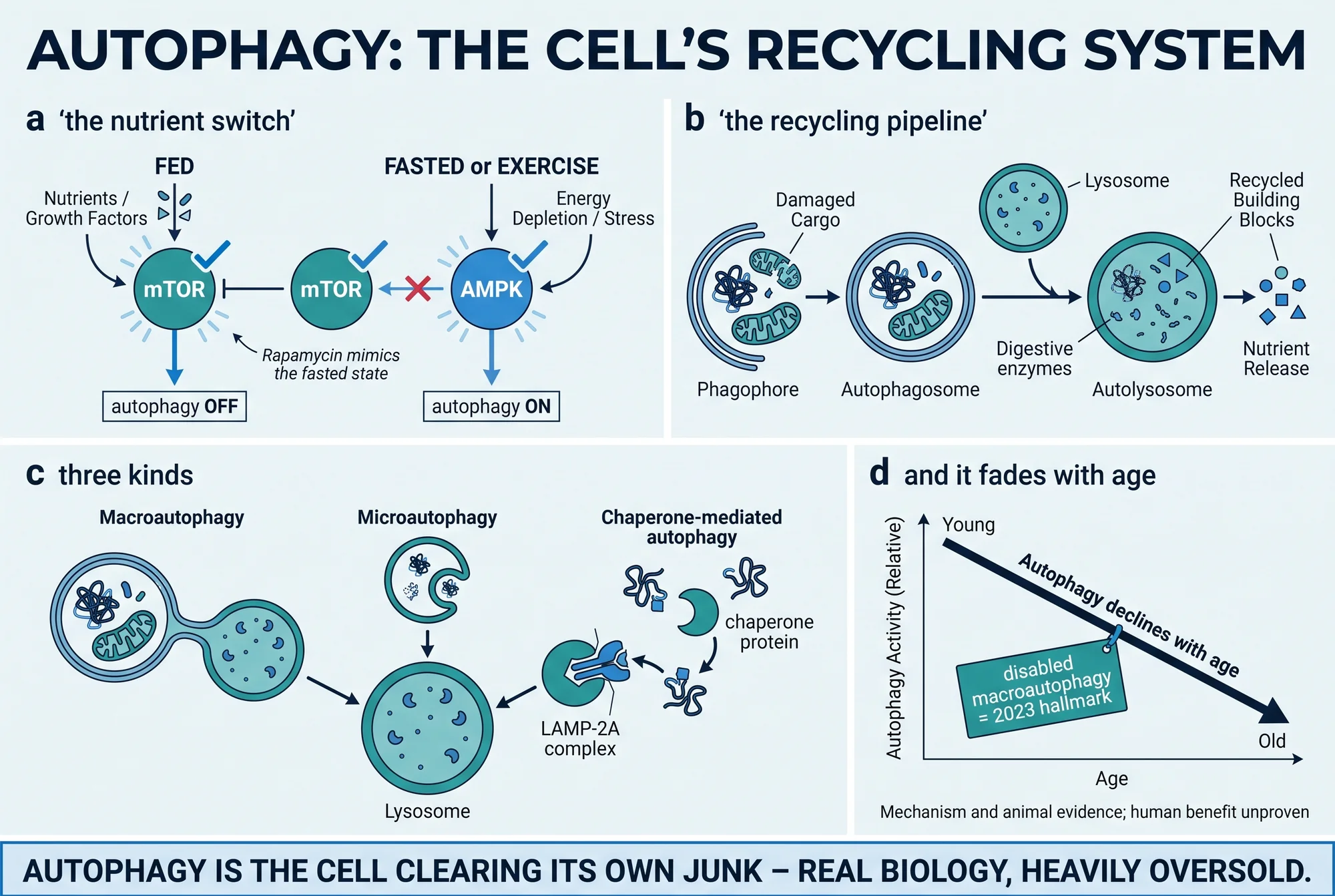
What autophagy actually is
Autophagy is, at bottom, bookkeeping for the inside of a cell: proteins misfold, mitochondria wear out, and aggregates pile up, and autophagy encloses such debris and delivers it to the lysosome, where enzymes reduce it to amino acids, fatty acids, and other components the cell reuses (Mizushima & Komatsu, 2011).
Two features make it distinctive. First, it is constant but adjustable — a basal level runs continuously as quality control, and cellular stress such as nutrient shortage dials it up (Dikic & Elazar, 2018). Second, it reaches what other systems cannot. The proteasome, the cell's other major degradation pathway, shreds individual tagged proteins but cannot swallow a whole organelle or a large insoluble clump; autophagy can, which makes it the only route capable of clearing, say, a damaged mitochondrion before it leaks (Parzych & Klionsky, 2014).
Three types, one destination
In everyday use "autophagy" means one specific process, but biologists distinguish three, and a serious account should not collapse them. A panel of leading researchers went to the trouble of defining the terms "based on specific biochemical features," precisely because "considerable confusion persists about the use of appropriate terms" (Galluzzi et al., 2017). All three end at the lysosome; they differ in how the cargo gets there.
Macroautophagy is the main, best-understood route, and the one almost everyone means by "autophagy." A fresh double membrane forms, curves around a portion of cytoplasm and its cargo, and seals into a vesicle called an autophagosome, which fuses with the lysosome so the contents can be digested (Mizushima & Komatsu, 2011; Parzych & Klionsky, 2014). This is the pathway behind the fasting response, the aging hallmark, and every other claim here unless stated otherwise.
Microautophagy skips the separate vesicle. Here the lysosome — or, in yeast, the vacuole — engulfs cargo directly, using "membrane dynamics to directly enwrap and transport cytosolic components into the lumen" by pinching in or protruding its own membrane (Oku & Sakai, 2018).
Chaperone-mediated autophagy (CMA) is the selective specialist, and among the three it is unique to mammals. There is no vesicle at all: a chaperone protein recognises a specific tag on an individual protein and threads it, one molecule at a time, across the lysosomal membrane through a dedicated translocation complex (Kaushik & Cuervo, 2018). CMA matters to aging in its own right — the same work notes that "CMA failure with age may aggravate diseases, such as ageing-associated neurodegeneration and cancer" (Kaushik & Cuervo, 2018).
How it was discovered: a yeast screen and a Nobel Prize
For most of the twentieth century autophagy was visible under the microscope but mechanistically a black box — no one knew which genes ran it. That changed with a single yeast experiment. In 1993, Yoshinori Ohsumi and Miki Tsukada starved baker's yeast of nitrogen and looked for mutants that could not digest their own proteins. They found strains that failed to break down protein under starvation and lost viability faster than normal cells. Seventy-five such mutants "fell into 15 complementation groups," implying that at least fifteen genes are needed for autophagy in yeast (Tsukada & Ohsumi, 1993). That screen is the origin of the entire autophagy-gene field.
The scattered early names those genes acquired were later consolidated into a single system — the ATG (autophagy-related) genes now used across species (Klionsky et al., 2003). Because the machinery is conserved from yeast to humans, naming it once let researchers study the same pathway in any organism.
The work became canonical in 2016, when the Nobel Prize in Physiology or Medicine was awarded solely to Yoshinori Ohsumi "for his discoveries of mechanisms for autophagy" (Nobel Foundation, 2016). It is worth dwelling on what kind of discovery this was: the core of human autophagy was worked out in budding yeast. That is a clean illustration of how model-organism biology becomes human-relevant — and a reminder, for everything that follows, that much of what we know about autophagy still comes from systems that are not human.
How autophagy is switched on and off
The control system is where mechanism and marketing most often part company. Autophagy is initiated by a kinase called ULK1 (the mammalian counterpart of yeast Atg1), and two opposing nutrient sensors set that switch.
The first is mTOR, the cell's "plenty" sensor. When nutrients and growth signals are abundant, mTOR is active, and active mTOR blocks autophagy — it phosphorylates ULK1 at a specific site (Ser757) and keeps it switched off (Kim et al., 2011). The second sensor is AMPK, the "low fuel" alarm. When energy runs short and glucose is scarce, AMPK switches on and activates autophagy by phosphorylating ULK1 at different sites (Ser317 and Ser777) (Kim et al., 2011). Fasting and energy stress therefore push the switch toward "on," abundant food toward "off." For more on the mTOR sensor and its wider role in growth and aging, see the mTOR explainer.
This is not only a cell-culture story. In transgenic mice engineered to make their autophagosomes glow, nutrient starvation visibly induced autophagy across most tissues, organ by organ — direct evidence that withdrawing food turns the pathway up in a living mammal (Mizushima et al., 2004). Downstream of the switch, the machinery runs as a relay: ULK1 initiates, a Beclin-1/VPS34 complex nucleates the new membrane, ATG proteins elongate it and tag it with lipidated LC3, the membrane closes around its cargo, and the autophagosome fuses with the lysosome to recycle the contents (Parzych & Klionsky, 2014).
Here is the careful line to hold. "Fasting turns on autophagy" is well supported as a mechanism and in animals. It is not the same statement as "fasting for a set number of hours boosts your autophagy and makes you healthier or longer-lived" — a claim that needs human outcome data the mechanism alone does not supply.
Autophagy, proteostasis, and aging
Keeping the cell's proteins in working order — folding them correctly, and clearing the ones that misfold — is called proteostasis, and autophagy is one of its pillars. When the influential "hallmarks of aging" framework was first set out, "loss of proteostasis" was one of the original nine hallmarks, with autophagy a central mechanism inside it (López-Otín et al., 2013). A decade later the framework was expanded, and autophagy was promoted to a hallmark in its own right: the 2023 update lists twelve hallmarks, and "disabled macroautophagy" is explicitly one of them (López-Otín et al., 2023). For the full framework, see The Hallmarks of Aging.
Two lines of evidence sit behind that promotion. The first is that autophagic capacity tends to fall with age. "Normal and pathological aging are often associated with a reduced autophagic potential," and genetically inhibiting autophagy "induces degenerative changes in mammalian tissues that resemble those associated with aging" (Rubinsztein et al., 2011); the CMA route specifically also declines with age (Kaushik & Cuervo, 2018). The honest caveat is that this decline is best documented in rodents and cells — there is no clean, decade-by-decade curve of autophagic activity in humans to put a number on it.
The second line is more dramatic. When researchers deleted an essential autophagy gene in the mouse nervous system, the animals developed neurodegeneration on their own. Removing Atg5 from neural cells "causes neurodegeneration even in the absence of any disease-associated mutant proteins," with progressive motor deficits and protein inclusion bodies (Hara et al., 2006); a companion study removing Atg7 from the central nervous system found behavioural deficits and "massive neuronal loss" while the proteasome kept working normally (Komatsu et al., 2006). Together they show that basal autophagy is required to keep neurons healthy. Two limits travel with that finding: these are mouse loss-of-function experiments, and "autophagy is necessary" is not the same as "more autophagy is better." The relationship between autophagy, aging, and disease is real and actively studied, but a recent synthesis calls the modulator evidence for age-related disease "preclinical" and states plainly that "the intricate relationship among autophagy, aging and disease remains unclear" (Aman et al., 2021).
The inducers, honestly
Four interventions come up again and again as ways to "boost autophagy": fasting or caloric restriction, exercise, the drug rapamycin, and the supplement spermidine. The essential point — the one the marketing omits — is that every strong causal result is in animals or non-mammalian models, and the organism belongs in the same breath as the finding.
Fasting and caloric restriction. Short-term fasting "induces profound neuronal autophagy" in mice, with a dramatic increase in cortical neurons and Purkinje cells and reduced neuronal mTOR activity (Alirezaei et al., 2010). The study's authors speculated that "sporadic fasting might represent a simple, safe and inexpensive means" to promote autophagy — but that is speculation, offered as such, in mice.
Exercise. Acute exercise induces autophagy in the skeletal and cardiac muscle of mice, and mice engineered so they cannot mount that response show reduced endurance and impaired glucose metabolism — so "autophagy induction may contribute to the beneficial metabolic effects of exercise" (He et al., 2012).
Rapamycin. Rapamycin is the pharmacological mirror of the mTOR brake: block mTOR and you release the switch. Fed to genetically diverse mice from 600 days of age — already old — it "extends median and maximal lifespan of both male and female mice," by roughly 14% in females and 9% in males at the 90%-mortality point, across three independent sites (Harrison et al., 2009). Those figures are mouse figures. Rapamycin is an approved immunosuppressant and cancer drug, not an approved longevity or autophagy therapy, and there is no completed equivalent human lifespan trial (see what longevity science actually shows).
Spermidine. Spermidine, a dietary polyamine, "markedly extended the lifespan of yeast, flies and worms, and human immune cells," inhibited oxidative stress in aging mice, and its effect depended on enhanced autophagy (Eisenberg et al., 2009). Read that precisely: the lifespan extension was in yeast, flies, worms, and cultured human immune cells; the mouse result was reduced oxidative stress, not a longer life. It is not a mammalian-lifespan or human-longevity result.
Why do these cluster together? In model systems, autophagy is a shared requirement downstream of several of the best-known longevity interventions: blocking it "compromises the longevity-promoting effects of caloric restriction, Sirtuin 1 activation, inhibition of insulin/insulin growth factor signaling, or the administration of rapamycin, resveratrol, or spermidine" (Rubinsztein et al., 2011). That convergence is why autophagy excites researchers — and why it is easy to oversell. But the gap that matters remains: none of these four inducers has a demonstrated human lifespan or healthspan outcome through autophagy. The human evidence is mechanistic, biomarker-level, associative, or absent. As a consumer proposition, "autophagy boosting" is largely unproven in people.
What remains uncertain
The distance between the biology and the marketing comes down to a few honest limits.
The first is measurement. Autophagy is genuinely hard to quantify — not a rhetorical hedge but a stated field consensus. The fourth edition of the community's assay guidelines, assembled by thousands of researchers, exists because "there continues to be confusion regarding acceptable methods to evaluate autophagy," and concludes that "no individual assay is perfect for every situation, calling for the use of multiple techniques" (Klionsky et al., 2021). For a reader this has a concrete consequence: a single static marker — a band of LC3 on a gel, say — is a snapshot, not a measure of flux. It cannot on its own prove autophagy went up or down, and it is especially hard to measure non-invasively in a living person. Claims about your personal autophagy level are, at present, claims no routine test can actually support.
The second is the species gap. Almost all of the causal evidence — the discovery genetics, the "autophagy is required" proofs, the lifespan and inducer results — comes from yeast, worms, flies, and mice (Aman et al., 2021). That is powerful biology, but not a demonstrated human outcome, which is why the most recent synthesis calls the age-and-disease evidence "preclinical" (Aman et al., 2021). The age-related decline of autophagy is likewise real but, in humans, qualitative: there is no verified human curve to quantify it (Rubinsztein et al., 2011; Kaushik & Cuervo, 2018).
The third is the fasting-hour myth, which deserves naming directly. The popular graphics that pin autophagy to a precise fasting window — that it "switches on" at twelve hours, or sixteen, or twenty-four — are not established by direct measurement of autophagy in fasting humans. That fasting induces autophagy is verified as a mechanism and in mice (Mizushima et al., 2004; Alirezaei et al., 2010); a specific human hour-threshold is not, and should be treated as an unproven popular claim, not a fact.
Finally, the biology resists the tempting "more is better" story. The mouse knockouts show autophagy is necessary (Hara et al., 2006; Komatsu et al., 2006), but the process is "tightly regulated" for a reason (Dikic & Elazar, 2018); it is a balance, not a dial you simply turn to maximum. The fair summary is the one the science supports on both sides: the mechanism is real, Nobel-validated, and central to aging biology, while the consumer promise — that you can reliably boost your own autophagy for a proven health or lifespan benefit — is not yet supported in humans.
This explainer covers what autophagy is, its three types, its discovery through Ohsumi's yeast genetics and the 2016 Nobel Prize, how the mTOR and AMPK nutrient sensors switch it on and off, its role in proteostasis and aging — including "disabled macroautophagy" as one of the twelve 2023 hallmarks — and the evidence behind the inducers most often marketed to "boost" it, drawn from primary studies and consensus statements retrieved from PubMed and PubMed Central, with the 2016 prize verified against the Nobel Foundation record. Evidence cutoff: sources as retrieved 24 August 2026. It is educational and is not medical advice, a diagnosis, or a treatment recommendation, and it names no dose, fasting schedule, or protocol for human use; every causal lifespan and healthspan result cited is from an animal or non-mammalian model, reported with the organism that produced it. For the wider framework, see The Hallmarks of Aging and what longevity science actually shows, and browse the Aging biology hub.
References
- 1.Alirezaei M, Kemball CC, Flynn CT, Wood MR, Whitton JL, Kiosses WB. Short-term fasting induces profound neuronal autophagy. Autophagy. 2010;6(6):702-710. doi:10.4161/auto.6.6.12376
- 2.Aman Y, Schmauck-Medina T, Hansen M, et al. Autophagy in healthy aging and disease. Nat Aging. 2021;1(8):634-650. doi:10.1038/s43587-021-00098-4
- 3.Dikic I, Elazar Z. Mechanism and medical implications of mammalian autophagy. Nat Rev Mol Cell Biol. 2018;19(6):349-364. doi:10.1038/s41580-018-0003-4
- 4.Eisenberg T, Knauer H, Schauer A, et al. Induction of autophagy by spermidine promotes longevity. Nat Cell Biol. 2009;11(11):1305-1314. doi:10.1038/ncb1975
- 5.Galluzzi L, Baehrecke EH, Ballabio A, et al. Molecular definitions of autophagy and related processes. EMBO J. 2017;36(13):1811-1836. doi:10.15252/embj.201796697
- 6.Hara T, Nakamura K, Matsui M, et al. Suppression of basal autophagy in neural cells causes neurodegenerative disease in mice. Nature. 2006;441(7095):885-889. doi:10.1038/nature04724
- 7.Harrison DE, Strong R, Sharp ZD, et al. Rapamycin fed late in life extends lifespan in genetically heterogeneous mice. Nature. 2009;460(7253):392-395. doi:10.1038/nature08221
- 8.He C, Bassik MC, Moresi V, et al. Exercise-induced BCL2-regulated autophagy is required for muscle glucose homeostasis. Nature. 2012;481(7382):511-515. doi:10.1038/nature10758
- 9.Kaushik S, Cuervo AM. The coming of age of chaperone-mediated autophagy. Nat Rev Mol Cell Biol. 2018;19(6):365-381. doi:10.1038/s41580-018-0001-6
- 10.Kim J, Kundu M, Viollet B, Guan KL. AMPK and mTOR regulate autophagy through direct phosphorylation of Ulk1. Nat Cell Biol. 2011;13(2):132-141. doi:10.1038/ncb2152
- 11.Klionsky DJ, Cregg JM, Dunn WA Jr, et al. A unified nomenclature for yeast autophagy-related genes. Dev Cell. 2003;5(4):539-545. doi:10.1016/s1534-5807(03)00296-x
- 12.Klionsky DJ, Abdel-Aziz AK, Abdelfatah S, et al. Guidelines for the use and interpretation of assays for monitoring autophagy (4th edition). Autophagy. 2021;17(1):1-382. doi:10.1080/15548627.2020.1797280
- 13.Komatsu M, Waguri S, Chiba T, et al. Loss of autophagy in the central nervous system causes neurodegeneration in mice. Nature. 2006;441(7095):880-884. doi:10.1038/nature04723
- 14.Lopez-Otin C, Blasco MA, Partridge L, Serrano M, Kroemer G. The hallmarks of aging. Cell. 2013;153(6):1194-1217. doi:10.1016/j.cell.2013.05.039
- 15.Lopez-Otin C, Blasco MA, Partridge L, Serrano M, Kroemer G. Hallmarks of aging: An expanding universe. Cell. 2023;186(2):243-278. doi:10.1016/j.cell.2022.11.001
- 16.Mizushima N, Yamamoto A, Matsui M, Yoshimori T, Ohsumi Y. In vivo analysis of autophagy in response to nutrient starvation using transgenic mice expressing a fluorescent autophagosome marker. Mol Biol Cell. 2004;15(3):1101-1111. doi:10.1091/mbc.e03-09-0704
- 17.Mizushima N, Komatsu M. Autophagy: renovation of cells and tissues. Cell. 2011;147(4):728-741. doi:10.1016/j.cell.2011.10.026
- 18.The Nobel Prize in Physiology or Medicine 2016. Yoshinori Ohsumi, 'for his discoveries of mechanisms for autophagy.' Nobel Foundation. Link
- 19.Oku M, Sakai Y. Three Distinct Types of Microautophagy Based on Membrane Dynamics and Molecular Machineries. Bioessays. 2018;40(6):e1800008. doi:10.1002/bies.201800008
- 20.Parzych KR, Klionsky DJ. An overview of autophagy: morphology, mechanism, and regulation. Antioxid Redox Signal. 2014;20(3):460-473. doi:10.1089/ars.2013.5371
- 21.Rubinsztein DC, Marino G, Kroemer G. Autophagy and aging. Cell. 2011;146(5):682-695. doi:10.1016/j.cell.2011.07.030
- 22.Tsukada M, Ohsumi Y. Isolation and characterization of autophagy-defective mutants of Saccharomyces cerevisiae. FEBS Lett. 1993;333(1-2):169-174. doi:10.1016/0014-5793(93)80398-e
Disclosures
Educational review of published evidence. Not medical advice, diagnosis, or a treatment recommendation, and it names no dose, fasting schedule, or protocol for human use. The autophagy mechanism is well established; the causal lifespan and healthspan data for every inducer discussed are from animal or non-mammalian models, not humans. Rapamycin is an approved immunosuppressant and cancer drug, and spermidine is a dietary supplement — neither is an approved longevity or 'autophagy' therapy.