
Muscle Is a Metabolic Organ
It is the largest place your blood sugar goes after a meal, an endocrine tissue that signals to the rest of the body, and a protein reserve you slowly spend as you age.
Abstract
Skeletal muscle is the body's largest site of insulin-stimulated glucose disposal — 80 to 90 percent under clamp conditions — and the tissue where the earliest defect on the road to type 2 diabetes appears. It is also an endocrine organ that secretes signaling proteins called myokines, and the body's principal amino-acid reserve. This review explains muscle's metabolic roles, the age-related loss of muscle known as sarcopenia and its observational links to disability and death, the lean-mass question raised by GLP-1 weight-loss therapies, and what the exercise evidence does and does not establish. Throughout, mechanism and animal findings such as myostatin and irisin are kept separate from demonstrated human outcomes, and observational associations are kept separate from the interventional and physiological anchors that license causal language.
Key findings
- Muscle is the body's largest glucose sink: 80–90% of insulin-stimulated glucose disposal occurs in skeletal muscle, most of it stored as glycogen, and impaired muscle glycogen synthesis is among the earliest detectable defects in type 2 diabetes (DeFronzo & Tripathy, 2009; Shulman et al., 1990).
- Muscle is an endocrine organ. It secretes myokines; the archetype, interleukin-6, can rise up to ~100-fold during exercise (Pedersen & Febbraio, 2008). Other signals sit at an earlier evidence stage — myostatin is mouse genetics (McPherron et al., 1997) and irisin's human magnitude is genuinely disputed (Albrecht et al., 2015; Jedrychowski et al., 2015).
- Muscle is lost with age (sarcopenia), and low muscle mass is associated with worse glucose regulation, disability, and roughly 1.6- to 3.6-fold higher mortality across meta-analyses — associations, not proof (Beaudart et al., 2017; Liu et al., 2017; Srikanthan & Karlamangla, 2014).
- GLP-1 weight loss includes some lean mass — reported at 40–60% of weight lost in some trials and ~15% or less in others — but lean mass is not muscle: it includes organs, bone, and fluid, and MRI suggests the muscle changes are largely adaptive (Neeland et al., 2024).
- Exercise measurably raises the muscle glucose sink in randomized trials (clamp-measured disposal, SMD 0.52; Rebello et al., 2023), and resistance training specifically tracks with lower diabetes risk in cohort data (Grontved et al., 2012) — though a resistance-training-only clamp effect has not been isolated.
Ask what skeletal muscle is for and the obvious answer is movement — the tissue that lets you stand, lift, and walk. That answer is correct and incomplete. Muscle is also one of the most metabolically active tissues in the body, and it earns the word organ three times over. It is the single largest destination for the sugar in your blood after a meal. It is an endocrine tissue, releasing signaling proteins that travel to fat, liver, bone, and brain. And it is the body's protein bank — the reserve of amino acids that vital organs draw on during illness and fasting, and a supplier of the raw material the liver uses to make new glucose (Wolfe, 2006). Seen this way, muscle is not the engine the metabolism drives; it is part of the metabolism itself.
That reframing has a sharp practical edge. Because muscle handles most of the body's glucose, it is where metabolic trouble tends to begin, and where the earliest signs of type 2 diabetes appear (DeFronzo & Tripathy, 2009). And because muscle is lost steadily with age, keeping it has moved from the gym toward the clinic — sharpened by weight-loss drugs that shed some muscle along with fat. What follows is what muscle does metabolically, why losing it matters, and how much of that is established versus merely suspected.
The body's largest glucose sink
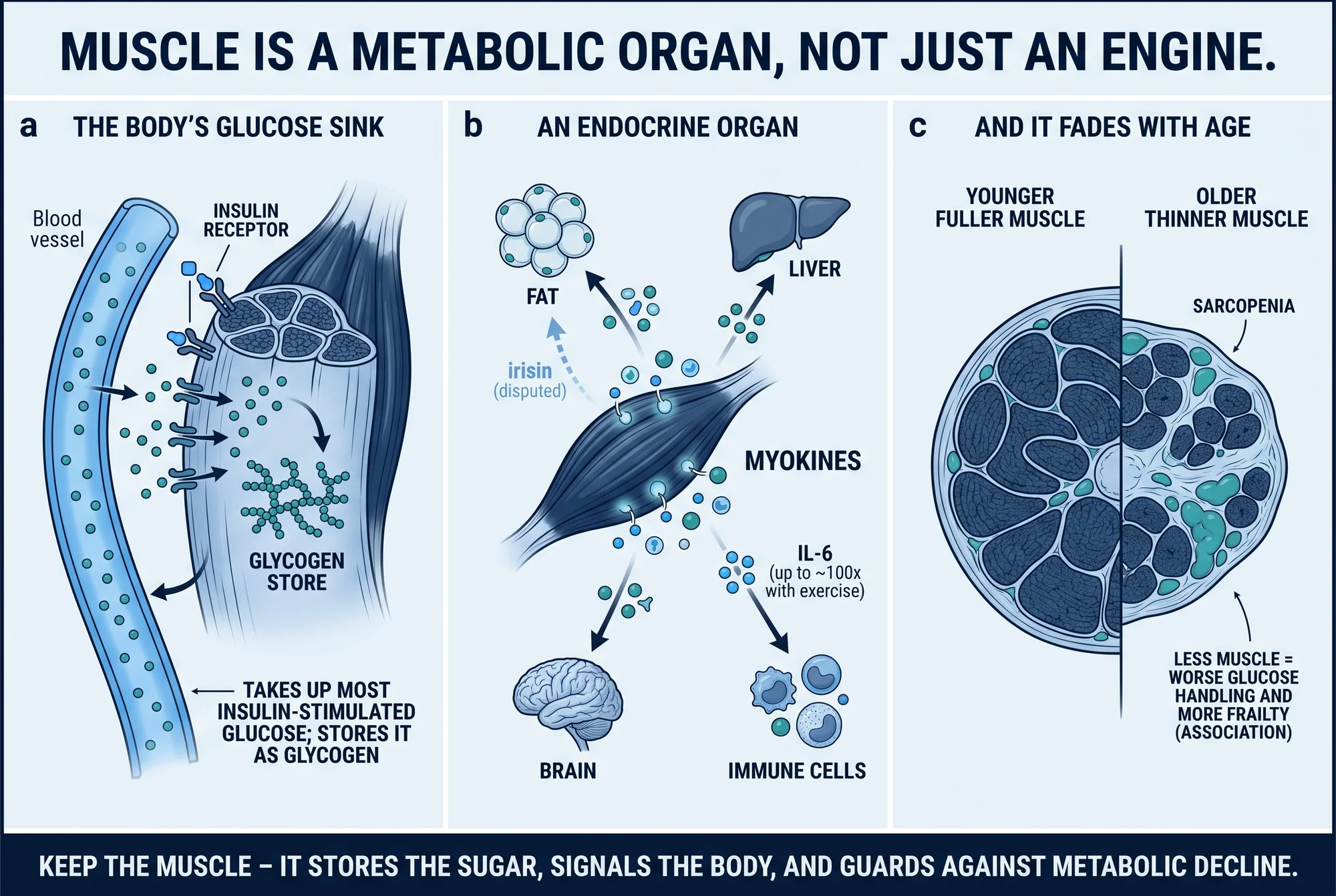
Start with the number that defines muscle's metabolic role. When insulin is doing its job — measured with a euglycemic-hyperinsulinemic clamp, the reference method for insulin sensitivity — 80 to 90% of the glucose the body disposes of is taken up by skeletal muscle (DeFronzo & Tripathy, 2009). No other tissue comes close. The contrast with fasting is the point: between meals only about a quarter of glucose uptake goes to insulin-sensitive tissue, while 70 to 75% is claimed by the brain, red blood cells, and gut, which need no insulin to take up sugar. Muscle's dominance is specifically an insulin-driven, after-a-meal phenomenon — striking enough that the sibling explainer on insulin resistance charts it directly.
Once inside the muscle, most of that glucose is filed away rather than burned. Roughly 75 to 80% of it is converted to glycogen, the branched storage form of sugar, making skeletal muscle the body's predominant site of glycogen synthesis (DeFronzo & Tripathy, 2009). That storage step is the fragile link: impaired muscle glycogen synthesis, traced to reduced activity of the enzyme glycogen synthase, is one of the earliest metabolic defects detectable in type 2 diabetes — present long before blood sugar climbs into a diagnostic range.
How large is that defect? One landmark study measured it directly, using a clamp with carbon-13 nuclear magnetic resonance to watch glycogen form inside the muscle. People with non-insulin-dependent diabetes made muscle glycogen at 78 versus 183 micromoles per kilogram of muscle per minute in matched controls, with glucose uptake of 30 versus 51 — less than half the normal rate on both counts (Shulman et al., 1990). Muscle glycogen synthesis, the authors concluded, is the principal pathway of glucose disposal, and its failure dominates the insulin resistance of that disease. Small — five patients, six controls — but a direct human measurement, not an inference.
The defect then spills over to other organs. When muscle cannot stow ingested carbohydrate as glycogen, the sugar goes to the liver instead. In young, lean, insulin-resistant volunteers given two high-carbohydrate meals, muscle made about 60% less glycogen than in insulin-sensitive controls, while the liver more than doubled its production of new fat; plasma triglycerides rose about 60% and HDL cholesterol fell about 20% (Petersen et al., 2007). Within a single day of eating, a muscle problem becomes a whole-body lipid problem — the common cellular thread being ectopic lipid, the diacylglycerols and ceramides that accumulate in muscle and liver and blunt the insulin signal (Samuel & Shulman, 2012).
This is why muscle sits upstream of so much else: skeletal-muscle insulin resistance is considered the initiating defect in type 2 diabetes, evident decades before the insulin-producing beta cells fail (DeFronzo & Tripathy, 2009) — a central thread in the broader account of metabolic health. It also hints that more muscle might help. The observational data are consistent: in more than 13,000 US adults, each 10% higher relative muscle mass was associated with an 11% lower level of insulin resistance (95% CI 6–15%) and a 12% lower prevalence of pre- or overt diabetes (95% CI 1–21%), adjusting for age, sex, ethnicity, and obesity (Srikanthan & Karlamangla, 2011). That is a cross-sectional association, not evidence that adding muscle lowers insulin resistance — the authors called for interventional studies — but it points the same way as the physiology.
An endocrine organ
Muscle does not only consume and store fuel; it talks. Contracting muscle secretes signaling proteins that act on distant tissues — the recognition of which earned it the label endocrine organ. These muscle-derived signals are called myokines (Pedersen & Febbraio, 2008).
The archetype is interleukin-6, and its behavior is dramatic: circulating IL-6 can rise up to about 100-fold during physical exercise, released from the working muscle itself (Pedersen & Febbraio, 2008). IL-6 also shows why context is everything here. Released acutely from contracting muscle, it is associated with enhanced insulin action in the hours after exercise; chronically elevated — the pattern seen in obesity — it is associated with the opposite, reduced insulin action and low-grade inflammation. The same molecule means different things depending on whether it spikes with activity or stays high all the time. The reframing matters more than any single pathway: maintaining muscle preserves a tissue that signals to the rest of the metabolism.
Two other myokines are worth naming precisely because they are so often overstated. The first is myostatin, a muscle-made protein of the TGF-beta family that acts as a brake on muscle growth. Delete its gene in mice and the animals become strikingly large, individual muscles weighing two to three times normal through a mix of more fibers and bigger ones (McPherron et al., 1997). It is a compelling demonstration that muscle mass is actively, genetically regulated — but a mouse gene-knockout experiment, to be read as biology, not a demonstrated human therapy. The "double-muscled" mouse shows that muscle has a built-in governor; it does not tell you what blocking that governor would do in a person.
The second is irisin, the cautionary tale of the field. In 2012 it was proposed as an exercise-induced myokine that turns energy-storing white fat toward calorie-burning brown-fat-like tissue and improves glucose handling — shown largely in mice and cultured cells, with limited human data (Bostrom et al., 2012). It became one of the most disputed findings in metabolism, and the dispute is specifically about magnitude and even existence in humans. On the skeptical side, a 2015 analysis found that the commercial antibody kits used to measure human irisin cross-reacted with unrelated proteins, and argued the data provided evidence against a physiological role for irisin in humans (Albrecht et al., 2015). The same year, a group using mass spectrometry rather than antibody kits reported that human irisin does circulate — at roughly 3.6 nanograms per milliliter at rest, rising to about 4.3 with aerobic interval training — and concluded it exists and is regulated by exercise (Jedrychowski et al., 2015). The honest reading: irisin almost certainly exists at low concentrations and responds to exercise, but its metabolic magnitude and any therapeutic relevance in humans remain unproven and actively debated — the science still arguing with itself, not a benefit to promise.
Muscle, aging, and sarcopenia
Muscle is not permanent. It is lost gradually with age, and when that loss becomes clinically meaningful it has a name: sarcopenia. A 2019 European consensus describes it as a muscle disease — muscle failure — rooted in adverse changes that accumulate across a lifetime, most common in older age but able to begin earlier. The consensus made low muscle strength the primary criterion, low quantity or quality confirmatory, and poor physical performance the marker of severe disease (Cruz-Jentoft et al., 2019).
An immediate complication is that the definition is not settled — itself a finding. Pooling 151 studies covering nearly 700,000 people, a global meta-analysis found sarcopenia prevalence ranging from 10% to 27% — and severe sarcopenia from 2% to 9% — depending purely on the diagnostic criteria and cut-points applied (Petermann-Rocha et al., 2022). There is no single correct prevalence figure, only a range and the reason for it; any "X% of older adults have sarcopenia" claim is incomplete without naming its definition.
What is more consistent is that less muscle tends to go with worse outcomes. A meta-analysis of 17 prospective studies found sarcopenia associated with substantially higher all-cause mortality — a pooled odds ratio of 3.60 (95% CI 2.96–4.37) — plus roughly threefold higher odds of functional decline (OR 3.03, 95% CI 1.80–5.12) and more falls and hospitalizations (Beaudart et al., 2017). A second meta-analysis, of community-dwelling older adults, put the mortality association lower, at a pooled hazard ratio of 1.60 (95% CI 1.24–2.06), shifting with how muscle mass was measured (Liu et al., 2017). Both numbers are verified; they differ because they use different metrics, inclusion criteria, and populations. The defensible statement is that sarcopenia is associated with roughly a 1.6- to 3.6-fold higher risk of death across analyses, not a single precise multiple.
The muscle-survival link is not confined to a sarcopenia diagnosis. In a general older US population, adults in the highest quartile of muscle-mass index had about 20% lower all-cause mortality than those in the lowest (adjusted hazard ratio 0.80, 95% CI 0.66–0.97), independent of central obesity (Srikanthan & Karlamangla, 2014). These are observational associations, measured with imperfect estimates of muscle mass and open to reverse causation — illness can both shrink muscle and raise mortality — so they show that muscle tracks with survival, not that preserving it guarantees a longer life. But they align with the physiological reason muscle matters with age: it is the body's amino-acid reserve, drawn on by vital tissues during the stress of illness and fasting (Wolfe, 2006). Losing it is not only losing strength.
Muscle in the GLP-1 era
The newest reason to think about muscle comes from the drugs reshaping obesity treatment. GLP-1-based therapies produce large, rapid weight loss, and a recurring worry is how much of it is muscle. The honest answer is that the evidence is unsettled, and the framing matters as much as the numbers.
Reported reductions in lean mass with these therapies are heterogeneous: 40 to 60% of total weight lost appears as lean mass in some trials, and roughly 15% or less in others (Neeland et al., 2024). The single most important caveat is definitional. "Lean mass" on a standard body-composition scan is not muscle; it includes organs, bone, connective tissue, and body water, all of which change during weight loss. When studies use MRI to look specifically at skeletal muscle, the changes appear largely adaptive — in line with what the degree of weight loss, aging, and baseline health would predict, and accompanied by improved insulin sensitivity and less fat infiltrating the muscle. A loss of strength or function is therefore not the default expectation for most people. The genuine open questions are at the edges: older patients and those with more advanced disease may face higher sarcopenia risk, which bears on who is a good candidate, and muscle-preserving combination agents are in development. What the evidence does not support is a single headline figure of the form "GLP-1 costs you X% of your muscle." The state of knowledge is a range, a measurement caveat, and an "appears largely adaptive, watch the high-risk groups" summary — explored more fully in what GLP-1 is and how it works.
What preserves it
If muscle does this much metabolic work, what keeps it functioning? The evidence divides cleanly into what controlled trials have demonstrated and what has only been observed.
The strongest interventional evidence is that exercise raises the muscle glucose sink itself. Pooling 25 randomized controlled trials that measured glucose disposal with the clamp method, exercise training produced a clear improvement — a standardized effect of 0.52 (95% CI 0.39–0.65) — maximized when training was paired with weight loss (Rebello et al., 2023). That is a measured human outcome, not a mechanism: exercise demonstrably increases how much glucose insulin can drive into the body. One honest limit travels with it — the meta-analysis pooled aerobic and resistance training together, and does not isolate a resistance-training-only effect size.
Resistance training on its own has a different kind of support — strong but observational. In a prospective study of 32,002 men followed for 18 years, weight training for at least 150 minutes a week was independently associated with 34% lower risk of type 2 diabetes (95% CI 7–54%), holding even after accounting for aerobic exercise (which was associated with 52% lower risk; doing both, 59%) (Grontved et al., 2012). That is a large, dose-responsive association — but a cohort study relying on self-reported activity in men only, evidence that resistance training tracks with lower diabetes risk, not proof that it causes it.
The gap between those two findings is worth stating plainly: the cleanest, clamp-measured evidence covers exercise broadly (Rebello et al., 2023), while the resistance-specific evidence is a prospective association (Grontved et al., 2012), with no dedicated resistance-only clamp trial isolated here. The reasonable summary is that exercise reliably improves the muscle's capacity to handle glucose, and resistance training in particular tracks with lower diabetes risk — reported as what the studies observed, not as a prescription.
What remains uncertain
Several honest caveats belong beside the confidence.
- Irisin is genuinely unresolved. Its existence at low circulating levels and its response to exercise are supported by mass spectrometry (Jedrychowski et al., 2015), but the earlier antibody-based human literature was undermined by cross-reactivity (Albrecht et al., 2015), and the large metabolic effects claimed from mouse work (Bostrom et al., 2012) are not established in humans. The correct description is "a real but small and still-debated signal," not "an exercise hormone that browns your fat."
- Sarcopenia thresholds are not standardized. The 2019 European consensus (Cruz-Jentoft et al., 2019) is one of several competing definitions; prevalence swings from roughly 10% to 27% purely on the criteria chosen (Petermann-Rocha et al., 2022), and the mortality association ranges from about 1.6-fold (Liu et al., 2017) to 3.6-fold (Beaudart et al., 2017) depending on the metric and how muscle mass is measured. Any figure must name its definition and method.
- Association is not causation — the central caveat of the whole subject. The links from more muscle to better glucose control (Srikanthan & Karlamangla, 2011), from more muscle to longer survival (Srikanthan & Karlamangla, 2014), and from weight training to lower diabetes risk (Grontved et al., 2012) are all observational, with reverse causation and confounding live. Causal language is licensed only by the interventional and physiological claims — exercise raising clamp-measured disposal in randomized trials (Rebello et al., 2023) and the direct human mechanism of muscle glucose disposal and glycogen storage (DeFronzo & Tripathy, 2009; Shulman et al., 1990; Petersen et al., 2007). The two registers should not be blurred.
- Mechanism is not human outcome. Myostatin biology is mouse genetics (McPherron et al., 1997) and irisin's browning effect is mouse and cell culture (Bostrom et al., 2012). They explain why muscle is regulated and secretory; they are not evidence of a clinical benefit in people.
This review summarizes published human, animal, and cell-culture evidence through 2024 on skeletal muscle as a metabolic organ — glucose disposal and glycogen storage, endocrine myokine signaling, age-related muscle loss, the GLP-1 lean-mass question, and the exercise evidence. Animal and in-vitro findings are labelled as such and are not presented as human outcomes; observational associations are kept separate from interventional and physiological evidence. It is educational and is not medical advice, a diagnosis, or a treatment plan. For related reading, see What Is Metabolic Health?, Insulin Resistance, and What Is GLP-1 and How Does It Work?, or browse the Metabolic health hub.
References
- 1.Albrecht E, Norheim F, Thiede B, et al. Irisin - a myth rather than an exercise-inducible myokine. Sci Rep. 2015;5:8889. doi:10.1038/srep08889
- 2.Beaudart C, Zaaria M, Pasleau F, et al. Health outcomes of sarcopenia: a systematic review and meta-analysis. PLoS One. 2017;12(1):e0169548. doi:10.1371/journal.pone.0169548
- 3.Bostrom P, Wu J, Jedrychowski MP, et al. A PGC1-alpha-dependent myokine that drives brown-fat-like development of white fat and thermogenesis. Nature. 2012;481(7382):463-468. doi:10.1038/nature10777
- 4.Cruz-Jentoft AJ, Bahat G, Bauer J, et al. Sarcopenia: revised European consensus on definition and diagnosis. Age Ageing. 2019;48(1):16-31. doi:10.1093/ageing/afy169
- 5.DeFronzo RA, Tripathy D. Skeletal muscle insulin resistance is the primary defect in type 2 diabetes. Diabetes Care. 2009;32(Suppl 2):S157-S163. doi:10.2337/dc09-S302
- 6.Grontved A, Rimm EB, Willett WC, et al. A prospective study of weight training and risk of type 2 diabetes mellitus in men. Arch Intern Med. 2012;172(17):1306-1312. doi:10.1001/archinternmed.2012.3138
- 7.Jedrychowski MP, Wrann CD, Paulo JA, et al. Detection and quantitation of circulating human irisin by tandem mass spectrometry. Cell Metab. 2015;22(4):734-740. doi:10.1016/j.cmet.2015.08.001
- 8.Liu P, Hao Q, Hai S, et al. Sarcopenia as a predictor of all-cause mortality among community-dwelling older people: a systematic review and meta-analysis. Maturitas. 2017;103:16-22. doi:10.1016/j.maturitas.2017.04.007
- 9.McPherron AC, Lawler AM, Lee SJ. Regulation of skeletal muscle mass in mice by a new TGF-beta superfamily member. Nature. 1997;387(6628):83-90. doi:10.1038/387083a0
- 10.Neeland IJ, Linge J, Birkenfeld AL. Changes in lean body mass with glucagon-like peptide-1-based therapies and mitigation strategies. Diabetes Obes Metab. 2024;26(Suppl 4):16-27. doi:10.1111/dom.15728
- 11.Pedersen BK, Febbraio MA. Muscle as an endocrine organ: focus on muscle-derived interleukin-6. Physiol Rev. 2008;88(4):1379-1406. doi:10.1152/physrev.90100.2007
- 12.Petermann-Rocha F, Balntzi V, Gray SR, et al. Global prevalence of sarcopenia and severe sarcopenia: a systematic review and meta-analysis. J Cachexia Sarcopenia Muscle. 2022;13(1):86-99. doi:10.1002/jcsm.12783
- 13.Petersen KF, Dufour S, Savage DB, et al. The role of skeletal muscle insulin resistance in the pathogenesis of the metabolic syndrome. Proc Natl Acad Sci U S A. 2007;104(31):12587-12594. doi:10.1073/pnas.0705408104
- 14.Rebello CJ, Zhang D, Kirwan JP, et al. Effect of exercise training on insulin-stimulated glucose disposal: a systematic review and meta-analysis of randomized controlled trials. Int J Obes (Lond). 2023;47(5):348-357. doi:10.1038/s41366-023-01283-8
- 15.Samuel VT, Shulman GI. Mechanisms for insulin resistance: common threads and missing links. Cell. 2012;148(5):852-871. doi:10.1016/j.cell.2012.02.017
- 16.Shulman GI, Rothman DL, Jue T, et al. Quantitation of muscle glycogen synthesis in normal subjects and subjects with non-insulin-dependent diabetes by 13C nuclear magnetic resonance spectroscopy. N Engl J Med. 1990;322(4):223-228. doi:10.1056/NEJM199001253220403
- 17.Srikanthan P, Karlamangla AS. Relative muscle mass is inversely associated with insulin resistance and prediabetes. Findings from the Third National Health and Nutrition Examination Survey. J Clin Endocrinol Metab. 2011;96(9):2898-2903. doi:10.1210/jc.2011-0435
- 18.Srikanthan P, Karlamangla AS. Muscle mass index as a predictor of longevity in older adults. Am J Med. 2014;127(6):547-553. doi:10.1016/j.amjmed.2014.02.007
- 19.Wolfe RR. The underappreciated role of muscle in health and disease. Am J Clin Nutr. 2006;84(3):475-482. doi:10.1093/ajcn/84.3.475
Disclosures
Educational review of published evidence. Not medical advice, diagnosis, or a treatment recommendation. Study parameters are reported with the population and design that produced them; animal and cell-culture findings are labelled as such and are not human outcomes.