
PNC-27 A fragment of the guardian gene, a door punched in the cancer-cell membrane, and how far a striking result reaches when no person has yet been treated
PNC-27 is a short piece of the human tumour-suppressor protein p53 tied to a molecular delivery tag. It was built to slip inside cancer cells and set their p53 free. Instead it was caught doing something stranger and more useful — tearing pores in the cancer-cell membrane by seizing a protein that, for reasons still unexplained, sits on the outside of cancer cells and almost nowhere else. This is the story of that accident, and of what a compelling result in dishes and mice is worth before a single human has been treated.
01The guardian and its jailer
Almost every human cancer has to solve the same problem. Our cells carry a protein called p53 whose job is to notice when something has gone badly wrong — DNA damage, garbled growth signals, the early moves of a cell turning malignant — and to respond by halting division or ordering the cell to kill itself. It is switched on precisely when a cell is becoming dangerous, which is why it earned the nickname “the guardian of the genome.” A cell that is trying to become a tumour cannot afford a working guardian.
Cancers dispose of p53 in two broad ways. Roughly half mutate the p53 gene directly, so the protein comes out broken. The rest often leave p53 intact but neutralise it, and the commonest tool for that job is a protein called MDM2 — in humans, HDM-2. HDM-2 is p53's designated jailer. It is an enzyme that grabs p53, attaches a molecular tag that marks it for the cell's protein-shredding machinery, and thereby keeps p53 levels low. In healthy tissue this is a sensible thermostat: p53 and HDM-2 hold each other in check. In many cancers the thermostat is broken the wrong way, with too much HDM-2 keeping a perfectly good guardian permanently locked up.
By the early 2000s this had made HDM-2 one of the most fashionable targets in cancer biology. The mainstream idea was elegant: if you could jam the exact spot where HDM-2 grips p53, the guardian would be released and could get back to work. Small molecules that do this — the nutlins and their descendants (Vassilev et al., 2004) — became a major research programme. But that strategy has a built-in limit: it only helps in cancers that still carry a working copy of p53. Where p53 is deleted or mutated, freeing it accomplishes nothing. PNC-27 came out of the same starting idea and then walked off in an entirely different direction.
02A decoy that misbehaved
In the laboratories of Matthew R. Pincus and Josef Michl, working at the State University of New York and collaborating institutions in New York City, a group had been using computer modelling to design short peptides — tiny protein fragments — that mimic functional pieces of larger proteins. Their broader project produced a numbered series of “PNC” peptides; some were copied from the cancer protein ras, and two, PNC-27 and its shorter sibling PNC-28, were copied from p53 (Pincus et al., 2011).
The design logic for PNC-27 was straightforward decoy chemistry. Take the exact stretch of p53 that HDM-2 grabs — residues 12 through 26 of the protein — and deliver it into the cancer cell in bulk. The reasoning: flood the cell with p53 look-alike fragments, let them occupy HDM-2, and the cell's own full-length p53 would be left alone and free to trigger apoptosis. A fragment that short cannot cross a cell membrane on its own, so the group fused it to a second peptide, a well-known “cell-penetrating” tag called penetratin, borrowed from a fruit-fly protein, that carries cargo across membranes. The p53 piece was the warhead; penetratin was the delivery truck.
What happened when they added it to cancer cells was not what the design predicted. The cells did not die slowly by apoptosis, the orderly self-destruct program p53 normally triggers. They died fast — within minutes to a few hours — and messily, spilling their contents in the unmistakable signature of necrosis, a cell bursting rather than quietly dismantling itself. Stranger still, PNC-27 killed cancer cells that had no p53 gene at all. Whatever it was doing, it was not the decoy trick it had been designed for. The molecule had a mechanism of its own, and the next fifteen years of work were largely an attempt to explain it.
The first careful look at the molecule itself came in 2004, when the group determined the three-dimensional shape of the 32-residue PNC-27 by nuclear magnetic resonance and published it under the peptide's now-standard name (Rosal et al., 2004). That structure — an amphipathic helix-loop-helix, described in the next section — was the first clue that PNC-27 behaved less like a decoy and more like the membrane-puncturing peptides found in venoms and antibiotics.
03What the molecule actually is
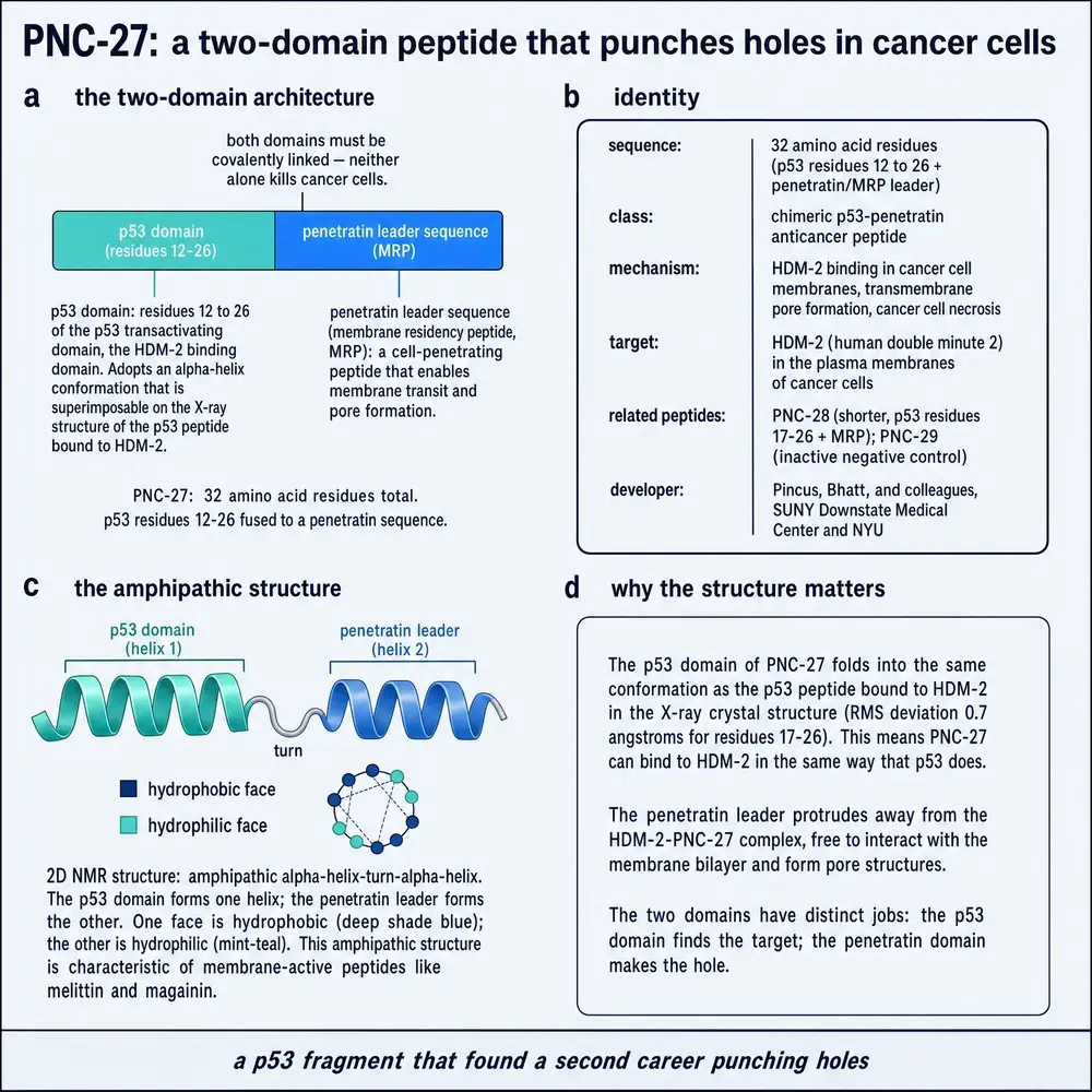
PNC-27 is a single chain of 32 amino acids in two functional halves (Sarafraz-Yazdi et al., 2022). The first fifteen residues are a verbatim copy of the human p53 protein's residues 12–26 — the exact sequence that grips HDM-2. The remaining seventeen residues are the penetratin-derived delivery tag, which the group calls the membrane-residency peptide, or MRP. The two halves are joined end to end into one continuous molecule.
That the two halves must be physically joined turned out to be essential, and the control experiments make the point cleanly. The p53 fragment on its own does nothing to cancer cells — it cannot get in. The delivery tag on its own does nothing. Even the two peptides added together, but not chemically linked, do nothing. Only the single fused molecule kills (Sarafraz-Yazdi et al., 2022; Michl et al., 2006). Two further peptides serve as the workhorse negative controls throughout the literature: PNC-29, an unrelated sequence from a liver enzyme carrying the same delivery tag, and PNC-26, the p53 fragment with the tag removed. Neither harms cancer or normal cells, which is what lets the researchers attribute effects specifically to PNC-27.
The shape of the molecule is the second half of its identity. By NMR, PNC-27 folds into an amphipathic helix-loop-helix: two short spiral segments joined by a bend, arranged so that the water-fearing (hydrophobic) side chains gather on one face and the charged, water-loving residues gather on the other (Rosal et al., 2004). This is not an exotic architecture — it is almost exactly the fold used by the natural membrane-puncturing peptides melittin (from bee venom) and magainin (an antimicrobial), which lyse cells by inserting into membranes. That resemblance was the tell. A molecule built to be a quiet intracellular decoy had the shape of a membrane weapon.
The commissioned plate that follows restates the same molecule as an identity card: the two domains, the inactive controls, the amphipathic fold, and why that fold matters for a membrane weapon.
04A protein in the wrong place
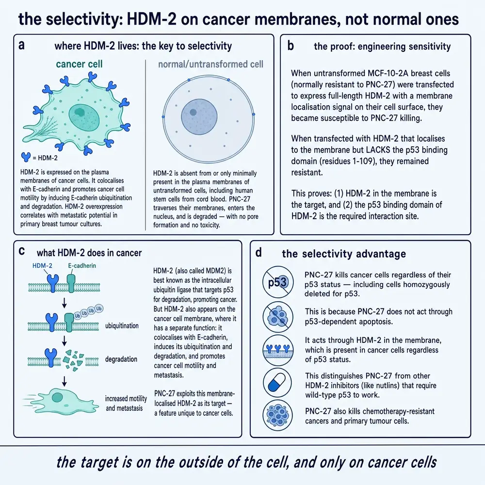
The explanation that eventually emerged is genuinely surprising, and it rests on one unexpected observation. HDM-2 is supposed to be an interior protein. It does its jailer's work on p53 inside the nucleus and cytoplasm; that is where a cell-biology textbook puts it. But the Pincus–Michl group, and later others, found significant amounts of HDM-2 sitting in the plasma membrane — the outer skin — of cancer cells, facing outward, where it has no business being. Just as importantly, they did not find it in the membranes of normal cells (Sarafraz-Yazdi et al., 2010).
This membrane-bound HDM-2 has now been reported on the surface of a long list of cancer cell types — pancreatic, breast, melanoma, colon, ovarian and leukaemic — and repeatedly found absent or minimal on matched normal cells (Sarafraz-Yazdi et al., 2010; Thadi et al., 2020; Wang et al., 2020; Krzesaj et al., 2025). Why HDM-2 should be mislocalised to the cell surface in cancer, and how it gets there, remains genuinely unexplained — it is one of the open questions the whole PNC-27 story hangs on. But if the observation holds, it hands a drug designer something rare: a molecular flag flown by cancer cells and almost no others.
The cleanest evidence that membrane HDM-2 is the actual target is a make-it-and-break-it experiment. Take a normal cell that PNC-27 leaves alone, and force it to display HDM-2 on its surface by adding a membrane-targeting signal to the gene. That formerly resistant normal cell becomes susceptible and is killed by PNC-27. Do the same with an empty gene, or with an HDM-2 that lacks the p53-binding pocket, and nothing changes — the cell survives (Sarafraz-Yazdi et al., 2010; 2022). Susceptibility travels with surface HDM-2. A monoclonal antibody aimed at HDM-2's p53-binding site blocks the killing in proportion to its dose (Krzesaj et al., 2024). The target is not a guess; it can be added, removed and masked, and the killing follows it each time.
The next plate expands that selectivity argument: where HDM-2 sits, the transfection proof that puts it there, what it does to E-cadherin, and why the mechanism still works when p53 itself is gone.
05Poptosis: punching the hole
What happens after PNC-27 binds surface HDM-2 has been imaged in unusual detail. Under electron microscopy, cancer cells treated with PNC-27 for only a few minutes develop discrete pores — ring-shaped holes — across their plasma membranes; untreated cells and treated normal cells show none (Sarafraz-Yazdi et al., 2022). Using antibodies tagged with gold particles of two sizes, the group could label PNC-27 and HDM-2 separately and watch where each ended up. The pores were rimmed by roughly one-to-one complexes of PNC-27 and HDM-2, with an inner diameter averaging about 35 nanometres (Sarafraz-Yazdi et al., 2022). The peptide was not merely near the pore; it was a structural part of it.
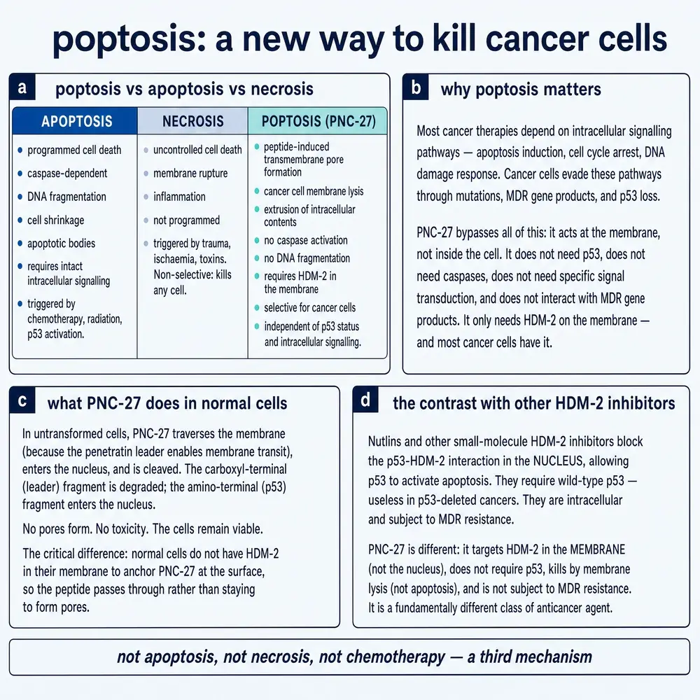
The killing that follows is fast and violent. The cell dumps lactate dehydrogenase — a large interior enzyme — into the medium almost immediately, the classic sign that the membrane has been breached. None of the markers of apoptosis appear: no DNA laddering, no annexin-V flip, no activated caspases (Bowne et al., 2008; Wang et al., 2020). The group named the process “poptosis” — a coinage meant to capture a death that is neither classical apoptosis nor undirected damage, but a specific, targeted rupture (Pincus et al., 2024). Because it does not route through p53, the apoptosis machinery, or the DNA-repair and drug-resistance programs that most chemotherapies fight, it works in cells that have deleted p53 and in cells that are multidrug-resistant — a point returned to in Part Three.
The same pore programme is laid out as a commissioned plate below: the 1:1 binding step, the temperature-dependent aggregation, the immuno-electron microscopy evidence, and the later mitochondrial hit. It is the visual companion to the schematic above, not a second mechanism.
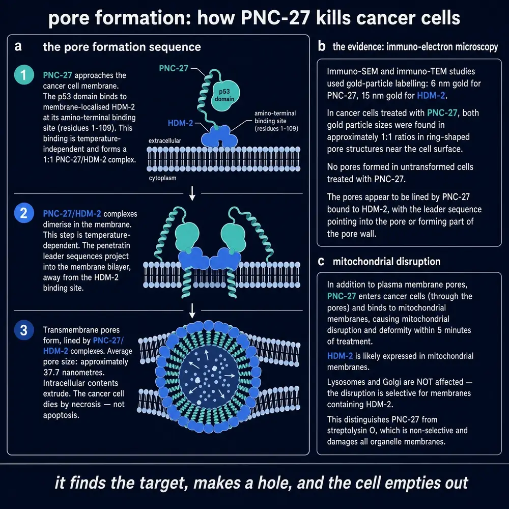
There is a second, later wrinkle. In 2024 the group reported that PNC-27 does not stop at the outer membrane: it also reaches the membranes of mitochondria — the cell's power plants — and disrupts them, with gold-labelled peptide visible on mitochondrial membranes and the organelles failing to hold a marker dye (Krzesaj et al., 2024). Whether this mitochondrial hit is a parallel kill mechanism or a downstream consequence of a cell already losing its membrane integrity is not settled, but it widens the picture beyond a single surface pore.
Before turning to the dissenting reading, one more commissioned plate sets poptosis beside apoptosis and necrosis, and shows what the peptide does — and does not do — in a normal cell.
06The dissenting reading
A monograph that only told the membrane-HDM-2 story would be doing the reader a disservice, because the mechanism is not universally settled and the strongest independent data complicate it. The most important qualification comes from a 2010 study by a separate group publishing in the Journal of Biological Chemistry (Yang et al., 2010). Working with PNC-27 and PNC-28 as model penetratin-carrying peptides, they measured a selectivity for tumour over normal cells of only about two- to threefold — real, but far more modest than the near-absolute sparing reported elsewhere. And they offered a different explanation for what selectivity there is: tumour cells over-produce a surface sugar polymer called chondroitin sulfate, which acts as a “molecular portal” that preferentially admits penetratin-tagged peptides. Strip the chondroitin sulfate away and the tumour-killing falls off. In this reading, part of PNC-27's apparent cancer specificity is a property of the delivery tag and the tumour cell surface, not solely of membrane HDM-2.
The mechanism has an internal tension too. When the City of Hope group studied PNC-27 in leukaemia, they found that the peptide drives HDM-2 to attack a third protein, the adhesion molecule E-cadherin, tagging it for destruction, and that this degradation is what precipitates the membrane damage in those cells (Wang et al., 2020). They argued the pores were not simply lined by PNC-27–HDM-2 complexes. The New York group's own high-resolution imaging, by contrast, shows the peptide physically built into the pore rim (Sarafraz-Yazdi et al., 2022). These are not necessarily contradictory — leukaemia and solid-tumour cells may porate by somewhat different routes — but they are not yet reconciled, and the honest summary is that the what (selective membrane rupture of HDM-2-bearing cancer cells) is far better supported than the precise how.
07In the dish: how broad, how selective
The in-vitro record is the broadest part of the PNC-27 literature and the most consistent. Across roughly two decades, PNC-27 (and PNC-28) have been reported to kill a wide range of human and animal cancer cell lines while sparing matched normal cells: pancreatic, breast, melanoma, ovarian, colon, cervical, non-small-cell lung, angiosarcoma and osteosarcoma among solid tumours, and several acute and chronic leukaemias among blood cancers (Pincus et al., 2024). The concentrations needed — the half-maximal killing dose, IC50 — cluster in the single- to low-tens of micromolar range: about 6–80 µM across solid-tumour lines, and roughly 6–32 µM in leukaemia lines and primary leukaemia cells (Pincus et al., 2024; Wang et al., 2020; Krzesaj et al., 2025).
Three features of the cell-culture data matter more than the breadth, because they distinguish PNC-27 from most anticancer agents. First, it kills cells that have deleted p53 entirely — osteosarcoma and leukaemia lines with no functional guardian at all — confirming that the effect does not run through p53 (Davitt et al., 2014). Second, it kills multidrug-resistant lines that shrug off conventional chemotherapy, such as drug-resistant ovarian OVCAR-3, because pore formation does not depend on the resistance machinery those cells rely on (Sarafraz-Yazdi et al., 2015). Third, and most relevant to any eventual clinical claim, it has been tested not only on immortalised laboratory lines but on primary cells freshly taken from patients — ovarian tumour cells removed at surgery, and leukaemia blasts from bone marrow — where it retained its selective killing (Sarafraz-Yazdi et al., 2015; Wang et al., 2020). It has also shown a capacity to hit the CD44-positive “cancer stem cell” subpopulations that are thought to seed relapse (Thadi et al., 2021).
How impressed to be depends on where the data come from, and here candour is owed. The large majority of these cell-culture reports come from one collaborating research network and appear in a small number of journals. That is not evidence of error — the experiments are internally consistent and use proper controls — but a mechanism this striking would carry more weight with more independent replication. The single most valuable counterweight, and the study that does the most to make the case credible, is an animal experiment from an outside group, discussed next.
08In animals: the two results that carry the weight
Two in-vivo studies do most of the work of turning a dish curiosity into a plausible drug lead. The first is the original pancreatic-cancer experiment. Rat pancreatic cancer cells that form aggressive, metastasising tumours in immune-deficient (“nude”) mice were implanted, and the animals were given PNC-28 by continuous infusion from an implanted mini-pump over two weeks. The treated tumours were eradicated and did not regrow in the fortnight after treatment stopped, while control-peptide tumours grew to large masses and spread (Michl et al., 2006). The treated mice gained weight normally, with no gross signs of toxicity — an early, coarse safety read, but a reassuring one.
The second study is the one that most changes the picture, because it is the most rigorous and the most independent. A team at the City of Hope cancer centre examined PNC-27 in acute myeloid leukaemia and published the results in the high-impact journal Leukemia (Wang et al., 2020). They first confirmed, in patient samples, that surface HDM-2 is present on AML blasts — including the treatment-resistant leukaemia stem-cell fraction — and absent on normal blood stem cells. They then treated mice carrying either mouse leukaemia or human patient-derived leukaemia with PNC-27 at 40 mg/kg by daily injection for two to three weeks. Treated animals lived significantly longer, carried fewer leukaemia cells in blood, marrow and spleen, and — the demanding test — when their marrow was transplanted into fresh mice, it seeded less leukaemia, meaning the treatment had reached the stem-cell population that drives relapse. Crucially, healthy mice given an even higher dose showed no damage to normal blood production, and their marrow reconstituted normally (Wang et al., 2020).
These are genuinely strong preclinical results, and the leukaemia study in particular — independent group, demanding secondary-transplant design, explicit test of normal-tissue toxicity, top-tier journal — is the reason PNC-27 deserves a serious hearing rather than a dismissal. Two other in-vivo findings round out the animal record: PNC-27 necrosed colon-tumour nodules in a live-imaging mouse model without harming normal tissue (Thadi et al., 2021), and, added to the chemotherapy drug paclitaxel in a mouse model of ovarian cancer, it curbed tumour growth more than paclitaxel alone — a synergy the authors traced to paclitaxel survivors up-regulating surface HDM-2 and so becoming more vulnerable to PNC-27 (Alagkiozidis et al., 2017). It must still be said plainly: every one of these results is in mice. None is a human outcome.
09In people: a blank, stated honestly
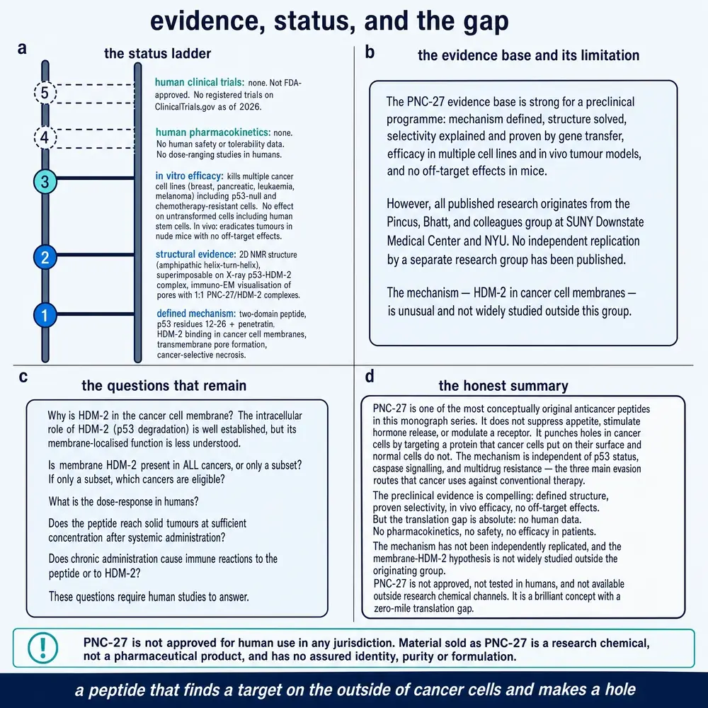
Here is the single most important fact in this monograph, and the one most often blurred when a compound like this is discussed outside the literature: PNC-27 has never been given to a human being in a registered clinical trial. There are no human pharmacokinetic data — nothing on how it is absorbed, distributed, broken down or cleared in a person. There are no human safety data. There are no efficacy data in patients. A search of the clinical-trials registry returns nothing for PNC-27.
It is worth being precise about what the “human”-sounding evidence actually is, because the distinction is exactly the one that gets lost. Two kinds of experiment involve human material. In the first, cancer cells taken from patients — ovarian tumour tissue, leukaemia blasts — are grown in a dish and treated with PNC-27 (Sarafraz-Yazdi et al., 2015; Wang et al., 2020). In the second, human cancer cells are implanted into mice and the mice are treated (Wang et al., 2020). Both are valuable; both use human cells. Neither is a test in a human body, with its immune system, circulation, liver, kidneys and the whole integrated physiology that decides whether a compound that kills cells in a dish can be given to a person safely and reach a tumour at a useful concentration. For a peptide — a molecule class notorious for being chewed up in blood and cleared in minutes — that gap is not a formality. It is the gap across which most promising anticancer agents fail.
10A second life as a targeting tag
One of the more interesting recent turns is that PNC-27's affinity for surface HDM-2 makes it useful not only as a weapon but as an address label. If cancer cells wear HDM-2 on the outside, a particle decorated with PNC-27 should home to them. Several groups have built on this. PNC-27 has been attached to the approved chemotherapy formulation Doxil (liposomal doxorubicin), improving its uptake and effect against HDM-2-positive colon tumours while leaving HDM-2-negative tumours unaffected (Darban et al., 2017). It has been used to steer gene-delivery carriers into cancer cells (Mokhtarzadeh et al., 2016), and to target iron-oxide nanoparticles as a potential cancer-imaging agent (Rahmani et al., 2022). A separate strand found that ketone bodies — the metabolic fuels produced on a ketogenic diet — lowered the concentration of PNC-27 needed to kill cancer cells in culture, raising the idea of metabolic co-treatment (Miller et al., 2023).
These are early, mostly in-vitro results, and several come from groups independent of the originators, which is useful. They should be read as evidence that the surface-HDM-2 idea is being taken up and built upon, not as evidence of clinical utility. Their main value here is corroborative: if PNC-27 reliably finds HDM-2-bearing cancer cells and largely ignores others, that is consistent with the selectivity claim at the centre of the whole story.
11What can and cannot be said
PNC-27 is one of the more intriguing compounds in the experimental-peptide world precisely because its story is coherent from top to bottom. A fragment of the body's own tumour-suppressor protein, delivered on a membrane-crossing tag, turns out to seize a protein that cancer cells — and, as far as anyone can tell, only cancer cells — display on their surface, and to punch that protein into a lethal pore. The idea is supported by structural models, by imaging of the pores themselves, by an add-and-remove test that ties killing to the target, by breadth across many cancer types, and by two solid animal studies, one of them independent and rigorous. For an experimental compound, that is a substantial and internally consistent body of work.
What cannot be said is nearly as important. The evidence is dominated by a single research network and concentrated in a few journals; independent replication, while it exists, is thin, and one independent study found the selectivity narrower than the flagship reports and offered a partly different explanation (Yang et al., 2010). The reason cancer cells mislocalise HDM-2 to their surface — the linchpin of the whole mechanism — is unknown. The precise structure of the pore, and whether it forms the same way in solid tumours and leukaemia, is unsettled (Wang et al., 2020; Sarafraz-Yazdi et al., 2022). And there is no human data of any kind. Weighing recency against the weight of evidence, the newest work (2020–2025) reinforces rather than overturns the core claim, and nothing recent contradicts it — but “not contradicted” is a long way from “proven in people.”
The status of that evidence, and the gap that remains, is the subject of the next plate.
The timeline below places the same arc on a calendar: two decades of progressive support for the membrane-HDM-2 mechanism, and a conspicuous absence of any human trial at any point on the line.
The most defensible reading is this. PNC-27 is a scientifically serious lead that has demonstrated a novel, selective, membrane-based way of killing a broad range of cancer cells in culture and of clearing tumours — including relapse-seeding stem cells — in mice, apparently without harming normal tissue. It is also a compound whose central biological premise is not yet independently nailed down and which has never entered human testing. Both halves of that sentence are true at once, and any responsible account of PNC-27 has to hold them together.
12References
Generated from verified NCBI records rather than from recall. Author lists, journal names, volumes, pages and identifiers are taken from the PubMed record for each citation and were checked against each title before this document was built.
- Alagkiozidis I, Gorelick C, Shah T, Chen YA, Gupta V, Stefanov D, et al.. Synergy between Paclitaxel and Anti-Cancer Peptide PNC-27 in the Treatment of Ovarian Cancer. Annals of clinical and laboratory science. 2017;47(3):271-281.
PMID 28667027 - Bowne WB, Sookraj KA, Vishnevetsky M, Adler V, Sarafraz-Yazdi E, Lou S, et al.. The penetratin sequence in the anticancer PNC-28 peptide causes tumor cell necrosis rather than apoptosis of human pancreatic cancer cells. Annals of surgical oncology. 2008;15(12):3588-600.
PMID 18931881 · doi:10.1245/s10434-008-0147-0 - Darban SA, Badiee A, Jaafari MR. PNC27 anticancer peptide as targeting ligand significantly improved antitumor efficacy of Doxil in HDM2-expressing cells. Nanomedicine (London, England). 2017;12(12):1475-1490.
PMID 28565974 · doi:10.2217/nnm-2017-0069 - Davitt K, Babcock BD, Fenelus M, Poon CK, Sarkar A, Trivigno V, et al.. The anti-cancer peptide, PNC-27, induces tumor cell necrosis of a poorly differentiated non-solid tissue human leukemia cell line that depends on expression of HDM-2 in the plasma membrane of these cells. Annals of clinical and laboratory science. 2014;44(3):241-8.
PMID 25117093 - Krzesaj P, Adler V, Feinman RD, Miller A, Silberstein M, Yazdi E, et al.. Anti-Cancer Peptide PNC-27 Kills Cancer Cells by Unique Interactions with Plasma Membrane-Bound hdm-2 and with Mitochondrial Membranes Causing Mitochondrial Disruption. Annals of clinical and laboratory science. 2024;54(2):137-148.
PMID 38802154 - Krzesaj PK, Seydafkan S, Miller AI, Chen HT, Premsrirut P, Shim A, et al.. HDM-2-Targeting Peptide PNC-27 Kills Cervical Cancer Cells but not Normal Cervical Cells. Annals of clinical and laboratory science. 2025;55(3):347-353.
PMID 40750238 - Michl J, Scharf B, Schmidt A, Huynh C, Hannan R, von Gizycki H, et al.. PNC-28, a p53-derived peptide that is cytotoxic to cancer cells, blocks pancreatic cancer cell growth in vivo. International journal of cancer. 2006;119(7):1577-85.
PMID 16688716 · doi:10.1002/ijc.22029 - Miller AI, Diaz D, Lin B, Krzesaj PK, Ustoyev S, Shim A, et al.. Ketone Bodies Induce Unique Inhibition of Tumor Cell Proliferation and Enhance the Efficacy of Anti-Cancer Agents. Biomedicines. 2023;11(9).
PMID 37760956 · doi:10.3390/biomedicines11092515 · PMC10526402 - Mokhtarzadeh A, Parhiz H, Hashemi M, Abnous K, Ramezani M. P53-Derived peptides conjugation to PEI: an approach to producing versatile and highly efficient targeted gene delivery carriers into cancer cells. Expert opinion on drug delivery. 2016;13(4):477-91.
PMID 26654047 · doi:10.1517/17425247.2016.1126245 - Pincus MR, Fenelus M, Sarafraz-Yazdi E, Adler V, Bowne W, Michl J. Anti-cancer peptides from ras-p21 and p53 proteins. Current pharmaceutical design. 2011;17(25):2677-98.
PMID 21728981 · doi:10.2174/138161211797416075 - Pincus MR, Silberstein M, Zohar N, Sarafraz-Yazdi E, Bowne WB. Poptosis or Peptide-Induced Transmembrane Pore Formation: A Novel Way to Kill Cancer Cells without Affecting Normal Cells. Biomedicines. 2024;12(6).
PMID 38927351 · doi:10.3390/biomedicines12061144 · PMC11201261 - Rahmani R, Darroudi M, Gharanfoli M, Chamani J, Gholamin M, Hashemi M. Conjugated PNC-27 peptide/PEI-superparamagnetic iron oxide nanoparticles (SPIONs) as a double targeting agent for early cancer diagnosis: In vitro study. Iranian journal of basic medical sciences. 2022;25(10):1234-1242.
PMID 36311203 · doi:10.22038/IJBMS.2022.65590.14430 · PMC9588323 - Rosal R, Pincus MR, Brandt-Rauf PW, Fine RL, Michl J, Wang H. NMR solution structure of a peptide from the mdm-2 binding domain of the p53 protein that is selectively cytotoxic to cancer cells. Biochemistry. 2004;43(7):1854-61.
PMID 14967026 · doi:10.1021/bi035718g - Sarafraz-Yazdi E, Bowne WB, Adler V, Sookraj KA, Wu V, Shteyler V, et al.. Anticancer peptide PNC-27 adopts an HDM-2-binding conformation and kills cancer cells by binding to HDM-2 in their membranes. Proceedings of the National Academy of Sciences of the United States of America. 2010;107(5):1918-23.
PMID 20080680 · doi:10.1073/pnas.0909364107 · PMC2836618 - Sarafraz-Yazdi E, Mumin S, Cheung D, Fridman D, Lin B, Wong L, et al.. PNC-27, a Chimeric p53-Penetratin Peptide Binds to HDM-2 in a p53 Peptide-like Structure, Induces Selective Membrane-Pore Formation and Leads to Cancer Cell Lysis. Biomedicines. 2022;10(5).
PMID 35625682 · doi:10.3390/biomedicines10050945 · PMC9138867 - Sarafraz-Yazdi E, Gorelick C, Wagreich AR, Salame G, Angert M, Gartman CH, et al.. Ex vivo Efficacy of Anti-Cancer Drug PNC-27 in the Treatment of Patient-Derived Epithelial Ovarian Cancer. Annals of clinical and laboratory science. 2015;45(6):650-8.
PMID 26663795 - Thadi A, Gleeson EM, Khalili M, Shaikh MF, Goldstein E, Morano WF, et al.. Anti-Cancer Tumor Cell Necrosis of Epithelial Ovarian Cancer Cell Lines Depends on High Expression of HDM-2 Protein in Their Membranes. Annals of clinical and laboratory science. 2020;50(5):611-624.
PMID 33067207 - Thadi A, Lewis L, Goldstein E, Aggarwal A, Khalili M, Steele L, et al.. Targeting Membrane HDM-2 by PNC-27 Induces Necrosis in Leukemia Cells But Not in Normal Hematopoietic Cells. Anticancer research. 2020;40(9):4857-4867.
PMID 32878773 · doi:10.21873/anticanres.14488 - Thadi A, Morano WF, Khalili M, Babcock BD, Shaikh MF, Foster DS, et al.. Molecular Targeting of H/MDM-2 Oncoprotein in Human Colon Cancer Cells and Stem-like Colonic Epithelial-derived Progenitor Cells. Anticancer research. 2021;41(1):27-42.
PMID 33419797 · doi:10.21873/anticanres.14749 - Vassilev LT, Vu BT, Graves B, Carvajal D, Podlaski F, Filipovic Z, et al.. In vivo activation of the p53 pathway by small-molecule antagonists of MDM2. Science. 2004;303(5659):844-8.
PMID 14704432 · doi:10.1126/science.1092472 - Wang H, Zhao D, Nguyen LX, Wu H, Li L, Dong D, et al.. Targeting cell membrane HDM2: A novel therapeutic approach for acute myeloid leukemia. Leukemia. 2020;34(1):75-86.
PMID 31337857 · doi:10.1038/s41375-019-0522-9 · PMC7951797 - Yang H, Liu S, Cai H, Wan L, Li S, Li Y, et al.. Chondroitin sulfate as a molecular portal that preferentially mediates the apoptotic killing of tumor cells by penetratin-directed mitochondria-disrupting peptides. The Journal of biological chemistry. 2010;285(33):25666-76.
PMID 20484051 · doi:10.1074/jbc.M109.089417 · PMC2919130
13How this document was assembled
The corpus was built against project 05, the Therapeutic Peptide Research Library. Every file with a document extension in the project's document stores — 45,962 files in all — was opened and its extracted text searched for explicit mentions of PNC-27; generic matches on p53, HDM-2/MDM2 or penetratin were recorded but never counted on their own. That sweep returned 184 raw matches, which collapsed to 174 after de-duplication.
Classifying those by kind of source is the step that matters. Not one was a peer-reviewed scientific full text: 173 were vendor catalogue and product-page archive copies of a research-chemical listing, and 1 was an internal compound dossier. Because the local snapshot held no primary science on this compound, the pipeline queried PubMed directly, retrieving 28 indexed records (2004–2025). Two of those are false positives — papers in which “PNC” means particle number concentration and postnatal care — leaving 26 records that genuinely concern PNC-27 or its analogue PNC-28. Nine records carried a PubMed Central identifier and were fetched as open-access full text (~75 printed-page equivalents); eight of those nine are relevant. Together with the abstracts of the remaining indexed records and one internal dossier, this is the reading corpus the monograph is written from.
Five commissioned plates were supplied in
Desktop\MONOGRAPH FIGJRES HIGGSFIELD\PNC-27\ with a caption
list. They were encoded as WebP data URIs at 1900 px / quality 88
(house style A8), interleaved into the one Figure series with the authored SVG
charts, and value-audited in assets/higgsfield/MAPPING.md. Plate 3
is a dark-ground plate and takes a navy mat.
| Stage | What it does | Result |
|---|---|---|
| 01b | Targeted scan of the project's document stores | 45,962 files opened |
| 02 | PubMed E-utilities harvest, complete publication record | 28 records |
| 03 | PubMed Central open-access full-text retrieval | 9 full texts |
| 04 | De-duplication, classification, inventory report | 174 local / 26 relevant indexed |
| 05 | Reference list generation from verified records | 22 citations |
| 09 | Commissioned plate encode (Higgsfield) | 5 plates |
| 06 | Assembly of this document | 1 deliverable / 13 figures |
The local library's PNC-27 holdings are almost entirely commercial: research-chemical product pages, archived many times over. That is a finding in itself. The actual science of this compound lives in the peer-reviewed literature, which is why the recency layer — a direct PubMed and PubMed Central harvest — does the real work here.
14Evidence handling
Findings in this document are labelled by the kind of study that produced them. Structural models, cell-culture measurements, animal experiments, ex-vivo work on patient-derived cells, and narrative reviews are different kinds of claim, and the differences are stated in the sentence that reports the result rather than left to the reader. Cell and animal findings are never phrased so as to imply a human outcome, because there is no human evidence for this compound to imply one from.
Where evidence conflicts, both sides are given. The flagship reports of
near-absolute cancer selectivity are presented alongside the independent 2010
study that measured a narrower margin and proposed a partly different mechanism;
the solid-tumour account of the pore is presented alongside the leukaemia
account that differs from it. Newer work is given weight where it is not
contradicted by the body of prior evidence — and here the newest work
reinforces rather than overturns the core mechanism — but recency is never
allowed to stand in for replication or for human data that do not yet exist.
Numbers printed on commissioned plates were checked against the corpus; riders
are in the captions and in MAPPING.md.

Continue exploring