
Bronchogen Four residues, two spellings, and a lung that has not been studied since 2017
Somewhere between a Soviet military laboratory and a twenty-milligram vial sold online, this molecule acquired a second name for itself — and the second name is wrong. Papers by the same group, in the same decade, sometimes with the same senior author, call it Ala‑Glu‑Asp‑Leu and Ala‑Asp‑Glu‑Leu. One of those is the family stem of the Khavinson short peptides plus a leucine. The other is a well-known sorting signal in fission yeast. They are not the same compound, the difference is one transposition, and the paper that carries the wrong order in its title is the one most often cited as evidence that the molecule does anything at all. This monograph is about a peptide whose identity has to be established before its evidence can be read, and about what is left once it has been.
Findings are labelled by the kind of study that produced them, in the sentence that reports them. A result in a dish of human cells is called that. A result in a rat lung, in a tobacco root, or in a computer is called that. Where a number appears, the species, the concentration and the duration travel with it.
Several molecules appear in these pages and they are not interchangeable. Bronchogen is the subject: the tetrapeptide Ala‑Glu‑Asp‑Leu, written AEDL, and also written — incorrectly, as Section 01 shows — as ADEL. Chonluten is a different synthetic peptide for the same organ, the tripeptide Glu‑Asp‑Gly, and much of what is sold as respiratory-peptide science belongs to it rather than to the subject. Epitalon (AEDG), Cortagen (AEDP), Cardiogen (AEDR) and Cartalax (AED) are the siblings that share this compound's stem; Epitalon is far more famous, and almost everything a reader may already believe about “Khavinson peptides” was measured with it. The peptides of the tracheal mucosa and of lung parenchyma are the natural tissue extracts that came first. Every finding below names the molecule that was actually studied, and where a claim about Bronchogen rests on work done with a relative, the sentence says so.
Concentrations appear only as reported experimental parameters. Nothing in this document is a recommendation, and no route or schedule for human use is described or endorsed anywhere in it.
Section 01A peptide that cannot agree on its own name
Start with the thing that ought to be simplest. A tetrapeptide is four amino acids in a row. Write them down and you have named the molecule completely: there is no folding to argue about, no isoform, no post-translational decoration. Four letters and you are done.
The literature on this compound cannot manage four letters.
In 2011 a group including Khavinson published a differential scanning calorimetry study in the Bulletin of Experimental Biology and Medicine under the title “Effect of the peptide bronchogen (Ala‑Asp‑Glu‑Leu) on DNA thermostability” (Monaselidze et al., 2011). In 2014 the same senior author published the compound's central biological experiment in Lung, and that paper calls it “Tetrapeptide Ala‑Asp‑Glu‑Leu (ADEL)” from its abstract to its conclusions (Khavinson et al., 2014). Those two papers are, between them, the mechanistic and the biological foundation of everything claimed for this molecule.
And in the same years the same laboratory wrote it the other way round. A 2011 study of nuclear penetration names “bronchogen (Ala‑Glu‑Asp‑Leu)” (Fedoreyeva et al., 2011). A 2012 differentiation study names “bronchogen (Ala‑Glu‑Asp‑Leu)” (Khavinson et al., 2012). A 2017 plant study names “bronchogen (Ala‑Glu‑Asp‑Leu)” (Fedoreyeva et al., 2017). A 2020 paper prints the identification in full — “AEDL (Ala‑Glu‑Asp‑Leu, Bronchogen)” — in a sentence listing four peptides of the family (Khavinson et al., 2020a).
A 2019 review manages both in a single paragraph. It states that “the ADEL (Ala‑Asp‑Glu‑Leu) peptide activated the expression of the proliferative markers Ki67 and Mcl‑1 in cultures of human bronchial epithelium”, and then, in the very next sentence, that “the AEDL peptide also regulated the expression of genes involved in the differentiation of bronchial epithelial cells” (Caputi et al., 2019). Two spellings, one paragraph, one molecule, no acknowledgement that anything has changed.
Which one is the molecule
This is settled, and it is settled cheaply. The chemical record resolves Bronchogen to PubChem CID 11690869: molecular formula C18H30N4O9, relative molecular mass 446.5, InChIKey OFZRHTIWKZWPQF‑BJDJZHNGSA‑N. Its systematic name reads outward from an alanyl group through a four‑carboxybutanoyl residue — that is glutamate — then a three‑carboxypropanoyl residue — aspartate — and terminates in 4‑methylpentanoic acid, which is leucine. Read from the amino end: alanine, glutamate, aspartate, leucine. AEDL.
Three independent lines agree. The family is built on a shared Ala‑Glu‑Asp stem, and every sibling extends it in the same direction: Cartalax is AED, Epitalon AEDG, Cortagen AEDP, Cardiogen AEDR. AEDL belongs to that series and ADEL does not. The four most recent primary studies of the molecule — all of them in plants, all between 2022 and 2025 — write it as “AlaGluAspLeu (AEDL)” or spell the residues out in that order, and the string ADEL does not appear in any of the three full texts this project retrieved. And the group's own 2020 paper, quoted above, prints the pairing explicitly.
Why the wrong order is not a harmless typographical slip
It would be tempting to treat this as a proofreading failure that changes nothing. It is not, for two reasons.
The first is chemical. Ala‑Glu‑Asp‑Leu and Ala‑Asp‑Glu‑Leu contain identical atoms in identical numbers. They are positional isomers: same formula, same mass, indistinguishable to a balance and to a low-resolution mass spectrometer, and different molecules. A supplier working from the wrong paper synthesises the wrong compound and every certificate of analysis based on mass agrees with itself.
The second is bibliographic, and it is the reason this section exists at the front of the document rather than in a footnote. A‑D‑E‑L is an X‑D‑E‑L string, and XDEL is the endoplasmic reticulum retention signal — the family that includes KDEL in mammals, HDEL in budding yeast and plants, and ADEL in the fission yeast Schizosaccharomyces pombe. This is not a curiosity. When this project searched the biomedical literature for “Ala‑Asp‑Glu‑Leu”, it retrieved a 1992 paper reporting that “the C‑terminal four amino acids of BiP are Ala‑Asp‑Glu‑Leu, a new variant of the XDEL sequence found at the C‑termini of luminal endoplasmic reticulum proteins” (Pidoux & Armstrong, 1992), and a 1998 paper describing a phosphatase whose “C‑terminal sequence Ala‑Asp‑Glu‑Leu … closely matches the consensus signal for ER retention, Lys‑Asp‑Glu‑Leu” (Romano et al., 1998). Both were admitted into this document's corpus on the first pass and both had to be thrown out. A researcher who searches on the spelling used in the compound's own most-cited biological paper retrieves cell biology from a different kingdom.
The four‑letter code fares no better. AEDL is also the Annual Effective Dose Level, a radiation-exposure quantity reported in millisieverts per year, and a radon survey of school buildings entered this project's corpus on the strength of a sentence about UK schools with “AEDL ranging between 0.58 and 3.07 mSv/y”. What makes that collision instructive rather than merely annoying is why it survived a subject-matter filter: radon dosimetry is concerned with dose to the lung, so the word “lung” sits a few hundred characters from every occurrence of the abbreviation. The organ that identifies this compound also identifies its homonym.
And the trade name has the crudest problem of the four. “Bronchogen” is a strict prefix of “bronchogenic” — as in bronchogenic carcinoma and bronchogenic cyst — a clinical literature orders of magnitude larger than this compound's. In the curated research library this project draws on, every single case-insensitive match for the string “bronchogen” was a bronchogenic carcinoma and not one was the peptide.
A document about a compound has to establish which molecule it is discussing before it can weigh a single result. For most compounds that is a formality. Here it is four separate problems — a superstring, a transposition that lands on a real sorting signal, a dosimetry unit and a protein motif — and none of them was predictable from the compound's name. Each was found by building the filter, running it, and reading what it refused.
Section 02What is a bioregulator?
The word is not a pharmacological classification. It appears in no regulatory nomenclature, it has no agreed definition outside the literature that coined it, and a reader who assumes it means something like “hormone” or “signalling peptide” will misread everything that follows. It is a term of art belonging to one research programme, and the honest way to introduce it is to say what that programme claims rather than to translate it into vocabulary it does not fit.
The claim has three parts, and each is unusual.
First, the mechanism is said not to be receptor binding. Every other compound class in this monograph series works by fitting a receptor: a growth hormone secretagogue finds the ghrelin receptor, a melanocortin agonist finds MC1R or MC4R, and the evidence that it does so is a dissociation constant, a displacement curve and a knockout animal. This class asserts something different. These peptides are said to cross the plasma membrane and the nuclear envelope, reach DNA and chromatin, and change transcription directly. There is no named receptor for any of them, no binding constant to any receptor, and no dose-response curve of the kind the rest of this series routinely reports.
Second, the concentrations are very low. The class is claimed to act at nanomolar and sub-nanomolar concentrations — 10−7 M in the plant experiments, 0.05 ng/mL in the explant work, a stated general range of 2 to 200 ng/mL in the group's own review (Khavinson et al., 2021). That is a real and interesting claim, because it is the concentration range at which a molecule is doing something informational rather than something stoichiometric. It also raises the obvious question, which the literature has not answered: how does a charged, water-soluble tetrapeptide get through two membranes to reach the DNA it is said to bind?
Third, there are two generations, and they are different kinds of object. The programme began with cytomedines — low-molecular-weight peptide fractions extracted from animal organs — and later produced cytogens, defined synthetic peptides of two to four residues, one assigned to each organ. The marketing treats these as a single technology. They are not. An extract is a mixture whose composition is nowhere fully specified; a tetrapeptide is one molecule with one formula. Evidence gathered on the first does not transfer to the second, and for the bronchopulmonary system almost all of the older evidence belongs to the first.
The vocabulary is worth pausing on, because it is used inconsistently in commerce and precisely in the literature. A cytomedine is the original tissue extract. A cytamin is that extract formulated as an oral supplement rather than an injectable preparation. A cytogen is a synthetic short peptide of the second generation. When a retailer describes a product as “a Khavinson peptide” without saying which of the three it is, the reader has been told almost nothing: the three differ in what is in the vial, in how they were made, and in what has been tested.
The contested part, said plainly
“Bioregulator” is a category proposed, populated and validated by one school. Outside that school and its collaborators it is not a recognised class of drug, and no regulator anywhere classifies compounds this way. The category does real work in the literature that uses it — it names a proposed mode of action that is genuinely distinct from receptor pharmacology — but a reader should understand that adopting the word is already adopting a claim.
That is not an argument that the claim is false. Peptides do get into cells; short peptides do bind nucleic acids; chromatin state does govern transcription. Each link in the chain is individually plausible. The question this document keeps returning to is whether the chain has been demonstrated for this molecule, at concentrations anything like those claimed, in the tissue it is sold for. That is a question about evidence, and Part Three is where it is answered.
Section 03A programme that began in 1973
The story usually told about these compounds is a good one, and parts of it are documented. Sorting which parts is worth doing carefully, because the undocumented parts are the ones doing the marketing.
The people and the institution are a matter of record. Vladimir Khatskelevich Khavinson was born on 27 November 1946 and died on 6 January 2024. He was a colonel of medical service, director of the Saint Petersburg Institute of Bioregulation and Gerontology, and vice-president of the Gerontological Society of the Russian Academy of Sciences. His collaborator Vladimir Morozov appears with him on the earliest work; Vladimir Anisimov, at the Petrov Institute of Oncology, ran much of the carcinogenesis and lifespan work that gave the class its geroprotective reputation.
The start date is citable and consistent. A 2022 paper by an Italian group collaborating with Khavinson states that these peptides were “characterized by Prof. Khavinson from 1973 onwards” and “were initially isolated from animal tissues and found to be organ specific” (Avolio et al., 2022). The biographical record gives the extraction programme as running 1973 to 2013, producing more than twenty organ-derived peptide complexes. Two independent framings, one date.
The military framing is partly documented and partly not. That the preparations were intended in the first instance for military medical service is stated in the biographical record. The much more specific story that circulates in commerce — a classified programme to protect submariners and cosmonauts from radiation and laser damage, declassified when the Soviet Union collapsed in 1991 — traces, in this project's searching, only to vendor and popular material. It also sits awkwardly with the programme's own chronology: Thymalin, the thymus extract, was registered as a medicine in the USSR in 1982, nine years before the supposed declassification. A programme can be founded for a military purpose and still publish and register medicines; but the secrecy story as usually told cannot accommodate a registered medicine in 1982, and this document does not repeat it.
What the programme produced, and what it did not
Six preparations from this programme became registered medicines in Russia: Thymalin, Epithalamin, Cortexin, Prostatilen (also sold as Samprost and Vitaprost), Retinalamin and Thymogen. That is a real regulatory achievement and it is worth stating without qualification.
It is also worth stating what is not on that list. No bronchopulmonary preparation is among the six. Neither the airway extract nor the synthetic tetrapeptide that is this document's subject has, so far as this project could establish, been registered as a medicine anywhere. The organ this compound is named for is one the programme did not carry through to approval.
The commercial structure that now sells these compounds is a separate matter from the science. The synthetic short peptides are distributed internationally as supplements and, in the grey research-chemical market, as lyophilised powder in vials. Section 21 returns to what is claimed at the point of sale.
Section 04The natural counterpart: peptides of the tracheal mucosa
Every synthetic short peptide in this class has an older relative — the tissue extract it descends from — and for most of them the extract has the broader evidence. Naming it, and comparing the two directly, is the single most useful thing a document like this can do, because commercial material almost never does it.
For the airway the counterpart is not one preparation but a small family of them, and the literature names them by abbreviation rather than by trade name. Between 1989 and 1994 the Khavinson group published four studies on peptides of the tracheal mucosa (PTM) and peptides of lung parenchyma (PLP), and a fifth study used a lung alveolar preparation called Pulmolin. The 1993 influenza paper is explicit about the provenance: these were “peptide preparations obtained from the tracheal mucosa (PTM) and lung parenchyma (PLP) of calves according to the technology used for obtaining thymalin” (Khavinson et al., 1993). Bovine tissue, by the thymus-extract process. That is the tissue and the species, which is a different and more useful statement than “derived from young animals”.
In commerce the bronchial cytomedine is sold as Taxorest, described as a natural bronchial peptide complex. This document treats the trade name and the research abbreviations as referring to the same kind of object — a bovine airway peptide extract — while noting that no paper in this corpus connects the commercial preparation to the specific extracts tested in the 1990s. A reader should not assume that a vial sold today contains what was tested in 1992.
What the extract's own record shows
The extract studies are more interesting than their obscurity suggests, because two of them contain controls that the synthetic literature lacks.
In 1992 the group measured relaxation of acetylcholine-constricted rat tracheal strips. Tracheal-mucosa and lung-parenchyma peptides both accelerated relaxation, dose-dependently across 10−6 to 10−3 g/mL, though less effectively than atropine. The useful part is the negative arm: preparations from the aortic wall and the thymus, tested in the same experiment, produced no significant effect (Khavinson et al., 1992a). That is an internal tissue-specificity control, and the class's specificity claim rests on experiments of exactly this shape.
In the same year, tracheal-mucosa peptides prolonged survival in hyperoxic lung injury, slowed the rise in lung coefficient, reduced malondialdehyde and raised glucose-6-phosphate dehydrogenase activity (Khavinson et al., 1992b). In 1994 they raised alveolar macrophage numbers and phagocytic activity in bleomycin-induced pulmonary fibrosis while normalising basal superoxide production (Khavinson et al., 1994).
Two results cut the other way, and they are the ones worth holding on to. The 1993 influenza study ranked the preparations against reference antivirals and concluded that none of them had direct antiviral activity; against influenza B, lung-parenchyma peptides did not change mortality at all (Khavinson et al., 1993). And in a 1989 carcinogenesis experiment, Pulmolin — the lung alveolar preparation — was deliberately chosen as the immune-inactive control against which thymogen's anti-tumour effect was measured, and it duly did nothing (Bespalov et al., 1989). The programme's own investigators, in 1989, used a lung peptide preparation as their example of a peptide preparation that does not work.
Isolated, or designed?
The load-bearing question about any compound in this class is whether the short peptide was isolated from the extract — sequenced out of it — or designed from it, assembled from knowledge of the extract's bulk amino-acid composition. These are entirely different epistemic objects wearing the same marketing language. If a tetrapeptide was purified from calf trachea and sequenced, it is a natural product with a claim to being the extract's active principle. If it was composed from an amino-acid analysis, it is a new molecule with a hypothesis attached.
For some members of the class the answer is known: Thymogen was isolated by chromatography from Thymalin, whereas Epitalon was designed from the bulk composition of Epithalamin rather than sequenced out of it. For Bronchogen, no paper in this document's corpus states which happened. The reviews describe the class as having moved from extracts to synthetic peptides without specifying the route for this compound, and no isolation or sequencing paper for an airway tetrapeptide was located.
This document therefore does not say that Bronchogen is the active fragment of the tracheal-mucosa extract, because nothing establishes that it is. Vendor material frequently implies it. The honest position is that the relationship between the extract and the tetrapeptide is, in the published record, an assertion of lineage rather than a demonstrated one.
Section 05Not receptor pharmacology
A reader arriving at this monograph from the sermorelin or the GHRP‑6 volume brings a mental model that will not survive contact with this compound, and it is worth dismantling it deliberately rather than letting it fail quietly.
The standard model runs: a peptide is a key, a receptor is a lock, and the science consists of measuring how well the key fits. You get a dissociation constant. You get a displacement curve against a labelled ligand. You get a knockout animal in which the effect disappears. You get a dose-response relationship with a slope and a maximum, and from that you get a rational basis for talking about potency at all.
None of that exists for this compound, and the class does not claim it should. The proposed mechanism skips the receptor entirely: the peptide is said to enter the cell, enter the nucleus, and act on DNA and chromatin directly. The evidence offered in its place is of a different kind — melting-curve shifts, fluorescence quenching constants, immunocytochemistry, transmission electron microscopy of chromatin, and molecular docking.
Two consequences follow, and both matter for how the rest of this document should be read.
The first is that potency has no well-defined meaning here. When a vendor or a review says these peptides are active at very low concentrations, that is a statement about the concentration at which an effect was observed in some assay, not a statement about affinity for anything. There is nothing to have affinity for. A concentration at which an effect appears in one cell culture is not comparable to a concentration in another, and neither is comparable to a dose.
The second is that the claim is unusually hard to refute. A receptor hypothesis makes sharp predictions: knock out the receptor and the effect goes. A direct-to-chromatin hypothesis with no named target makes almost none. That is not a reason to dismiss it — it is a reason to be careful about what counts as confirmation, and to notice when a study has been designed in a way that cannot come out negative.
Section 06The route in
Everything in the proposed mechanism depends on a step that is rarely examined: the peptide has to get inside. A charged tetrapeptide in the medium outside a cell is a long way from the DNA it is supposed to bind. How does it cross?
There are two candidate answers in the literature, and neither has been tested in the relevant tissue.
Passive entry, imaged in the wrong cells
The class's standard citation for cell penetration is a 2011 study in which fluorescein-labelled short peptides were incubated with HeLa cells and observed in the cytoplasm, the nucleus and the nucleolus. It is a clean result. But read the peptide list: the peptides actually imaged were epithalon (Ala‑Glu‑Asp‑Gly), pinealon (Glu‑Asp‑Arg) and testagen (Lys‑Glu‑Asp‑Gly) (Fedoreyeva et al., 2011). Bronchogen appears in the same paper — but in the cell-free binding experiments, not in the imaging. The demonstration that this compound enters a human cell nucleus does not exist in that paper. Its penetration is inferred from siblings.
The only direct imaging of this molecule entering a cell and accumulating in a nucleus is in tobacco root, where fluorescein-labelled AEDL was seen in cell wall, cytoplasm and nucleus after twenty hours, with the authors reporting that it accumulates in the nucleus to a greater extent than in the cytoplasm (Fedoreyeva et al., 2022). That is a real result and it is the best evidence of nuclear entry anywhere in this corpus. It is also in a plant, by a symplastic route through plasmodesmata that has no animal equivalent, and at 10−5 M — a hundredfold higher than the 10−7 M used in the growth experiments it is invoked to explain.
Transporters, modelled but not measured
The more testable proposal is that these peptides use existing peptide carriers. A 2023 study docked twenty-six ultrashort peptides into the LAT1, LAT2, PEPT1 and PEPT2 transporters and reported binding scores for each (Khavinson et al., 2023). This is the class's most falsifiable mechanistic claim, and it deserves to be read closely.
Three observations, in ascending order of importance.
First, this compound is not a standout. Among twenty-six peptides it ranks twentieth against LAT1 and twenty-second against LAT2. Its best result is sixth against PEPT1. Against PEPT2 its score is positive — +12.13 — where negative scores indicate favourable binding; twenty-one of the twenty-six peptides do better.
Second, the paper frames these peptides as inhibitors at least as readily as substrates. Its own discussion proposes that several of them, this one included, may be more potent inhibitors of amino-acid transport than the known reference inhibitors. A molecule that occupies a transporter is not necessarily a molecule the transporter carries.
Third, and most consequentially: PEPT1 is a di- and tripeptide transporter, and this compound has four residues. The paper says so itself, describing PEPT1 in its introduction as a low-affinity di- and tri-peptide transporter before docking tetrapeptides into it. The transporter literature outside this school is firmer still: the POT/PEPT family carries di- and tripeptides, and tetrapeptides are not substrates. The compound is one residue too long for the door it scores best against.
And the study is entirely computational. There is no uptake assay, no transfected cell line, no competition experiment — nothing that would convert a docking score into a measurement of transport. The authors are appropriately careful, describing the PEPT2 results as preliminary because the available structure was not representative. The caution belongs to the whole paper.
Section 07What it does to DNA
The most physical evidence about this molecule is a melting curve. In 2011 a Georgian and Russian group put calf thymus DNA and mouse liver DNA into a differential scanning microcalorimeter with the peptide and measured how much harder the double helix became to pull apart (Monaselidze et al., 2011).
The result is clear and modest. The melting temperature rose by 3.1 °C, across a narrow band of peptide-to-base-pair molar ratio — from about 0.01 to 0.055 — and then stopped rising. Adding twenty times more peptide did nothing further. The enthalpy of melting did not change at all across the whole range tested, staying at 11.4 cal/g for thymus DNA and 12.7 cal/g for liver DNA.
So the peptide binds DNA and stabilises it. That is established, in a cell-free system, by a direct physical measurement, and this document treats it as one of the firmer facts about the compound.
What the same paper says next is the part that commercial summaries omit. The authors conclude that bronchogen is neither AT-specific nor GC-specific, that binding occurs with both strands and mainly with the nitrogen bases, and — in their own words — that the binding is of a type best described as “strong and occasional”.
An occasional, base-non-specific, saturating interaction is what a small charged molecule does to DNA in general. It is a long way from selecting a promoter. The calorimetry establishes contact; it does not establish choice, and the paper does not claim it does.
A preference, from a different instrument
The claim that the peptide discriminates between sequences comes from a different experiment in the same year. Measuring fluorescence quenching against labelled deoxyribo-oligonucleotides, the group reported that epithalon, pinealon and bronchogen all bind preferentially to oligonucleotides containing CNG sequences — the sites at which cytosine methylation occurs in eukaryotes — and that within that class epithalon, testagen and pinealon prefer CAG-containing sequences while bronchogen prefers CTG (Fedoreyeva et al., 2011).
This is the single most important specificity result in the corpus, because it is the only place where the subject and its siblings are reported doing demonstrably different things at the molecular level. It deserves both its weight and its caveats: it is a fluorescence-quenching constant against short synthetic oligonucleotides in a cuvette, not a footprint on a genome, and the distance from “prefers CTG” to “regulates NKX2‑1” is not travelled by any experiment in this literature.
Section 08Histones, chromatin, and a motif two peptides share
If the peptides do not select genes by sequence alone, the alternative is that they change chromatin. Two lines of evidence bear on this.
The first is binding to histones. In 2013 the group measured fluorescence quenching of labelled wheat histones H1, H2B, H3 and H4 by six short peptides including this one, and reported site-specific binding to the N-terminal tails — the regions whose modification governs chromatin accessibility (Fedoreyeva et al., 2013). The 2021 review restates the finding and adds the detail that matters: the peptide-binding motif reported for AEDL and AEDG is the same one, an amino-acid sequence the review renders as kaakakk, while lysine- and arginine-containing peptides bind a different motif (Khavinson et al., 2021).
It is worth being clear about what a fluorescence-quenching measurement can and cannot establish, because three of the specificity results in this corpus rest on one. The method attaches a fluorescent tag to a target — a histone, an oligonucleotide — and watches how much its glow dims as a candidate binder is added. Dimming means the two came close enough to interact, and the rate at which it dims gives a binding constant. What it does not give is a location. It cannot say which residues of the histone tail were touched, or whether the peptide sat in the major groove or merely stuck to the outside of the duplex. A quenching constant is evidence of association and silence about geometry, which is why a difference in constants between two peptides is suggestive and a shared binding motif between them is awkward.
Read that carefully. Bronchogen is assigned to the bronchi and Epitalon to the pineal gland, and the claim that one residue selects an organ is the class's central assertion. On the histone evidence, these two peptides bind the same motif on the same histone tails. Whatever distinguishes their tissue effects, on this evidence it is not their interaction with histones.
The second line is direct imaging of chromatin state, and it exists only in plants. Transmission electron microscopy of tobacco root nuclei found that the peptide increased condensed chromatin by about 22 per cent and decreased decondensed chromatin by the same amount, redistributing condensed chromatin into a network across the nuclear surface (Fedoreyeva et al., 2022). That is a real structural observation, and it is the closest anything in this corpus comes to showing the proposed mechanism happening. It was made in a tobacco root at 10−7 M, and the numbers are given as approximations throughout.
The docking study, and a sibling that is a fragment
A 2016 paper modelled DNA-peptide complexes for nineteen short peptides and assigned each a preferred tetranucleotide (Khavinson et al., 2016). Bronchogen was assigned the site ctcc. So was EDL — which is Bronchogen minus its first residue. Two peptides differing by one alanine were predicted onto the same site, which is either a real finding about how little the first residue contributes or an artefact of the method. The paper does not resolve it, and no experiment in the corpus tests it.
Section 09One residue apart: the specificity claim
This is the claim on which the whole class stands or falls, and it is extraordinary enough to deserve a section of its own.
The assertion is that Ala‑Glu‑Asp‑Gly acts on the pineal gland, Ala‑Glu‑Asp‑Leu on the bronchi, Ala‑Glu‑Asp‑Pro on the cerebral cortex and Ala‑Glu‑Asp‑Arg on the myocardium — that a single terminal residue, on an otherwise identical three-residue backbone, selects an organ. Nothing in ordinary pharmacology behaves like this. Receptor selectivity across a peptide family is normally the product of extensive contacts across many residues, and single-residue substitutions usually shift potency rather than redirect a molecule to a different organ.
What is the evidence?
The strongest design is a 2006 explant study, and it is the right shape: four peptides, four organs, one experiment. Organotypic explant cultures of heart, lung, prostate and pancreas from three-week-old and eighteen-month-old Wistar rats were exposed to cardiogen, bronchogen, prostamax and pancragen at 0.05 ng/mL, and each peptide is reported to have stimulated its own tissue relative to control (Zakutskiĭ et al., 2006).
The diagonal is the result. It is also, in this corpus, essentially the whole of the direct evidence for organ specificity in a mammal.
Three cautions belong beside it, and none of them is a technicality. The report is a four-page paper in Advances in Gerontology, a conference-adjacent Russian-language journal, and no full text is retrievable, so a reader cannot see the effect sizes, the replication or the statistics behind the word “stimulated”. The measured endpoint in organotypic explant culture is typically the area of the outgrowth zone, which is a coarse readout. And the paper closes by recommending clinical use for stimulating reparative processes in ageing tissue — a conclusion its design cannot support, and one this document does not adopt.
A second experiment tests the same idea in cell culture, and is better. In 2012 the group measured differentiation factors in three human cell types — embryonic pancreatic cells, embryonic bronchial cells and prostatic fibroblasts — and reported that pancragen stimulated CXCL12 and Hoxa3 in pancreatic cells, bronchogen in bronchial epithelial cells, and vesugen in fibroblasts, with the effect more pronounced in aged cultures (Khavinson et al., 2012). Three peptides, three tissues, one paper: the design is right, and the abstract reports the diagonal again.
Against that stands the evidence in the other direction. AEDL and AEDG are reported binding the same histone motif. AEDL and EDL are docked onto the same DNA site. And the 2021 systematic review's own summary of the field lists AEDL and KEDW together as inducers of “lung and pancreatic cell differentiation” respectively (Khavinson et al., 2020b) — a pairing that presupposes the specificity it is describing.
A head-to-head is worth more than any number of single-peptide studies, because it is the only design that can support a specificity claim at all. The corpus contains two, both from the originating group, both with the diagonal as their result, and neither with a retrievable full text.
What it does not contain is the obvious modern experiment: run AEDL beside AEDG and AEDP on the same bronchial gene panel used in the 2014 study. That experiment would take one technician a fortnight, it would test the central claim of the class in the tissue this compound is sold for, and in twenty years nobody has published it.
There is one near-miss worth recording. In 2022 an Italian group, working with Khavinson, tested five of these peptides side by side in a human monocyte line — Epitalon, Vilon, Thymogen, Thymalin and Chonluten (Avolio et al., 2022). It is the class's most substantial independent head-to-head. Bronchogen was not among the five; the respiratory peptide they chose was Chonluten.
Section 10The central experiment
One paper carries most of the weight. In 2014, in the journal Lung, Khavinson and eleven co-authors reported what the peptide does to human bronchial epithelial cells, and every subsequent review of this compound — including the ones that make clinical claims — traces back to it (Khavinson et al., 2014).
The design is a passage experiment. Human embryonic bronchoepithelial cell cultures were sampled at the first, seventh and fourteenth passages, and the peptide's effect was measured at each. Passage number is being used here as a proxy for cellular ageing: late-passage cultures are the “old” condition. Five proteins were measured by immunocytochemistry — Ki67, Mcl‑1, p53, CD79 and NOS‑3 — and eight genes by real-time polymerase chain reaction: NKX2‑1, SCGB1A1, SCGB3A2, FOXA1, FOXA2, MUC4, MUC5AC and SFTPA1. Separately, in the same paper, the peptide's interaction with DNA was studied by spectrophotometry, viscometry and circular dichroism, with the binding region reported in the major groove at the N7 position of guanine.
The headline results, as reported: the peptide regulates the levels of all five proteins, and its strongest activating effect on proliferation — through Ki67 and Mcl‑1 — was in the “old” cell cultures. It regulates the five differentiation genes. And it activates MUC4, MUC5AC and SFTPA1, which the authors describe as genes whose reduced expression correlates with pathological lung development.
One word in that summary is doing a great deal of work, and it is worth pausing on it. The paper says the peptide regulates these proteins and genes. Regulation is not a direction: it accommodates an increase, a decrease, and a change that runs one way early and the other way late. For Ki67 and Mcl‑1 the direction is given and it is upward in aged cultures. For several of the others the abstract does not say which way the needle moved, and without the full text a reader cannot find out. That is not a criticism of the experiment; it is a limit on what can responsibly be built on top of it, and it is why this document reports the panel rather than the effect sizes.
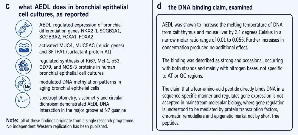
This is a real experiment on human cells of the right tissue, and it is the reason this compound has a monograph rather than a paragraph. It is also a single experiment, from the originating laboratory, at one concentration, with no comparator peptide, and no full text deposited in any open archive — so what is reported above is what the abstract states, and this document makes no claim about anything the abstract does not contain.
Section 11The genes, and a direction worth questioning
The gene panel is well chosen, and worth explaining, because the names carry the argument.
NKX2‑1 is the master transcription factor of lung identity: it is what makes a cell in the developing foregut become lung rather than something else, and it remains active in the adult airway. FOXA1 and FOXA2 are forkhead factors that work with it. SCGB1A1 encodes the club-cell secretoglobin often called CC16, an anti-inflammatory protein of the small airway whose circulating level falls in chronic obstructive pulmonary disease and in smokers; SCGB3A2 is a related airway secretoglobin. SFTPA1 encodes surfactant protein A1, part of the innate immune and surface-tension machinery of the alveolus. A compound that genuinely moved this panel in the right direction would be doing something interesting to airway epithelial identity.
Then there are the mucins, and here the argument needs pressing.
MUC5AC is the principal gel-forming mucin of the airway, and its over-production is one of the defining lesions of chronic bronchitis. Goblet cell hyperplasia — more mucin-secreting cells making more MUC5AC — is a textbook feature of obstructive airway disease. The 2014 paper reports the peptide activating MUC4, MUC5AC and SFTPA1, on the stated reasoning that reduced expression of these genes correlates with pathological lung development.
That reasoning is defensible in a narrow frame. In a senescent culture losing its differentiated phenotype, restoring mucin gene expression may indeed represent recovery of epithelial identity rather than pathological hypersecretion, and there is a real literature on mucin loss in damaged airway epithelium. But hold it against the same research programme's other work. The rat model reports in Sections 13 count goblet-cell hyperplasia as damage that the peptide reverses. So in the cell-culture paper more mucin gene expression is the therapeutic direction, and in the animal papers less mucin-secreting tissue is the therapeutic direction.
These are not formally contradictory — gene expression per cell in a senescent culture is a different quantity from the number of goblet cells in a diseased airway, and both could move usefully. But the pair is not reconciled anywhere in this literature, and a reader is entitled to notice that the compound is credited with a beneficial effect whichever way the mucin axis moves. A claim that is confirmed by both directions of a measurement is not being tested by it.
Section 12The mechanism that explains half the result
The most interesting paper in this corpus is the one that tried hardest to break its own hypothesis, and partly succeeded.
If the peptide changes gene expression epigenetically, the most direct prediction is that it changes promoter methylation. In 2015 Ashapkin, Linkova, Khavinson and Vanyushin tested exactly that, in the same bronchial cell system and in a pancreatic system alongside it (Ashapkin et al., 2015).
Promoter methylation is the right thing to have tested, and it is worth explaining why. A gene's promoter is the stretch of DNA immediately upstream of it where the transcription machinery assembles. Attaching methyl groups to cytosines in that stretch generally makes the gene harder to read; removing them makes it easier. It is one of the few chromatin marks that is stable, inheritable through cell division, and straightforwardly measurable, which makes it the natural first candidate for a compound claimed to change gene expression epigenetically. If the peptide works the way the class says it does, methylation is where the fingerprints should be.
The design has one feature that deserves more attention than it usually gets: the same paper ran a pancreatic cell system alongside the bronchial one, and tested the pancreatic peptide KEDW on it. That makes the study a two-tissue, two-peptide comparison rather than a single-arm experiment — which is the design Section 09 says the specificity claim needs and rarely gets. It is the closest thing in the corpus to a controlled test of the class's central assertion, and it was run for a different purpose.
The result splits cleanly in two.
For NKX2‑1 and SCGB1A1 the hypothesis held. Promoter methylation changed as the cultures aged, and it changed in correlation with expression. The authors conclude that altered promoter methylation might be the cause of the age- and peptide-induced changes in expression of these genes.
For FOXA1, SCGB3A2 and SFTPA1 it failed. The methylation patterns of these promoters do not change with culture age and are unaffected by the peptide — while their expression levels change in both cases. And for FOXA2 the result is starker still: in bronchial cells the promoter region is completely unmethylated, irrespective of culture age and irrespective of the peptide. There is no methylation there to modulate.
The authors' own summary is unusually direct: expression of FOXA1, FOXA2, SCGB3A2 and SFTPA1 “seem to be controlled by some other mechanisms”.
That sentence is the most valuable in the mechanistic literature on this compound, and it comes from the originating group. The proposed epigenetic route accounts for two of the six bronchial genes it was invoked to explain. For the other four, the peptide is reported to change expression by a mechanism that has not been identified.
This is what a partially-supported hypothesis looks like when it is honestly reported, and it should raise a reader's confidence in the group's other work rather than lower it. It should also stop anyone from writing that this compound works by regulating promoter methylation, which is a claim its own test does not support.
Section 13Rats, nitrogen dioxide, and a repaired epithelium
The only whole-animal disease experiments on this compound come from a pulmonology group in St Petersburg, reported twice: in 2015 in the Bulletin of Experimental Biology and Medicine, and in 2017 in a Russian-language physiology journal (Kuzubova et al., 2015; Titova et al., 2017).
The model is a reasonable one. Chronic obstructive pulmonary disease was induced in Wistar rats by sixty days of intermittent nitrogen dioxide exposure, which produces a recognisable phenotype: goblet cell hyperplasia, squamous metaplasia, lymphocytic infiltration and emphysema. The tetrapeptide was then given for one month, and the animals were assessed by bronchoalveolar lavage cytology, cytokine and enzyme profiling, secretory immunoglobulin A, surfactant protein B, and histology.
The reported outcome is a broad reversal. Bronchial epithelial structure was restored and ciliated cells returned; the hyperplasia, metaplasia and emphysema characteristic of the model were eliminated; secretory IgA rose, which the authors read as restored local immune function; surfactant protein B rose; and neutrophilic inflammation fell, with the cell composition and pro-inflammatory cytokine profile of the bronchoalveolar space normalising.
It is worth being precise about what this model does and does not stand in for. Sixty days of intermittent nitrogen dioxide produces a chemically induced airway injury with several features of human chronic obstructive disease — mucus-cell excess, squamous change, airspace enlargement, neutrophil influx. It does not reproduce the disease's cause, its decades-long course, or its defining functional deficit, which is progressive and largely irreversible airflow obstruction. A compound that repairs an epithelium after a chemical insult has done something real and has not thereby been shown to alter a human disease that is driven by a different exposure over a different timescale.
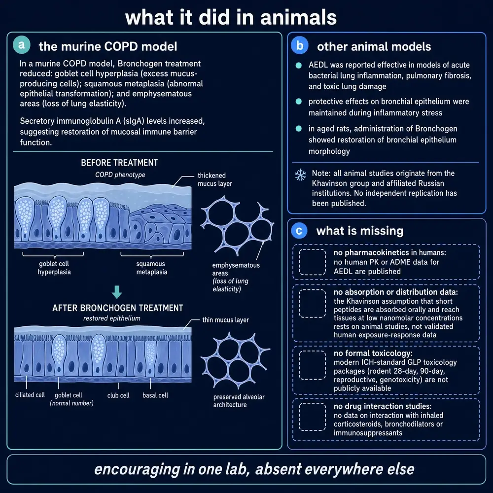
Taken at face value this is the most therapeutically suggestive result in the corpus. Several things should be held alongside it.
Both reports come from the same group, and neither has a retrievable full text, so the effect sizes, the statistics, the group sizes and the blinding are not available to a reader. One dose was tested, so there is no dose-response. There is no comparator peptide, so nothing in these experiments bears on tissue specificity. And the outcomes are structural and biochemical rather than functional: no measurement of airway resistance, gas exchange or exercise capacity is reported in either abstract.
The route and schedule used in these animals are not reproduced in this document. They are experimental parameters in a rat model of a chemically induced lesion, they have no established relationship to any human situation, and printing them would invite exactly the inference this series exists to avoid.
Section 14What the mammalian evidence adds up to
It is worth stopping to count, because the literature reads as larger than it is.
Strip out the reviews, the plant work, the sibling compounds and the tissue extracts, and the mammalian evidence that this specific tetrapeptide does something in an airway consists of four primary reports: one human cell-culture study of a gene and protein panel (2014), one methylation study in the same system that partly contradicts the proposed mechanism (2015), and two rat COPD reports from one pulmonology group (2015, 2017). To those can be added one three-tissue differentiation study (2012) and one four-organ explant study (2006) that include the compound among others.
Six reports, one institute, twenty years, and not one of them with a retrievable full text.
That is not a reason to disbelieve them. Russian-language and Russian-published biomedical work is systematically under-represented in open-access archives, and the absence of a deposited full text says something about publishing economics rather than about the quality of an experiment. But it does bound what a document like this can responsibly say. Everything reported in Sections 10 to 13 is drawn from indexed abstracts, and no claim in this monograph rests on material an abstract does not contain.
It also means the most basic quality questions cannot be answered. Whether the 2014 cell cultures were run blind. How many animals were in each arm of the rat studies. Whether the explant measurements were made by an observer who knew the treatment. These are not exotic demands; they are the ordinary furniture of a methods section, and for this compound a reader cannot reach them.
Section 15A tetrapeptide in a tobacco root
Here is the fact that reorganises everything else in this document. The lung literature on this compound stopped in 2017. The literature that has continued — four primary papers between 2017 and 2025, including the two most recent studies of the molecule anywhere — is about tobacco.
A plant-biology group in Moscow, led by Larisa Fedoreyeva and Neonila Kononenko and including Boris Vanyushin, has been studying AEDL in Nicotiana tabacum for nearly a decade. The connection to the Khavinson programme is real — Fedoreyeva and Vanyushin co-authored the 2011, 2013 and 2017 mechanism papers with Khavinson himself — but the recent plant papers stand oddly apart from it. None of the three full texts retrieved for this document mentions Bronchogen, or Khavinson, or the word bioregulator. The molecule has quietly become a plant growth regulator in a literature that no longer cites its own origin.
The experiments are consistent in design and modest in scale: seedlings of one cultivar grown from seed on hormone-free medium, a single peptide concentration of 10−7 M supplied continuously, a harvest at twenty-eight days, three biological replicates, one-way analysis of variance. Across all three papers there is no dose-response and no time course.
What they report is substantial. The 2022 paper found that the peptide doubled taproot length and increased shoot height by about a fifth (Fedoreyeva et al., 2022). The 2024 paper reported total wet weight up 1.7-fold and dry weight up 1.4-fold, with endogenous glutathione in roots raised 3.24-fold and manganese superoxide dismutase expression up 1.85-fold (Kononenko & Fedoreyeva, 2024). The 2025 paper found the peptide switching root cells from lytic vacuoles to protein-storage vacuoles and starch-filled amyloplasts, with the autophagy gene ATG8c up more than 1.7-fold (Lazareva et al., 2025).
These groups also report their nulls, which is worth crediting. The peptide alone did not change expansin expression — a result the 2022 authors use to rule out cell-wall loosening as the mechanism of root elongation. In leaves, glutathione reductase and S-transferase were unchanged. The autophagy gene ATG5 was unchanged, and the authors describe their own observed decrease in TOR expression as not significant. Several of the reported fold-changes are 1.1 to 1.2, which is close to the noise floor of the method.
Section 16What the plant work shows that the lung work does not
It would be easy to treat the tobacco papers as a curiosity — a respiratory peptide wandering into agronomy. They are more useful than that, for three reasons, and one problem.
They contain the only direct evidence that this molecule enters a cell nucleus. Section 06 established that the famous HeLa penetration experiment imaged the siblings, not this compound. The tobacco work imaged this compound: fluorescein-labelled AEDL in cell wall, cytoplasm and nucleus, accumulating in the nucleus more than in the cytoplasm. For the central premise of the entire class — that these peptides get to the DNA — the best evidence involving this specific molecule is in a plant.
They contain the only direct evidence that it changes chromatin structure. The electron-microscopic measurement of condensed versus decondensed chromatin, described in Section 08, is a plant result. Again, the mechanism the class asserts has been observed for this molecule in Nicotiana and not in a mammal.
And they are the only place where uptake is shown to be spatially selective. The labelled peptide concentrates in the elongation and root-hair zones and is barely detectable in the meristem — and the authors build a mechanistic argument on that exclusion. Whatever one makes of the argument, tissue-differential uptake within one organ is a more sophisticated observation than anything in the mammalian literature.
The problem
Plant and animal cells are not interchangeable, and the mechanism these papers propose is specifically a plant mechanism. The 2022 and 2024 papers hypothesise that AEDL binds CLV1 — a leucine-rich-repeat receptor kinase of the CLAVATA pathway that governs stem-cell number in plant meristems, and which has no animal counterpart. The proposed route into the tissue is symplastic, through plasmodesmata, which animals do not have. The functional analogy drawn is to CLE40 and CLV3, plant signalling peptides — and the 2022 authors note the awkwardness themselves, observing that AEDL is being compared to CLV3 “although they differ significantly in molecular weights”. AEDL has four residues; CLV3 and CLE40 have twelve or thirteen.
It should also be said that the CLV1 binding is hypothesised and never assayed in any of the three papers. There is no binding experiment, no mutant receptor, no competition. It is a plausible story attached to a real phenotype — which is, structurally, the same situation as the mammalian literature.
So the plant work is genuinely the strongest primary evidence that this molecule does something to a living cell, and it is evidence in a system whose signalling architecture the molecule's proposed lung mechanism cannot borrow from. Both halves of that sentence are true and neither cancels the other.
Section 17How a clinical sentence gets made
In 2020 a review in International Journal of Molecular Sciences, surveying peptides that might be useful against COVID‑19, contains this sentence (Khavinson et al., 2020c):
“Oral administration of EDG tripeptide (Chonluten) and AEDL tetrapeptide (Bronchogen) is effective for the treatment of bronchopulmonary pathology (chronic obstructive pulmonary disease, chronic bronchitis with an asthmatic component).”
This is, so far as this project could establish, the strongest clinical claim made for this compound anywhere in the indexed literature. It is worth following what stands behind it, because the answer is instructive about how this class is written about.
The sentence carries no citation to a human trial of Bronchogen. In the surrounding passage, the citations attached to this compound are to the 2014 bronchial cell-culture study and the 2015 methylation study — both discussed in Part Three, both in human cells in a dish, neither involving a patient. The clinical-sounding material immediately following the sentence — an increase in a physical performance index, normalisation of functional state under low oxygen partial pressure, enhanced effectiveness of standard therapy in chronic bronchitis — is attributed in the text to the EDG tripeptide, which is Chonluten, a different molecule.
The paper's own concluding section is more careful than the sentence above: it lists “peptides-bronchoprotectors (EDG, AEDL)” among substances “suitable for further research”, which is a proposal rather than a report. But the earlier sentence is the one that reads like a finding, and it is the one that has propagated.
It is worth asking why a sentence like that propagates, because the mechanism is not dishonesty. A review is written for readers who want the state of a field compressed, and compression removes qualifiers first. “Oral administration is effective for the treatment of bronchopulmonary pathology” is what remains after “in open-label observation, in one country's clinical literature, for a combination that includes a different peptide, without a control group” has been squeezed out. The next document to cite it inherits the compressed version, and by the third citation the qualifiers are unrecoverable without going back to the source. That is why this monograph quotes the sentence rather than paraphrasing it, and then walks back to what stands behind it.
This document therefore records the position plainly. No controlled human trial of Bronchogen was located in any source consulted for this monograph. ClinicalTrials.gov returns no registration for this compound, for Chonluten, for Taxorest, or for any compound in this class. Where human effectiveness is asserted for this molecule, the assertion is a review sentence, its citations point to cell culture, and its clinical texture belongs to a neighbouring compound.
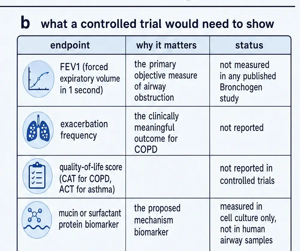
Two peptides are assigned to the same organ. One of them — Chonluten — has the human-flavoured observations. The other — Bronchogen — has the molecular experiments. A review discussing both in one paragraph produces a sentence in which each lends the other its strengths, and a reader takes away a compound with both a mechanism and a clinical result. Neither has both.
The same conflation runs the other way in commerce, where vendor material credits Chonluten with regulating mucin genes and ciliary function — which is the readout panel of the 2014 study of Bronchogen.
Section 18What is absent
For most compounds in this series, a section on gaps is a short list of the studies that would strengthen an existing case. Here the absences are large enough that they are the finding, and they belong in the body of the document rather than in a late caveat.
Three of these deserve saying in words as well as in a table.
There is no pharmacokinetic measurement of any kind. Not a plasma concentration-time curve, not a half-life, not a bioavailability figure, not a tissue distribution study, by any route, in any species. This is unusual even for a research chemical, and it has a practical consequence: there is no basis on which anyone can reason about what concentration a given exposure produces at the bronchial epithelium, which is the tissue every claim concerns. The concentrations at which effects have been observed — 10−7 M in plants, 0.05 ng/mL in explants — cannot be connected to any exposure in a whole organism because the connecting measurements have never been made.
There is no dose-response for this compound anywhere in the corpus. Every study located tested a single concentration. Dose-response is not a refinement; it is the primary evidence that an observed effect is caused by the substance rather than by something correlated with its administration, and its complete absence across twenty years of work on a molecule is a substantive weakness rather than a bibliographic one.
There is no toxicology. No dedicated safety or toxicity study of this compound was located. That is not evidence of safety. The class is frequently described in commercial material as harmless — and the word “harmlessness” appears in the 2014 paper's own abstract as a property of peptides generally — but an untested compound is untested, and the absence of reported harm from a literature that has not looked for harm carries no information.
Section 19One school, and what that costs
Almost every mammalian result about this compound traces to the Saint Petersburg Institute of Bioregulation and Gerontology, to Khavinson personally, or to immediate collaborators. That is a statement about the evidence base, not an accusation, and it has specific consequences worth naming rather than gesturing at.
The first is that replication and origination are not independent here. When the 2015 methylation study examines the 2014 study's gene panel, it is the same group revisiting its own result with a new method — which is valuable, and is not the same thing as an outside laboratory reproducing the finding. The corpus contains no instance of a group unconnected to the programme testing this compound and reporting what it found.
The second is that the class's reputation was not earned by this compound. When a reader arrives believing that Khavinson peptides extend lifespan, suppress tumours and reset telomerase, they are recalling a body of work done almost entirely with other members of the family. Epitalon increased mean survival in CBA mice and reduced spontaneous tumour incidence (Anisimov et al., 2001); Epitalon reduced tumour number and size in HER‑2/neu transgenic mice, where in the same experiment Vilon increased mammary cancer incidence and shortened the latent period (Anisimov et al., 2002); Epithalon extended the proliferative life of human fetal fibroblasts past the Hayflick limit (Khavinson et al., 2004). The immunological work belongs to Thymalin and its dipeptides (Khavinson et al., 2020d; Linkova et al., 2023). None of that is evidence about Bronchogen, and the Vilon result is a useful corrective to the assumption that membership of this family is itself reassuring: one member of it made a cancer model worse.
The third is that the negative results that do exist are internal, and they are the most credible things in the corpus. The 1993 influenza study concluding that no preparation had direct antiviral activity; the 1989 study using a lung peptide preparation as its inactive control; the 2015 methylation study finding that four of six genes are governed by something else; the plant papers reporting unchanged expansins and unchanged ATG5. A research programme that publishes its own nulls is behaving properly, and each of those results raises the credibility of the group's positive claims more than another positive claim would.
The fourth is the one that most affects a reader: the near-total absence of retrievable full texts. Six mammalian primary reports, none deposited. This is a structural feature of Russian-language biomedical publishing rather than a choice by any author, but its effect is that the ordinary apparatus of critical reading — group sizes, blinding, statistical tests, effect sizes with confidence intervals — is unavailable for the studies that matter most.
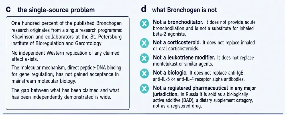
There is one genuine external engagement, and it is worth its own sentence. In 2022 a group at the University of Chieti‑Pescara, working with Khavinson, tested five of these peptides in a human monocyte line and reported that all five modulated proliferative signalling and that Chonluten inhibited tumour necrosis factor production by lipopolysaccharide-stimulated monocytes (Avolio et al., 2022). It is a real, independent-laboratory, head-to-head experiment on the class. It did not include this compound.
Section 20Recency, and what it points at
Where the evidence is thin, the newest evidence deserves particular weight, provided it is not contradicted by a preponderance of older work. Applying that rule to this compound produces an uncomfortable result.
The four most recent primary studies of this molecule — 2022, 2023, 2024 and 2025 — are, respectively, a tobacco root experiment, a molecular docking study, a tobacco root experiment, and a tobacco root experiment. The most recent primary work on the bronchial system is from 2017, and it is a Russian-language rat paper. The most recent human-cell experiment is from 2014.
Nothing newer contradicts the bronchial findings. Nothing newer supports them either, and nothing newer has tested them. On a recency-weighted reading of this literature, the best-evidenced thing about this molecule in 2026 is what it does to a plant root — where it has been imaged entering nuclei, observed changing chromatin structure, and shown to double the length of a taproot.
That is a genuinely interesting scientific position and a genuinely poor commercial one. The compound is sold for lungs; the science has moved to roots.
Section 21What is sold
The compound is offered by research-chemical retailers in twenty-milligram vials; two such offerings are recorded in this project's vendor data, and the market is wider than that. It is also sold internationally as a supplement in capsule form, within a product family covering most organs of the body.
The distance between what is claimed at the point of sale and what this document could establish is worth setting out precisely, because most of the individual claims are not fabrications — they are real findings with their qualifiers removed.
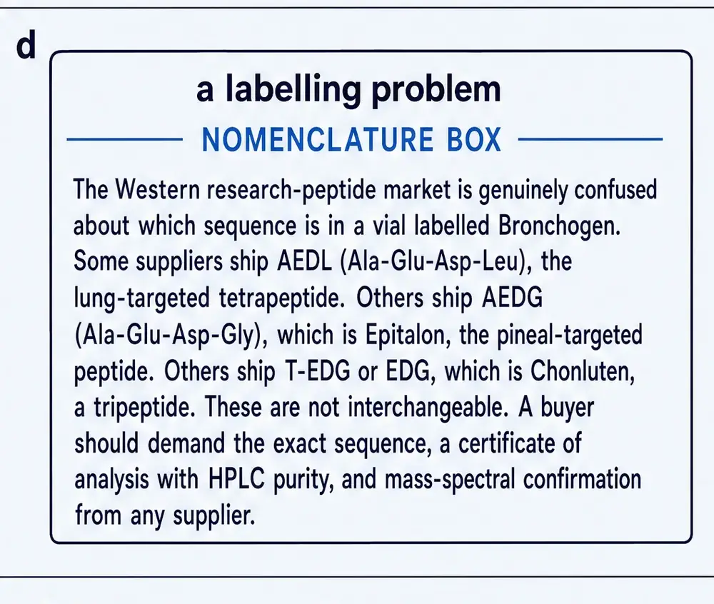
Three patterns recur and are worth naming.
A cell-culture result becomes a clinical indication. “Tunes gene expression in bronchial epithelial cells” is a fair description of the 2014 study. “Used for acute and chronic bronchitis and COPD” is a different kind of statement, and nothing in the corpus supports it.
A neighbouring compound's properties migrate across. Vendor material describing Chonluten as the regulator of mucin synthesis and ciliary function is describing the readouts of the 2014 study of Bronchogen. The two products are sold as a complementary pair with a division of labour — parenchyma versus mucosa — that no experiment in this corpus establishes.
Pharmacokinetic parameters appear from nowhere. Reconstitution and half-life guidance is published for this compound. There is no pharmacokinetic study of it in any species. A half-life quoted for a molecule that has never had one measured is not a rounded figure or a species extrapolation; it is a number with no referent.
Section 22What would settle it
It is easy for a document like this to end in a shrug. It should not, because the experiments that would move this evidence base are neither expensive nor technically demanding, and listing them is a fairer summary of the situation than any adjective.
The striking thing about that list is how cheap it is. A dose-response in an existing cell model, a transport assay in a transfected line, a three-peptide comparison on a gene panel that has already been assembled, a single-dose pharmacokinetic curve in a rat. None of this is frontier science. All of it could have been done at any point in the last decade by the laboratory that generated the original findings.
That it has not been done is itself a datum, and a reader is entitled to weigh it. A research programme that continues to publish mechanism papers and reviews about a compound for twenty years without ever measuring its dose-response or its pharmacokinetics is telling you something about what the programme is for.
Section 23Where this leaves the compound
Bronchogen is a real molecule with a settled structure and an unsettled name. It is Ala‑Glu‑Asp‑Leu; a substantial part of its own clinical literature, including the title of one paper and the whole of another, calls it Ala‑Asp‑Glu‑Leu, which is a different compound and also a well-known cell-biological motif. Establishing that took a chemical database, a family pattern and four recent papers, and no reader should have to do it.
What is genuinely established about it is narrow and real. It binds DNA and raises its melting temperature by about three degrees, without base preference, saturating at a low ratio. It binds histone tails, at a motif it shares with its pineal sibling. In one human bronchial cell system it moves a panel of airway identity genes, most strongly in aged cultures. In rats with chemically induced airway damage, one group reports structural and inflammatory repair. In tobacco it enters nuclei, condenses chromatin and doubles a root.
What is not established is nearly everything a person choosing to use a substance would want to know. There is no receptor, no binding constant, no dose-response, no pharmacokinetics, no toxicology, no controlled human trial, no independent replication, and no registration anywhere. The proposed epigenetic mechanism was tested by its own authors and explained two of the six genes it was invoked for. The transporter route is a docking score against a carrier that takes shorter peptides than this one. The organ-specificity claim — the reason the compound exists as a separate product at all — rests on two experiments from one institute, neither with a retrievable full text, against which stands the group's own finding that this peptide and its pineal sibling bind the same histone motif.
The compound is not a fraud and its literature is not junk. It is a small, internally honest, geographically concentrated body of work that has been stretched, by reviews and by commerce, over claims it does not reach. The useful thing a reader can take from this document is where the stretching starts.
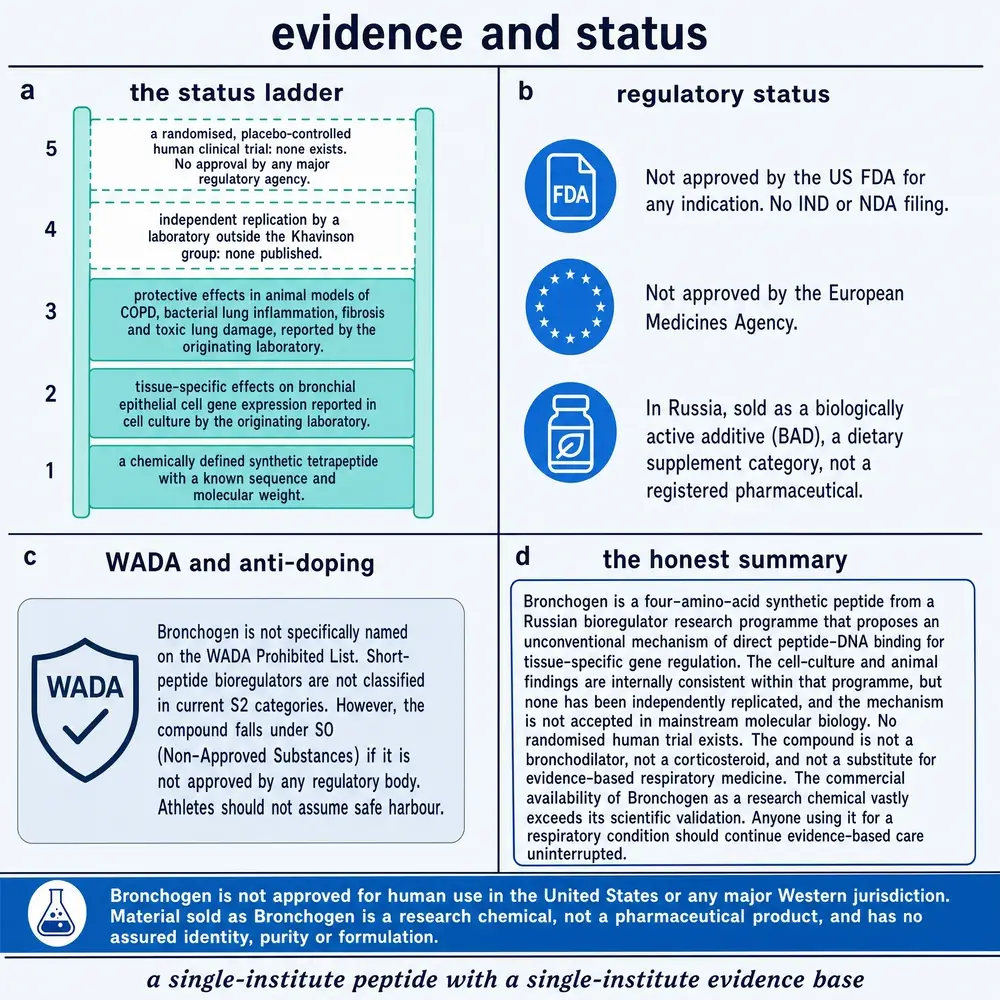
This monograph describes published research. It does not recommend human use of Bronchogen or of any compound in this class, and it specifies no dose, route or schedule for any person. Concentrations, exposures and durations appear only as parameters of the experiments that reported them, always attached to the species and system in which they were measured. Several of the studies discussed above conclude by proposing clinical application; those conclusions belong to their authors and are reported as claims, not adopted. No controlled human trial of this compound was located, and no regulatory authority has assessed it. This document is not medical advice.
Section 24References
- Anisimov VN, Khavinson VK, Mikhalski AI, Yashin AI. Effect of synthetic thymic and pineal peptides on biomarkers of ageing, survival and spontaneous tumour incidence in female CBA mice Mech Ageing Dev 2001;122(1):41-68. PMID 11163623 · doi
- Anisimov VN, Khavinson VK, Provinciali M, Alimova IN, Baturin DA, Popovich IG et al.. Inhibitory effect of the peptide epitalon on the development of spontaneous mammary tumors in HER-2/neu transgenic mice Int J Cancer 2002;101(1):7-10. PMID 12209581 · doi
- Ashapkin VV, Linkova NS, Khavinson VKh, Vanyushin BF. Epigenetic mechanisms of peptidergic regulation of gene expression during aging of human cells Biochemistry (Mosc) 2015;80(3):310-22. PMID 25761685 · doi
- Avolio F, Martinotti S, Khavinson VK, Esposito JE, Giambuzzi G, Marino A et al.. Peptides Regulating Proliferative Activity and Inflammatory Pathways in the Monocyte/Macrophage THP-1 Cell Line Int J Mol Sci 2022;23(7). PMID 35408963 · doi · PMC8999041
- Bespalov VG, Troian DN, Petrov AS, Morozov VG, Khavinson VKh. [Inhibiting effect of thymogen on the development of tumors of the esophagus and forestomach induced by N-nitrososarcosine ethyl ester in rats] Eksp Onkol 1989;11(4):23-6. PMID 2759010
- Caputi S, Trubiani O, Sinjari B, Trofimova S, Diomede F, Linkova N et al.. Effect of short peptides on neuronal differentiation of stem cells Int J Immunopathol Pharmacol 2019;33:2058738419828613. PMID 30791821 · doi · PMC6376556
- Fedoreyeva LI, Kireev II, Khavinson VKh, Vanyushin BF. Penetration of short fluorescence-labeled peptides into the nucleus in HeLa cells and in vitro specific interaction of the peptides with deoxyribooligonucleotides and DNA Biochemistry (Mosc) 2011;76(11):1210-9. PMID 22117547 · doi
- Fedoreyeva LI, Smirnova TA, Kolomijtseva GY, Khavinson VKh, Vanyushin BF. Interaction of short peptides with FITC-labeled wheat histones and their complexes with deoxyribooligonucleotides Biochemistry (Mosc) 2013;78(2):166-75. PMID 23581987 · doi
- Fedoreyeva LI, Dilovarova TA, Ashapkin VV, Martirosyan YT, Khavinson VK, Kharchenko PN et al.. Short Exogenous Peptides Regulate Expression of CLE, KNOX1, and GRF Family Genes in Nicotiana tabacum Biochemistry (Mosc) 2017;82(4):521-528. PMID 28371610 · doi
- Fedoreyeva LI, Baranova EN, Chaban IA, Dilovarova TA, Vanyushin BF, Kononenko NV. Elongating Effect of the Peptide AEDL on the Root of Nicotiana tabacum under Salinity Plants (Basel) 2022;11(10). PMID 35631778 · doi · PMC9147445
- Khavinson V, Diomede F, Mironova E, Linkova N, Trofimova S, Trubiani O et al.. AEDG Peptide (Epitalon) Stimulates Gene Expression and Protein Synthesis during Neurogenesis: Possible Epigenetic Mechanism Molecules 2020;25(3). PMID 32019204 · doi · PMC7037223
- Khavinson V, Linkova N, Diatlova A, Trofimova S. Peptide Regulation of Cell Differentiation Stem Cell Rev Rep 2020;16(1):118-125. PMID 31808038 · doi
- Khavinson V, Linkova N, Dyatlova A, Kuznik B, Umnov R. Peptides: Prospects for Use in the Treatment of COVID-19 Molecules 2020;25(19). PMID 32987757 · doi · PMC7583759
- Khavinson VK, Lin'kova NS, Tarnovskaya SI. Short Peptides Regulate Gene Expression Bull Exp Biol Med 2016;162(2):288-292. PMID 27909961 · doi
- Khavinson VK, Linkova NS, Rudskoy AI, Petukhov MG. Feasibility of Transport of 26 Biologically Active Ultrashort Peptides via LAT and PEPT Family Transporters Biomolecules 2023;13(3). PMID 36979488 · doi · PMC10046148
- Khavinson VK, Popovich IG, Linkova NS, Mironova ES, Ilina AR. Peptide Regulation of Gene Expression: A Systematic Review Molecules 2021;26(22). PMID 34834147 · doi · PMC8619776
- Khavinson VK, Linkova NS, Kvetnoy IM, Polyakova VO, Drobintseva AO, Kvetnaia TV et al.. Thymalin: Activation of Differentiation of Human Hematopoietic Stem Cells Bull Exp Biol Med 2020;170(1):118-122. PMID 33237528 · doi · PMC7686446
- Khavinson VKh, Tendler SM, Vanyushin BF, Kasyanenko NA, Kvetnoy IM, Linkova NS et al.. Peptide regulation of gene expression and protein synthesis in bronchial epithelium Lung 2014;192(5):781-91. PMID 25015171 · doi
- Khavinson VKh, Linkova NS, Polyakova VO, Kheifets OV, Tarnovskaya SI, Kvetnoy IM. Peptides tissue-specifically stimulate cell differentiation during their aging Bull Exp Biol Med 2012;153(1):148-51. PMID 22808515 · doi
- Khavinson VKh, Kozhemiakin AL, Fedin AN, Fomichev SN. [Relaxant effects of oligopeptides isolated from the tracheal mucosa and pulmonary parenchyma on the smooth muscle of isolated rat trachea] Biull Eksp Biol Med 1992;113(5):483-6. PMID 1421259
- Khavinson VKh, Kozhemiakin AL, Kuleshov VI, Okuneva NE. [Effect of peptides of the tracheal mucosa and vascular wall in acute hyperoxic lung injury] Patol Fiziol Eksp Ter 1992;(3):7-10. PMID 1480431
- Khavinson VKh, Kozhemiakin AL, Volgarev AP, Platonov VG. [The protective effect of peptides from the thymus and tracheal mucosa in an experimental respiratory influenzal infection] Zh Mikrobiol Epidemiol Immunobiol 1993;(1):68-73. PMID 8067076
- Khavinson VKh, Kozhemiakin AL, Letunova AV, Valeev RI, Danilov LN. [Effect of peptides, isolated from the tracheal mucosa, on alveolar macrophages and the nature of the course of fibrosing alveolitis in the rat] Patol Fiziol Eksp Ter 1994;(4):38-41. PMID 7700699
- Khavinson VKh, Bondarev IE, Butyugov AA, Smirnova TD. Peptide promotes overcoming of the division limit in human somatic cell Bull Exp Biol Med 2004;137(5):503-6. PMID 15455129 · doi
- Kononenko NV, Fedoreyeva LI. Peptide AEDL and Glutathione Stimulates Root Development Nicotiana tabacum Int J Mol Sci 2024;26(1). PMID 39796141 · doi · PMC11720632
- Kuzubova NA, Lebedeva ES, Dvorakovskaya IV, Surkova EA, Platonova IS, Titova ON. Modulating Effect of Peptide Therapy on the Morphofunctional State of Bronchial Epithelium in Rats with Obstructive Lung Pathology Bull Exp Biol Med 2015;159(5):685-8. PMID 26468022 · doi
- Lazareva EM, Kazakov EP, Dilovarova TA, Kononenko NV, Fedoreyeva LI. Peptide AEDL Activates Metabolism and Autophagy in Root Cells of Nicotiana tabacum Int J Mol Sci 2025;26(22). PMID 41303518 · doi · PMC12652066
- Linkova N, Khavinson V, Diatlova A, Petukhov M, Vladimirova E, Sukhareva M et al.. The Influence of KE and EW Dipeptides in the Composition of the Thymalin Drug on Gene Expression and Protein Synthesis Involved in the Pathogenesis of COVID-19 Int J Mol Sci 2023;24(17). PMID 37686182 · doi · PMC10488166
- Monaselidze JR, Khavinson VKh, Gorgoshidze MZ, Khachidze DG, Lomidze EM, Jokhadze TA et al.. Effect of the peptide bronchogen (Ala-Asp-Glu-Leu) on DNA thermostability Bull Exp Biol Med 2011;150(3):375-7. PMID 21240358 · doi
- Pidoux AL, Armstrong J. Analysis of the BiP gene and identification of an ER retention signal in Schizosaccharomyces pombe EMBO J 1992;11(4):1583-91. PMID 1373379 · doi · PMC556607
- Romano PR, Wang J, O'Keefe RJ, Puzas JE, Rosier RN, Reynolds PR. HiPER1, a phosphatase of the endoplasmic reticulum with a role in chondrocyte maturation J Cell Sci 1998;111 ( Pt 6):803-13. PMID 9472008 · doi
- Titova ON, Kuzubova NA, Lebedeva ES, Preobrazhenskaya TN, Surkova EA, Dvorakovskaya IV. [ANTIINFLAMMATORY AND REGENERATIVE EFFECT OF PEPTIDE THERAPY IN THE MODEL OF OBSTRUCTIVE LUNG PATHOLOGY] Ross Fiziol Zh Im I M Sechenova 2017;103(2):201-8. PMID 30199201
- Zakutskiĭ AN, Chalisova NI, Ryzhak GA, Aniskina AI, Filippov SV, Zeziulin PN. [The tissue-specific effect of synthetic peptides-biologic regulators in organotypic tissues culture in young and old rats] Adv Gerontol 2006;19:93-6. PMID 17152728
- National Center for Biotechnology Information. PubChem Compound Summary CID 11690869, L-alanyl-L-alpha-glutamyl-L-alpha-aspartyl-L-leucine (Ala-Glu-Asp-Leu). PubChem, Bethesda MD. Molecular formula C18H30N4O9, monoisotopic mass 446.5, InChIKey OFZRHTIWKZWPQF-BJDJZHNGSA-N. Queried 3 August 2026. link
- European Molecular Biology Laboratory, European Bioinformatics Institute. ChEMBL database, compound search for “Bronchogen”. Zero records returned. Queried 3 August 2026. link
- United States National Library of Medicine. ClinicalTrials.gov, intervention search for Bronchogen, AEDL, Chonluten, Taxorest and peptide bioregulators. No registration of any compound in this class was returned. Queried 3 August 2026. link
Section 25How this document was assembled
The corpus was built in three named arms and screened in two stages. The numbers below are reported as separate figures rather than pooled, because a single corpus figure would claim coverage this document does not have.
Retrieval
Three query arms were run against PubMed and PubMed Central: a subject arm covering the compound's trade name, both residue spellings and both four-letter codes; a class arm covering the Khavinson programme and the peptide-bioregulator literature; and a counterpart arm covering the bronchial and lung tissue extracts. Together they returned 234 PubMed records, reducing to 227 unique after de-duplication across arms, and a PubMed Central full-text body surface of 163 documents for the subject arm.
The identity gate
Section 01 describes the four collisions this compound's retrieval keys suffer. The gate that separates them is a single shared module imported by every stage, so the discovery scan and the corpus prune cannot drift apart. It admits on an unambiguous designation — the trade name, or the spelled-out Ala‑Glu‑Asp‑Leu — and requires subject-matter corroboration for a bare four-letter code. It carries two inverted arms, which refuse a match outright: one for the endoplasmic-reticulum retention-signal context, one for radiation dosimetry.
Four properties of the gate were arrived at by running it and reading what it refused, and each corrected a real error in the first version:
- Text is de-hyphenated before matching. A word boundary does not survive a typesetting hyphen: extracted text contains “bronchogen‑[line break]ic”, and a boundaried matcher admits it as the compound.
- Ala‑Asp‑Glu‑Leu is not an unambiguous designation. It was written into the first version of the gate as one, on the reasoning that a spelled-out four-residue sequence cannot be a homograph. It can: two ER retention-signal papers were admitted before the arm was inverted.
- Words describing a peptide's size do not corroborate anything. The first corroboration set included “tetrapeptide” and “short peptide”, which corroborate every four-residue sequence ever published — precisely the set the bare code collides with.
- Co-occurrence with the family admits. Tightening corroboration to genuine subject matter correctly ejected the ER papers and also ejected a genuine Khavinson review that names no organ and no person. A text naming AEDL beside AEDG, KEDW or EDP is in this programme's literature by construction.
The gate is break-tested against thirty-eight cases, including every observed collision and every hyphenation form. Of the 227 unique records, 16 were admitted as being about the compound and 211 refused. The refusals are reported by cause rather than pooled, because they mean different things: 119 carried no subject form at all; 69 belong to a named sibling (63 of them Epitalon); 6 were bare four-letter codes without corroboration; 5 were bronchogenic carcinoma or a bronchogram; 2 were the ER retention signal; 1 was radiation dosimetry; and 1 was corroborated only by a peptide-size word.
The substantive-use screen
The PubMed Central body-text surface of 163 documents was small enough to retrieve in full, so the screen was applied on the far side rather than in front of the fetch. Ninety-seven returned no machine-readable body — they are indexed without a deposited full text — and of the 66 that did, 55 never named the compound anywhere in the retrieved body. The remaining eleven were classified by document structure rather than by title: four name the compound inside a Methods section, two are experimental papers naming it elsewhere, two are reviews with three or more mentions, and three are passing mentions of one or two occurrences and were dropped.
One property of that classifier had to be corrected during the build, and it is worth recording. Ordering the document-structure test before the mention-count test admitted two papers that cite the compound exactly once in a discussion — a cricket-protein hydrolysate study and a radon survey — purely because they are experimental papers with a Methods section. Document structure establishes that a paper is primary; it says nothing about whether it is primary about this compound.
The reading corpus
Eight full texts, about 94 printed-page equivalents. Four are the tobacco papers; two are reviews by the originating school; one is the transporter docking study; one is a sibling-compound paper that supplies the sequence identification. Twelve further subject records exist as an indexed abstract only, and they include the central 2014 bronchial study, the methylation study, both rat COPD reports, the explant grid and the DNA calorimetry — that is, most of the evidence that bears on the lung.
This split is the single most important thing to know about this document's foundations, and it is why the class arm is reported separately: the compound's own decisive experiments are abstract-only, while its full-text corpus is dominated by plants and reviews. Every claim in Parts Three and Four that rests on an abstract is drawn only from what that abstract states.
Stage outputs
| Stage | What it did | Output |
|---|---|---|
| 01 | Ungated survey of the curated research library, to map the collision landscape before writing a filter | 4 raw matches, all bronchogenic carcinoma |
| compound | Shared identity module; admit, refuse, sibling and inverted arms | 38/38 break-test cases pass |
| 02 | PubMed and PMC harvest in three named arms; identifiers read only from the article identifier list | 234 records, 227 unique |
| 03 | PMC full-text fetch and substantive-use screen | 163 surface, 66 with body, 8 kept |
| 04 | Corpus inventory; surface and corpus reported separately | 94 printed-page equivalents |
| 05 | Reference list generated from verified NCBI records | build refuses on any unresolved identifier |
| 06 | Assembly, with gates on markers, figure continuity, section-number uniqueness, palette and SVG integrity | body fragment |
| 07 | Chrome render, both editions | light and dark PDF |
| 11 | Page density, margins and artwork integrity | gate results at release |
| 12 | Contrast audit, both themes | WCAG ratios per pair |
| 14 / 15 | Release manifest and format verification | saved, current and correct in kind at every location |
Databases queried directly
PubChem, for the structure and the sequence order, returning CID 11690869. ChEMBL, for bioactivity, returning no record for this compound. ClinicalTrials.gov, for registrations of this compound, of Chonluten, of Taxorest and of peptide bioregulators generally, returning none. All three were queried on 3 August 2026 and each is cited for what it does not contain as much as for what it does.
Section 26Evidence handling
Study type is named in the sentence that reports the finding. A result in a human bronchial cell culture is called that; a result in a rat lung, a tobacco root, an organotypic explant or a docking simulation is called that. Animal, plant and in-silico results are never phrased so as to imply a human outcome. Where this document reports a number, the species, the concentration and the duration travel with it.
Where a claim rests on a relative rather than on the subject, the sentence says so. This is the recurring failure mode in writing about this class, because the compounds differ by a single residue and share their entire vocabulary. The nuclear-penetration result most often cited for this compound was obtained with three siblings; the clinical-sounding observations in the COVID‑19 review belong to Chonluten; the geroprotective reputation of the class was largely earned by Epitalon. Each of those is flagged where it appears.
Conflicting evidence is presented as conflict. The methylation study partly contradicts the mechanism it was designed to support, and that is reported in the section that describes the mechanism rather than quarantined. The histone motif shared between this compound and Epitalon is reported in the section that makes the specificity claim. The mucin-direction problem is reported beside the gene panel it concerns.
Absence is reported as a finding. For a compound in this class the gaps are not an artefact of a thin search; they are the shape of the evidence base. Where a database returned nothing, this document says which database, on what date, and for which query.
No dose, route or schedule for human use appears anywhere in this document. Several of the studies discussed conclude by recommending clinical application. Those recommendations are reported as claims made by their authors and are not adopted, and the experimental parameters that would allow a reader to construct a human regimen are deliberately not assembled here.
Figures. No third-party published figure has been reproduced. Most of the figures in this document are authored SVG generated from the values cited here; each one's canvas is computed from its own drawn content rather than asserted, so no artwork can be clipped by its own frame, and every colour resolves through the series token set so both editions render from one source.
The commissioned artwork, and what was withheld
Five commissioned illustrations were supplied for this monograph after the document had been gated and filed. Each was audited before use, beginning with the cheapest test — whether a plate contradicts its own other panels or this document's own prose — and then against the sources for every printed value. Six panels were withheld and one label was corrected. The dispositions are recorded in each caption and in the project's artwork mapping file, so a reader can see which values were checked rather than assumed.
| Panel | Outcome | What the check found |
|---|---|---|
| Cell-culture findings; DNA-binding claim | admitted | every printed value correct — the full gene and protein panel, the major-groove N7-guanine site, and all six calorimetry figures |
| Endpoints a trial would need | admitted | all four status cells accurate against this corpus |
| Single-source problem; what the compound is not | admitted | agrees with Sections 19 and 21; regulatory status correct |
| Buyer guidance on nomenclature | admitted | the three sequences correctly distinguished; the market claim shown as supplied, not verified |
| Animal findings | corrected | the plate says “murine”; both obstructive-lung reports are in Wistar rats. Corrected in the caption |
| Status ladder and regulatory position | caveated | the S0 reasoning is sound; “not specifically named on the Prohibited List” was not verified against the List itself |
| The AED family table | withheld | printed Cartalax as AEDL targeting cartilage. Cartalax is the tripeptide Ala‑Glu‑Asp, C12H19N3O8. Shipping it would have put a wrong sequence two pages from Figure 2 |
| Identity table | withheld | gave the designation “T‑33”, which appears in no source in this corpus, and asserted an origin by hydrolysis that Section 04 shows the literature does not settle |
| The lineage from tissue extract | withheld | built the derivation on a preparation called “Broncholin”, a name found in no source located for this document |
| Theory of peptide bioregulation | withheld | sound, but superseded by the authored comparison in Figure 8, which carries the contrasts |
| Russian clinical observations | withheld | credited this compound with results its own source attributes to Chonluten — the error Section 17 exists to expose |
Three of those withholdings were detectable without opening a single source, because the plate disagreed with this document's own finished prose. That is the fastest test available on supplied artwork and it was applied first.
Continue exploring