
How Peptides Act on the Human Body Receptors, signaling cascades, and physiological response
A peptide does not carry a response. It carries a bias — a shift in the probability that a receptor sits in one shape rather than another. Everything a reader would recognise as the drug's effect is contributed by the cell that receives it: which receptor it makes, how many, wired to which machinery, with how much gain, against what it was already doing, and with what capacity to stop listening. This monograph follows one molecule from the moment it touches a receptor to the moment a body does something differently, and argues that the interesting part of that journey is almost all downstream of the binding event.
01A poison that acts where no poison should
Nobody saw a receptor until 2007. The idea is a century older than the observation, and for most of that century it was held on the strength of experiments that could not, in principle, show the thing they were about. That history is worth a Part of this document not out of ceremony but because the reasoning is still the reasoning: almost everything in modern receptor pharmacology is an inference from a response, and knowing what kind of inference it is turns out to be the difference between reading a table correctly and reading it wrongly.
The cleanest early example is John Newport Langley's. In a series of papers in the Journal of Physiology across 1907 and 1908, Langley worked on the fowl and the frog with nicotine and curare, and reported something that the physiology of the day could not accommodate: nicotine still made a muscle contract after the nerve supplying it had been cut and allowed to degenerate (Langley, 1908). If the nerve was gone and the response was not, the drug was not acting on the nerve. Langley proposed that the muscle itself carried what he called a receptive substance — something distinct from the contractile machinery, which received the chemical and passed the instruction on.
Two features of that argument survive into everything that follows. The first is that a receptor is inferred from a response, which means every inference about a receptor is an inference through whatever machinery lies between the receptor and the thing being measured. The second is that the argument is essentially about limits: the drug's effect can be blocked by another drug, and a blockable effect implies a finite number of somethings to be occupied. Saturability is the whole foundation. It is what makes a dose-response curve bend over, and everything in Part Two is an elaboration of what happens on the way to that bend.
02Counting, and the equation that was borrowed
Alfred Joseph Clark did the arithmetic. In 1926, working on the frog heart and the rectus abdominis muscle with acetylcholine, Clark measured the relationship between concentration and effect and found it described by a rectangular hyperbola (Clark, 1926a). In a companion paper he showed that atropine shifted the acetylcholine curve to the right without lowering its maximum, which is the signature of two agents competing for one site (Clark, 1926b).
The equation Clark used was not derived for cells. It is the adsorption isotherm Irving Langmuir had published for gas molecules binding to a metal surface, and Clark's contribution was the argument that a drug meeting a cell was formally the same problem. It was an enormously productive borrowing and it imported four assumptions, not all of which survive:
- One ligand binds one site. Mostly true for small molecules; often false for peptides, which frequently engage two receptor domains in sequence, and false in a different way for receptors that dimerise.
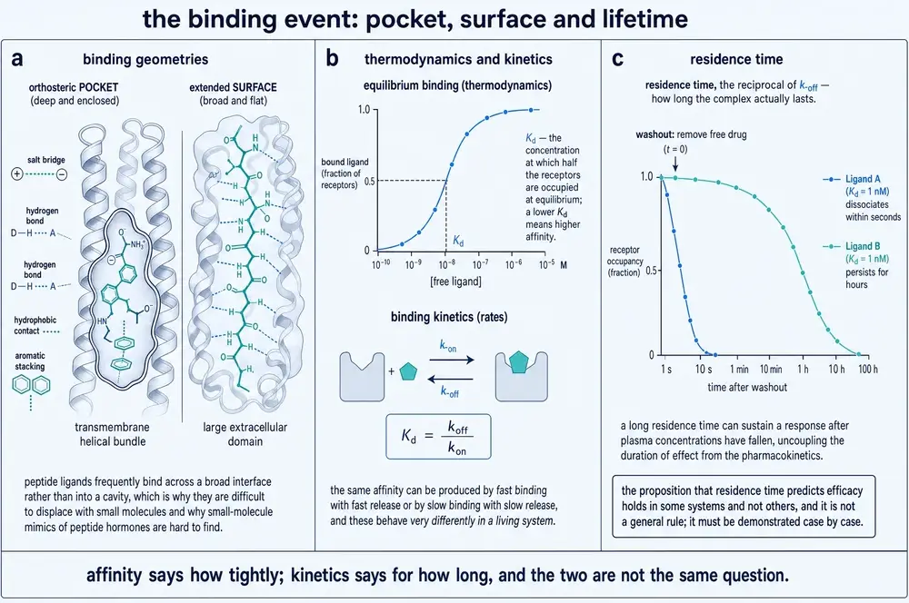
- Binding is reversible and rapidly equilibrating. True often enough to be useful, and untrue in exactly the cases that matter clinically — see section 07 on residence time.
- All sites are equivalent and independent. Negative cooperativity in insulin binding, reported by de Meyts and colleagues in 1973 in cultured human lymphocytes and in rat liver membranes, was the first widely accepted demonstration that they are not (de Meyts et al., 1973).
- Response is proportional to occupancy. This one is simply wrong, and correcting it took the next thirty years. It is the subject of section 03.
What Clark's hyperbola does establish, and what no later correction has disturbed, is the shape of the occupancy relationship itself. If a single ligand reversibly occupies a single site, the fraction of sites occupied at equilibrium is the concentration divided by the concentration plus the dissociation constant. The curve is steep at low concentrations and flattens as sites run out; on a logarithmic concentration axis it becomes the sigmoid that every pharmacology paper prints. Half the sites are occupied when the concentration equals the dissociation constant, which is what that constant means and the only thing it means.
03The gap between binding and doing
The correction came in three steps, and it is the most important sequence of ideas in this document, because every modern controversy about peptide drugs — bias, partial agonism, therapeutic window — is a dispute about where in this scheme a particular observation belongs.
Step one: efficacy is not affinity. Everard Ariëns, working in Nijmegen and publishing in 1954, pointed out that two compounds could occupy the same receptor equally well and produce different maximal effects (Ariens & de Groot, 1954). He gave the difference a name, intrinsic activity, and a number between zero and one. A compound with an intrinsic activity of one was a full agonist; zero made it an antagonist; anything between made it what would later be called a partial agonist. Nothing about affinity had changed. The molecule was doing two separable things: getting there, and mattering once there.
Step two: maximal response does not require maximal occupancy. R. P. Stephenson's 1956 paper in the British Journal of Pharmacology and Chemotherapy is titled, with characteristic understatement, A modification of receptor theory (Stephenson, 1956). Working with alkyltrimethylammonium compounds on guinea-pig ileum, Stephenson showed that a potent agonist could produce its full effect while occupying a small fraction of the available receptors. He replaced Ariëns's bounded intrinsic activity with an unbounded efficacy term, and separated the receptor event from the tissue's translation of it. The consequence is the one this whole document turns on: there is no fixed relationship between how much of a receptor population is occupied and how large the response is. The relationship is a property of the tissue.
Step three: the reserve can be measured. Robert Furchgott's work on adrenergic receptors, reviewed in Pharmacological Reviews in 1959, supplied the experiment (Furchgott, 1959). Inactivate a known fraction of the receptor population irreversibly, then re-measure the agonist curve. If the maximum survives, the tissue had receptors it was not using. The size of the surviving maximum against the fraction inactivated is a direct measurement of the reserve, and it is still the method.
James Black and Paul Leff completed the scheme in 1983 (Black & Leff, 1983). Their operational model stopped treating efficacy as a dimensionless fudge factor and made it what it physically is: a ratio between the receptor concentration in a tissue and the concentration of occupied receptor needed to produce half the tissue's maximal response. That ratio — the transducer ratio, conventionally written τ — is the amplification of the system, and it is the quantity the preceding figure varies. The model matters here for a specific reason: it is the only widely used framework that separates a ligand property from a system property with two independent numbers, and every attempt in section 11 to establish whether a compound is genuinely biased is an attempt to do that separation correctly.
04Why a peptide changes the problem
The framework just described was built almost entirely with small molecules: acetylcholine, atropine, catecholamines, alkylammonium compounds. Peptides break several of its working assumptions, and it is worth being precise about which ones, because the ways in which peptide pharmacology departs from classical pharmacology are not arbitrary.
A peptide is large, and it buries a large surface. A classical small -molecule drug occupies a pocket within a transmembrane bundle. A peptide agonist of a class B1 receptor is thirty to forty residues long and contacts both an extracellular domain and the bundle, burying an interface an order of magnitude larger. The consequence is not simply higher affinity: a large, distributed interface can be modified at many points, and a modification at one end of it can change what happens at the other. That is the structural precondition for biased agonism, and it is why bias is easier to find at peptide receptors than at aminergic ones.
A peptide is flexible. Most peptide hormones are substantially disordered in solution and acquire their bound conformation on binding. The lock-and-key image fails immediately here, because there is no key until the lock is met. This also means the entropic cost of binding is large and is paid differently by different analogues, which is one reason peptide structure-activity relationships are less transferable between series than small-molecule ones.
A peptide often binds in two stages. This is the subject of section 05 and is the single largest departure from the one-ligand-one-site assumption.
These three facts together explain something that looks at first like a failure of medicinal chemistry. Peptide receptors are abundant, well characterised and physiologically central, and yet small-molecule agonists for them are rare. The reason is geometric rather than chemical: a compound of a few hundred daltons can occupy a pocket, and a pocket is what a small molecule needs, but a class B1 peptide agonist does not act by occupying a pocket. It acts by organising an interface spread across two receptor domains, and there is no cavity for a small molecule to fill in a way that reproduces that organisation. Where small molecules have succeeded at these receptors they have usually done so as antagonists, which need only obstruct, or as allosteric modulators, which act somewhere the peptide does not. Both routes are consistent with the argument of this section, and neither is a route to a small-molecule agonist of a peptide hormone.
One more difference is worth stating before Part Two, because it shapes what the rest of the document can claim. Peptide receptors are a large fraction of the druggable receptor surface and a small fraction of the structurally characterised one. A census of approved drugs against G protein-coupled receptor targets by Sriram and Insel found that G protein-coupled receptors are the targets of approximately 34 per cent of FDA-approved drugs, dominated by aminergic receptors rather than peptide ones (Sriram & Insel, 2018). The structural revolution reached peptide receptors late: the first structure of a receptor in complex with its G protein, the β2-adrenergic receptor–Gs complex, was published in 2011 (Rasmussen et al., 2011), and the first class B peptide receptor in an active, transducer-coupled state — the GLP-1 receptor — followed in 2017 (Zhang et al., 2017). Much of what this document says about peptide receptor activation therefore rests on a literature under a decade old, and several of its generalisations are extrapolations from a handful of receptors. Where that is the case, the text says so.
05Surfaces, not keyholes
The lock-and-key image comes from Emil Fischer and it was about enzymes and sugars. Applied to a peptide meeting its receptor it fails at the first step, because most peptide hormones have no key shape until they arrive. They are substantially disordered in solution and fold as they bind, which means the binding event is not a fitting but a joint construction.
For the class B1 receptors — the family that receives glucagon, GLP-1, GIP, parathyroid hormone, calcitonin, CGRP, secretin and corticotropin-releasing hormone — the construction happens in two stages, and the two stages do different jobs. The receptor carries a large extracellular domain above the membrane. The peptide's C-terminal half, which is helical or readily becomes so, is captured by that domain. That first event contributes most of the binding energy and almost none of the activation. What it achieves is geometric: it tethers the peptide to the receptor and raises the effective local concentration of the still-free N-terminus by orders of magnitude, so that the N-terminus finds and inserts into the transmembrane bundle. The second event contributes little binding energy and nearly all of the activation.
This is not a model inferred from mutagenesis alone. A 2025 study in Biochemistry characterised the two-domain mechanism directly for the human CGRP receptor, resolving the separate contributions of the extracellular domain interaction and the bundle interaction to the overall binding energetics in purified and cell-based systems (Babin et al., 2025). The structural counterpart arrived with the cryo-EM era: the first active, G protein-coupled class B structure — the GLP-1 receptor bound to peptide and Gs — showed the peptide N-terminus deep in the bundle with the C-terminal helix held by the extracellular domain, exactly the arrangement two decades of biochemistry had predicted (Zhang et al., 2017), and the CGRP receptor structure followed (Liang et al., 2018). A crystal structure of the parathyroid hormone type 1 receptor added a second class B1 view of the same arrangement (Ehrenmann et al., 2019), and a direct comparison of peptide against small-molecule ligand binding at one receptor has since set out what the two modes demand of a binding site (Williams et al., 2024).
It is worth being explicit about what "disordered in solution" means here, because it is an experimental statement and not a figure of speech. A peptide described this way gives no interpretable signal in the structural methods that require an ordered specimen: no diffracting crystal on its own, broad and uninformative nuclear magnetic resonance spectra, and in cryo-EM no density that can be traced. The ordered conformation appears only in the complex. One practical consequence is that the bound conformation of a peptide hormone is frequently the only conformation anyone has seen, so a reader looking at a structure is looking at a shape the receptor helped make. A second is that the entropic cost of ordering the chain is part of the binding energy, and it is paid differently by analogues that differ only in flexibility — which is why constraining a peptide, by cyclisation or by a helix-stabilising substitution, so often changes affinity far more than the substitution's contacts can account for.
Not every peptide receptor works this way, and the exceptions matter to the argument. Chemokine receptors use a two-site mechanism of a different geometry, with the chemokine core docking on the receptor N-terminus and its own N-terminus reaching into the pocket; structural work on this family has shown that a single receptor can accommodate several chemokines through substantial structural mimicry rather than through a single complementary shape (Saha et al., 2025; Kayastha et al., 2024). The relaxin receptor RXFP1 turns out to be held in an autoinhibited state that the ligand relieves rather than a resting state that the ligand switches (Erlandson et al., 2023). And several receptors do not act alone at all: the melanocortin-4 receptor's signalling and oligomerisation state are modified by the accessory protein MRAP2 (Sohail et al., 2025), and the amylin receptors are constructed from a calcitonin receptor plus a receptor activity-modifying protein, with the subunit interaction itself modulated by which agonist is bound (Gostynska et al., 2025). A receptor, in other words, is not always a stable object that a ligand meets. Sometimes the ligand determines what the receptor is.
The two classical accounts — the ligand moulds the receptor, or the ligand picks a shape the receptor was already visiting — are usually presented as alternatives. For peptide receptors the honest answer is that both occur in the same binding event, and the question is which dominates at which step. A disordered peptide folding onto an extracellular domain is closest to induced fit; the subsequent transition of the transmembrane bundle is closest to conformational selection, because the bundle samples active-like states even when empty, which is what constitutive activity is.
The document uses both terms where the evidence supports them and does not adjudicate between them in general, because the general question is not well posed.
06Affinity, potency, efficacy: three numbers, routinely conflated
Three quantities, three meanings, and a great deal of confusion.
Affinity is how tightly a ligand binds. It is measured in a binding assay and reported as a dissociation constant, or as the concentration of competitor that displaces half a labelled ligand. It says nothing whatever about what the ligand does once bound. Efficacy is what it does once bound — how strongly it stabilises the receptor conformations that engage a transducer. It cannot be measured in a binding assay at all. Potency is the concentration producing half the maximal response in a particular system, and it is a composite: it depends on affinity, on efficacy, and on how much the system amplifies. Potency is not a property of a molecule. It is a property of a molecule in a preparation.
The practical consequences are large and they are routinely missed:
- A compound can be more potent and less efficacious than another. A high-affinity partial agonist will sit to the left of a low-affinity full agonist on a concentration axis while reaching a lower maximum. Ranking compounds by potency alone reverses the ordering that matters clinically.
- An EC50 from an overexpressing cell line is a statement about the cell line. Raising receptor density raises the transducer ratio, which shifts the response curve left and lifts partial agonists toward full agonism. This is not a subtle laboratory artefact: a 2023 study showed that apparent biased signalling at a G protein-coupled receptor could be lost when the same ligands were examined in systems with different expression levels, precisely because amplification differences between pathways scale with receptor number (Li et al., 2023).
- The ratio between binding and functional potency is itself informative. A 2025 analysis in Frontiers in Pharmacology proposed using the Kd/EC50 ratio of a full agonist as a direct gauge of the amplification a system provides (Buchwald, 2025). The larger the ratio, the more amplification lies between the binding event and the readout — which is the same quantity Stephenson called efficacy and Black and Leff called τ, arrived at from the measurement side.
07Residence time, and why the clock matters
Every constant in the previous section describes a system at equilibrium. A body is not at equilibrium. Plasma concentrations rise and fall, tissues are perfused unevenly, and a receptor sees a concentration that changes on a timescale comparable to the binding reaction itself.
The quantity that matters under those conditions is not the dissociation constant but the dissociation rate — how long the complex lasts once formed, conventionally its reciprocal, the residence time. Two ligands with identical dissociation constants can have residence times differing by orders of magnitude, because the constant is a ratio and the same ratio is reached by fast-on/fast-off and slow-on/slow-off pairs. Where dissociation is slow relative to clearance, duration of action decouples from plasma exposure: the drug is gone and the effect is not. A 2025 review in Pharmacological Reports surveys the concept and its limits, including the substantial difficulty of demonstrating that a residence-time advantage seen in a purified system survives into a tissue where rebinding, local partitioning and target turnover all intervene (Kordylewski et al., 2025).
Kinetics also produces one of the cleaner explanations of an old pharmacological puzzle. Buprenorphine behaves as an insurmountable antagonist at the human μ-opioid receptor — it depresses the maximum of an agonist curve rather than shifting it in parallel — and the reason is not a separate non-competitive site. A 2025 study in the European Journal of Pharmacology attributes it to hemi-equilibrium: the antagonist dissociates so slowly that the assay never reaches equilibrium within its measurement window, so a competitive interaction is read as a non-competitive one (Wedemeyer et al., 2025). The mechanism is in the clock, not in the site.
08Selectivity and specificity are different claims
Specificity is an absolute: this ligand acts at this receptor and no other. Almost nothing has it. Selectivity is a ratio: this ligand acts at this receptor at a concentration n-fold below the concentration at which it acts at that one. Every real compound has a selectivity profile and no real compound has specificity, and the distinction is not pedantry — it determines whether an off-target effect appears within the therapeutic range or outside it.
Two structural facts make selectivity hard for peptide receptors in particular. The first is paralogue similarity: receptor families that evolved by duplication share their binding surfaces, and the endogenous ligands often cross-react by design. The relaxin family receptors RXFP3 and RXFP4 recognise relaxin-3 essentially without discrimination, and structural work has shown that this is not a failure of evolution but a consequence of the two receptors presenting near-identical recognition determinants (Chen et al., 2025). The second is promiscuity by design in the chemokine system, where many ligands share many receptors, and structural analysis of one receptor's binding of multiple chemokines shows the same pocket accommodating different ligands through mimicry rather than through distinct sub-pockets (Saha et al., 2025).
There is a third fact, and it is methodological rather than structural. A selectivity ratio measured at equilibrium narrows in a tissue. If the on-target receptor has large reserve and the off-target one has none, a compound that is a hundred-fold selective in a binding assay may produce on-target and off-target responses only ten-fold apart, because reserve amplifies the on-target arm and not the other — or the reverse, if the reserve lies with the off-target tissue. Selectivity established in binding is a hypothesis about function, not a finding about it.
09Agonists, partial agonists, antagonists, inverse agonists
These four words name positions on one continuum, not four kinds of chemical. The continuum is the degree to which a ligand shifts the receptor's conformational equilibrium toward transducer-engaging states. A full agonist shifts it as far as the system allows; a partial agonist shifts it part way; a neutral antagonist occupies the receptor and shifts nothing; an inverse agonist shifts it the other way, below whatever the receptor was doing unoccupied.
Inverse agonism only exists because receptors are active without ligands. Constitutive activity was formalised in the extended ternary complex model after a mutated β2-adrenergic receptor was found to signal in the absence of agonist (Samama et al., 1993), building on the ternary complex model that De Lean, Stadel and Lefkowitz had introduced to explain agonist-specific binding behaviour (De Lean et al., 1980). The structural basis has now been visualised: cryo-EM structures of the κ-opioid receptor with inverse agonists bound show how the ligand stabilises an inactive arrangement rather than simply failing to stabilise an active one (Tyson et al., 2025).
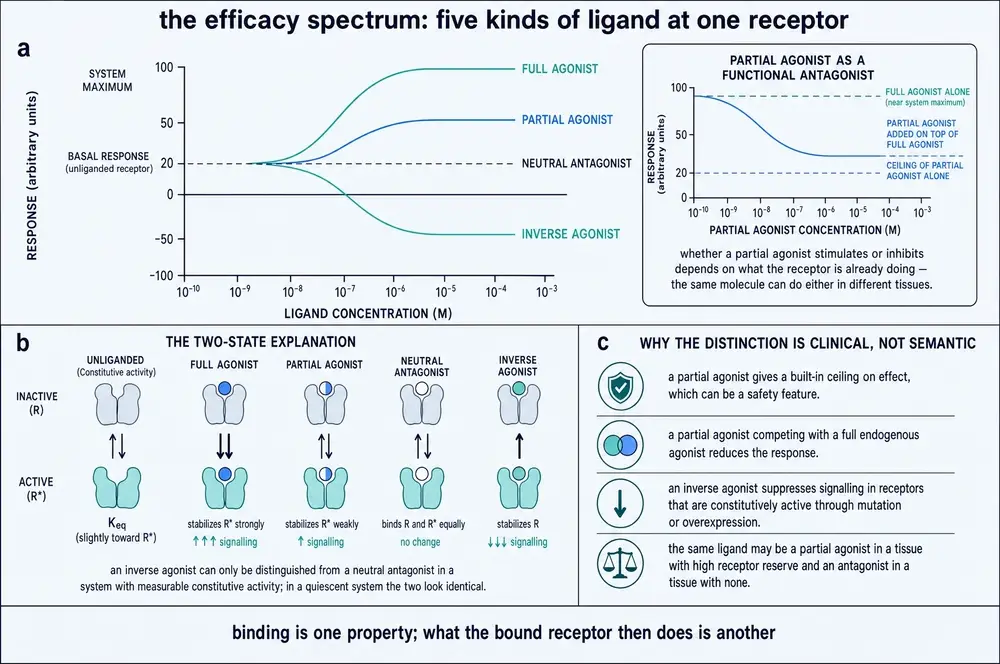
The most consequential point in this section is that "partial agonist" is not a property of a molecule. A ligand of fixed efficacy produces a sub-maximal response in a tissue with little reserve and a full response in a tissue with plenty. The same compound is therefore accurately described as a partial agonist in one preparation and a full agonist in another, with no change to the compound. Every table of intrinsic activities is implicitly a table about the assay it was run in.
Antagonism itself comes in two experimentally distinguishable forms. A competitive antagonist binds the same site and can be outcompeted, so increasing agonist restores the full maximum and the curve shifts rightward in parallel; the size of the shift against antagonist concentration gives the Schild relation and, from it, the antagonist's affinity. A non-competitive or insurmountable antagonist depresses the maximum. The important caution is the one section 07 introduced: a competitive antagonist can produce an insurmountable-looking result if it dissociates too slowly for the assay to equilibrate. And the same antagonist can produce a parallel shift in a tissue with high receptor reserve and a depressed maximum in a tissue without it, because reserve is what allows the surviving receptors to carry the full response.
10Allosteric modulation
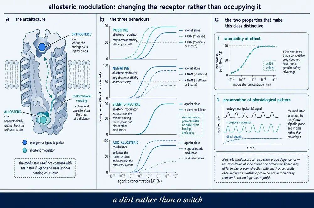
An allosteric ligand binds somewhere other than the orthosteric site and changes what happens there. It can raise or lower the affinity of the orthosteric ligand, its efficacy, or both, and it may or may not do anything on its own. The mechanisms are structurally diverse — sites have been described in the transmembrane bundle, at the lipid interface, in the intracellular vestibule, and on the extracellular face — and a 2023 review catalogues that diversity across the G protein-coupled receptor superfamily (Shpakov, 2023). The opioid receptors have become the best-worked example, with distinct modulatory sites resolved by combined structural and dynamic methods and a mechanistic account now available at the level of individual receptor motions (Wang et al., 2026; Zhang et al., 2026).
Two properties make allosteric modulation strategically interesting, and one makes it treacherous.
Saturability is a safety property. An orthosteric agonist's effect grows with concentration until the receptor population is occupied. An allosteric modulator's effect grows until its own site is occupied, and then stops — the modulator has changed the receptor as much as it can. The result is a built-in ceiling. A positive allosteric modulator of the μ-opioid receptor has been shown to act cooperatively with naloxone in a way that would be difficult to achieve orthosterically (O'Brien et al., 2024), and positive allosteric modulators have now been described for a receptor guanylyl cyclase as well, where the orthosteric ligand is a peptide and small-molecule orthosteric agonism is essentially unavailable (Ma et al., 2024).
Subtype selectivity is easier allosterically. Orthosteric sites are conserved within a receptor family because the endogenous ligand is shared; allosteric sites are under no such constraint and diverge more freely. The principle is not confined to the opioid receptors: allosteric modulation has been reviewed across the gonadotrophin receptors (Lazzaretti et al., 2023), and the allosteric communication pathway within the glucagon receptor has been mapped directly (van der Velden et al., 2025).
And probe dependence is the trap. A modulator's effect is defined relative to the orthosteric ligand it is modulating, and the same modulator can enhance one agonist and inhibit another at the same receptor. A modulator characterised against a synthetic probe may behave differently against the endogenous peptide, and a result reported without naming the probe is not interpretable. The same applies to what the modulator is measured on: a μ-opioid receptor modulator has been reported to confer agonist-dependent G protein bias (Grieble et al., 2025), which is to say that its selectivity for a pathway is a property of the pair, not of the modulator.
11Biased agonism, and the argument about it
A receptor can engage more than one transducer. If a ligand stabilises a conformation that engages one better than another, relative to a reference ligand, it is biased. The therapeutic promise is obvious and has been stated for two decades: if the wanted effect runs through one transducer and the unwanted effect through another, a biased ligand should separate them (Violin & Lefkowitz, 2007; Drake et al., 2008).
The evidence that ligands can be biased at the level of receptor conformation is now strong and structural. Distinct conformational changes in β-arrestin were reported to report biased agonism at seven-transmembrane receptors as early as 2008 (Shukla et al., 2008). More recently, angiotensin receptor conformations stabilised by different biased ligands have been shown to modulate β-arrestin engagement differentially (Elgeti et al., 2026), phosphorylation "barcodes" laid down by different kinases have been shown to direct biased chemokine signalling at CXCR3 (Eiger et al., 2023), and biased agonists at the angiotensin II type 1 receptor promote distinct subcellular β-arrestin conformations rather than merely different amounts of recruitment (Chundi et al., 2025).
What is contested is not whether bias exists but how often a reported bias is real, and whether it translates. Three artefacts produce apparent bias with no underlying ligand bias, and all three are common.
How much does this matter in practice? The most instructive case in pharmacology is the μ-opioid receptor, because the biased-agonism hypothesis was tested there about as hard as a hypothesis can be tested. The proposal was that analgesia is G protein-mediated while respiratory depression and tolerance are β-arrestin-mediated, so a G protein-biased agonist should be a safer analgesic. Two lines of evidence have substantially revised that account. Mice expressing phosphorylation-deficient μ-opioid receptors — genetically G protein-biased — showed improved analgesia and diminished tolerance but worse respiratory depression, the opposite of the prediction for the effect that mattered most (Kliewer et al., 2019). And a systematic operational analysis found that the compounds described as G protein-biased have low intrinsic efficacy, and that their apparent bias is largely explained by that low efficacy interacting with differing pathway amplification — artefact (a) and artefact (c) of the figure above, operating together (Gillis et al., 2020a). The subsequent critical assessment by the same group set out the case in full (Gillis et al., 2020b), and an IUPHAR themed review has since summarised the state of the field as one in which efficacy, bias and selectivity are much harder to separate than the first decade of work assumed (Ramos-Gonzalez et al., 2023).
Ligand bias is real at the level of receptor conformation; the structural evidence for distinct ligand-stabilised states is good and is getting better. Whether a reported bias is real requires that pathway amplification, assay time and receptor reserve have been controlled, and in a large fraction of the published literature they have not been. Whether a real bias translates into a separated therapeutic profile in a human being is a separate question again, and the one case tested most rigorously — the opioid receptor — returned a negative answer for the endpoint that mattered.
The document therefore treats bias as a live and promising mechanism with a poor translational record to date, and says so wherever it is invoked. Peptide receptors are not exempt: biased agonism at the GLP-1 receptor is under active investigation (Fletcher et al., 2018; Tran et al., 2026), and the same three artefacts apply.
12The GPCR activation sequence
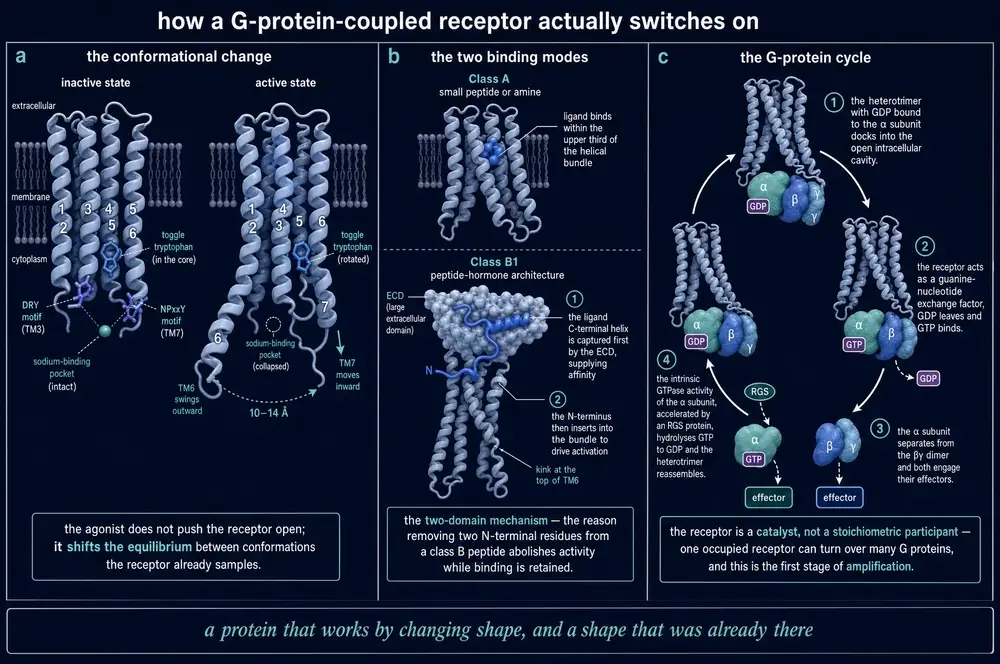
What happens between a peptide arriving and a G protein turning over is now visible, and it is worth stating precisely what "visible" means. Structures resolve states, not transitions. A cryo-EM map of an active complex is a population average of particles that were vitrified in a similar configuration, frequently with the help of a nanobody, a fusion protein or a nucleotide-free G protein that traps the state long enough to be seen. The sequence below is therefore assembled from several structures of several receptors, and its order is an inference from spectroscopy and mutagenesis as much as from the maps.
The receptor bundle in the unliganded state samples a range of conformations, mostly inactive. Agonist binding shifts that distribution. The change that matters is a large outward movement of the cytoplasmic end of transmembrane helix 6, which opens a cavity on the intracellular face that did not previously exist, together with rearrangement of conserved motifs that couple the ligand pocket to that face. The G protein's C-terminal helix inserts into the cavity. That insertion — not the agonist — is what destabilises the nucleotide-binding site, releasing GDP and leaving the G protein empty until GTP arrives. The nucleotide-free ternary complex is the state the 2011 β2-adrenergic receptor–Gs structure captured (Rasmussen et al., 2011), and it is short- lived in a cell.
Two points about this sequence are frequently lost. The first is that the receptor is the nucleotide-exchange factor: the energy for signalling comes from GTP, not from the peptide, and the peptide's contribution is catalytic. One occupied receptor can activate many G proteins in succession, which is the first amplification stage of section 21. The second is that the activating conformational change can be reached by routes other than the orthosteric one. A 2023 structure showed a class B1 receptor activated by an agonist binding on the intracellular side (Kobayashi et al., 2023), which is difficult to reconcile with any account in which the extracellular ligand pocket is the switch. The receptor is not a switch. It is an equilibrium that many things can lean on. The family resemblance that made this generalisation possible was visible long before any structure: cloning the β2-adrenergic receptor revealed homology with rhodopsin, and so revealed that a light receptor and a hormone receptor are the same machine (Dixon et al., 1986). The structural and pharmacological state of the field has since been reviewed comprehensively (Cheng et al., 2023).
13Which G protein, and what it does
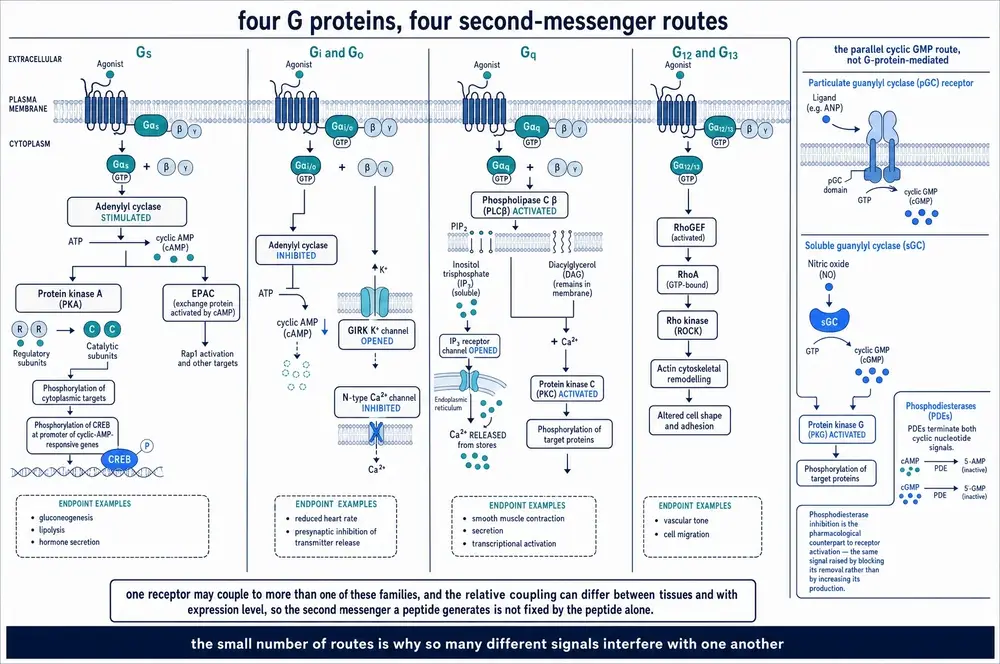
The heterotrimeric G proteins fall into four families by their α subunit, and the family determines what the receptor's activation actually means for the cell. Gs stimulates adenylyl cyclase and raises cyclic AMP. Gi/o inhibits it, and its released βγ subunits are signals in their own right. Gq/11 activates phospholipase C. G12/13 engages Rho guanine-nucleotide exchange factors and acts on the cytoskeleton. The purification of the first of these regulatory components in 1980 (Northup et al., 1980) followed a decade after the demonstration that hormone-stimulated adenylyl cyclase has an obligatory requirement for guanine nucleotides (Rodbell et al., 1971), which is the observation the whole G protein field grew from.
Peptide receptors couple promiscuously, and this is not sloppiness. A receptor that engages two G protein families produces two different intracellular consequences from one extracellular event, and which of them dominates depends on the relative abundance of the transducers in the cell being signalled to. This is one of the concrete mechanisms behind the document's central claim, and it is why tissue identity changes the meaning of a message. Structural work on the kisspeptin receptor has resolved determinants of G protein subtype selectivity directly, showing that the choice is encoded in the receptor's intracellular face and can be shifted by ligand (Wu et al., 2024).
14cAMP and PKA
Cyclic AMP was the first second messenger and it remains the best characterised. Sutherland and Rall isolated it from liver preparations and characterised it in 1958 (Sutherland et al., 1958), and the concept it established — that a hormone acting at the cell surface produces an intracellular chemical which does the actual work — is the foundation of everything in this Part.
The chain is short. Activated Gsα stimulates adenylyl cyclase; the cyclase makes cyclic AMP from ATP; cyclic AMP binds the regulatory subunits of protein kinase A and releases its catalytic subunits; those phosphorylate substrates, including the transcription factor CREB, which is how a surface event reaches the genome. A parallel effector, EPAC, is activated by the same messenger and does not involve PKA at all, which matters because experiments that block PKA do not block cyclic AMP signalling.
Two refinements have changed how this pathway should be read, and both work against the textbook picture of a cell filling with a messenger.
Compartmentation. Cyclic AMP does not distribute freely. It is generated at defined membrane locations, degraded by phosphodiesterases positioned to shape local gradients, and read by PKA held near its substrates by anchoring proteins. A whole-cell measurement can therefore be flat while the signal that produces the response is confined to a nanodomain, and two ligands producing identical total cyclic AMP can produce different outcomes. A 2026 study of oppositely biased GLP-1 receptor agonists in β-cells found spatially diffuse cyclic AMP signalling that did not follow the pattern the bias classification predicted (Chen et al., 2026), which is a useful caution about inferring location from pathway labels.
The signal continues after internalisation. The membrane was long assumed to be where signalling happens and the endosome where it stops. For several class B peptide receptors that is wrong. Parathyroid hormone receptor signalling generates cyclic AMP from endosomes, under Gq/11-dependent regulation (White et al., 2020), and the emergent signalling modes this implies have been reviewed structurally (Sutkeviciute & Vilardaga, 2020). More recently the effect of endocytosis on the cyclic AMP/PKA cascade has been shown to be receptor-selective rather than general (Blythe et al., 2026) — some receptors' signals are prolonged by internalisation and others' are terminated by it. Section 25 returns to this, because it is the mechanism that makes duration of action partly a trafficking property.
15Calcium and the phospholipase pathways
A Gq/11-coupled peptide receptor activates phospholipase Cβ, which cleaves the membrane lipid PIP2 into two messengers that go in different directions. Inositol trisphosphate diffuses into the cytoplasm and opens calcium channels on the endoplasmic reticulum; diacylglycerol stays in the membrane and, with calcium, activates protein kinase C. Streb, Irvine, Berridge and Schulz demonstrated the first arm in permeabilised rat pancreatic acinar cells in 1983, showing that inositol trisphosphate releases calcium from a non-mitochondrial store (Streb et al., 1983), and the second messenger role was set out the following year (Berridge & Irvine, 1984).
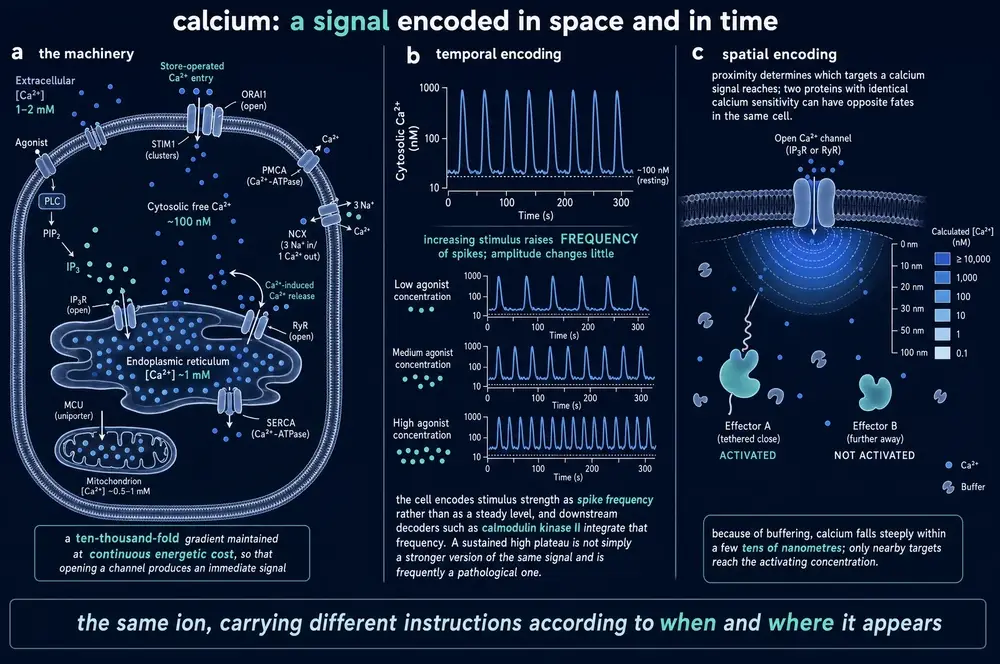
Calcium differs from cyclic AMP in a way that matters for peptide pharmacology. It is not primarily an amplitude signal. Cells encode information in the frequency of calcium oscillations, and downstream decoders — calmodulin-dependent kinases, calcineurin — respond to frequency rather than to mean concentration. A peptide that raises mean cytosolic calcium and a peptide that changes oscillation frequency are not doing the same thing, and an assay that integrates a calcium signal over thirty seconds cannot tell them apart. This is one of the concrete reasons an apparent equivalence between two agonists in a plate-reader assay can fail in a tissue.
16Receptor tyrosine kinases, and the insulin exception
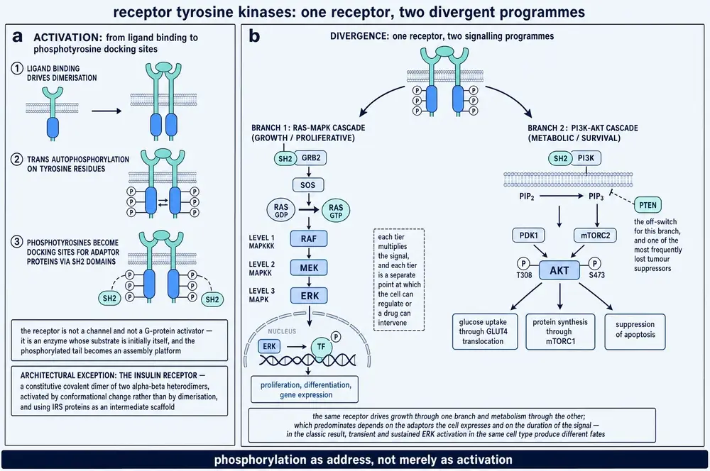
A second architecture. Instead of recruiting a separate transducer, the receptor is an enzyme: a single membrane pass with an extracellular ligand-binding region and an intracellular tyrosine kinase domain. The canonical mechanism, set out by Schlessinger and Ullrich, is that ligand binding brings two receptor molecules together, the two kinase domains phosphorylate each other on tyrosines, and the resulting phosphotyrosines become docking sites for proteins carrying SH2 domains, which assemble a signalling complex (Schlessinger & Ullrich, 1992). The output is not a small diffusible messenger but a pattern of phosphorylation, and which proteins dock determines which pathways run.
The insulin receptor is drawn wrongly in most textbook figures. It is not two separate chains that a ligand brings together. It is already a covalent dimer, disulphide-linked, before any insulin arrives. Insulin does not assemble it — insulin rearranges it, converting an autoinhibited, roughly inverted-V arrangement into a compact one that brings the intracellular kinase domains into position. Cryo-EM of the fully liganded receptor established the arrangement directly (Uchikawa et al., 2019), and structures of the complete ligand-saturated ectodomain followed (Gutmann et al., 2020).
Two older observations fall into place once the architecture is right. Cuatrecasas established that the insulin receptor is a membrane protein separable from the cell's metabolic machinery (Cuatrecasas, 1971), and de Meyts and colleagues found negative cooperativity in insulin binding (de Meyts et al., 1973) — the first insulin molecule makes the second bind less well, which is exactly what a two-site rearranging dimer predicts and what a simple one-site model forbids.
Bias is not confined to G protein-coupled receptors. Different fibroblast growth factors acting at the same receptor, FGFR1, produce different signalling outputs, and a 2024 analysis attributes the difference to ligand bias rather than to potency (Karl et al., 2024). On the engineering side, an insulin analogue with dual insulin and IGF-1 receptor agonism and a distinct signalling profile has been reported (Selicharová et al., 2026), and an allosteric pocket in the insulin receptor kinase domain itself has been described from conformational-dynamics work (Verma et al., 2026) — a molecular-dynamics result, and labelled as such.
17Cytokine receptors and JAK-STAT
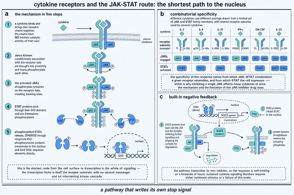
A third architecture, and the shortest route from the cell surface to the genome. Cytokine receptors have no catalytic activity of their own. They borrow it, from Janus kinases held constitutively on their intracellular tails. Ligand binding brings two receptor chains into the right relative geometry, the associated JAKs phosphorylate each other and then the receptor tails, STAT proteins dock on the resulting phosphotyrosines, are themselves phosphorylated, dimerise, and translocate to the nucleus to act as transcription factors. Darnell, Kerr and Stark set out the pathway in 1994 (Darnell et al., 1994).
Two peptide hormones use this machinery and are worth naming, because they are usually filed under endocrinology rather than immunology: growth hormone and leptin. The growth hormone receptor's mechanism is the crispest demonstration in the family that geometry, not merely proximity, is the signal. A single growth hormone molecule carries two distinct binding faces and recruits two receptor chains in sequence, which the 1994 X-ray structure of a growth hormone–prolactin receptor complex resolved (Somers et al., 1994). Because one ligand engages two receptors, excess ligand converts productive 1:2 complexes into unproductive 1:1 complexes, and the concentration-response curve is bell-shaped rather than sigmoid — more is less, above a point. That is a nonlinearity produced by stoichiometry alone, and section 23 returns to it.
The pathway also carries its own brake. Among the genes STATs transcribe are the suppressor-of-cytokine-signalling proteins, which bind the receptor and the JAKs and shut the pathway down. Feedback is not bolted on here; it is part of the output. Section 30 generalises the point.
Two recent findings illustrate how much the specific geometry matters. Naturally occurring variants of the interferon lambda receptor produce differential signal transduction rather than simply more or less of it (Novotny et al., 2026), and engineered interleukin-12 mimetics built on bispecific antibody scaffolds have been used to produce deliberately biased agonism at a cytokine receptor (Lipinski et al., 2025) — that is, bias is being engineered outside the G protein-coupled receptor family. A structural caution belongs beside them: some current models of cytokine-receptor-JAK2 assemblies are computational predictions from AlphaFold-Multimer rather than experimental structures (Pogozheva et al., 2023), and this document labels them as modelled wherever they are used.
18A receptor that is its own enzyme
The fourth architecture is the simplest and the least discussed. A receptor guanylyl cyclase has an extracellular ligand-binding domain, one transmembrane pass, a kinase-homology domain that is catalytically dead, and a guanylyl cyclase domain. Peptide binding relieves the kinase-homology domain's inhibition of the cyclase, and the receptor makes cyclic GMP directly. There is no G protein and no separate effector. Singh and colleagues established that a membrane guanylate cyclase is itself a cell-surface receptor in 1988 (Singh et al., 1988).
The peptides that use it are a coherent physiological group: the natriuretic peptides at GC-A and GC-B, and guanylin and uroguanylin at GC-C in the intestine. Cyclic GMP activates protein kinase G, and phosphodiesterases — PDE5 most famously — set the signal's lifetime, which is why inhibiting a phosphodiesterase amplifies a peptide signal without touching its receptor. The therapeutic consequences in heart failure have been reviewed extensively (Mangmool et al., 2023; Mishra et al., 2025).
Two cautions belong here rather than in a late section. First, this family has now yielded positive allosteric modulators — small molecules acting at GC-B (Ma et al., 2024) — which is notable precisely because the orthosteric ligand is a peptide and orthosteric small-molecule agonism has been essentially unavailable. Second, and more soberingly for translation: the receptor guanylyl cyclase GC-C has diverged evolutionarily to an extent that has implications for preclinical models (Mishra et al., 2024). A mechanism established in a rodent GC-C is not automatically a mechanism in the human one, and this is a specific instance of the general limit stated in the Evidence handling section.
19MAPK and PI3K-AKT: where everything converges
Below the receptor-specific layer sit two kinase cassettes that almost everything reaches. The MAPK cassette — Ras to Raf to MEK to ERK, with parallel p38 and JNK arms — and the PI3K-AKT-mTOR axis are reached from G protein-coupled receptors, from receptor tyrosine kinases, and from cytokine receptors, by several different routes each. They are the shared downstream, and their outputs are proliferation, survival, metabolic adjustment and transcriptional change.
The biology of this convergence is well described. What is under-stated is its epistemic consequence, and this document states it as a rule:
20Channels, and the much weaker case for intracellular targets
Peptides interact with ion channels in three well-supported ways. They modulate channels indirectly through the pathways already described — G protein βγ subunits acting directly on potassium and calcium channels is the fastest of all the mechanisms in this Part, requiring no diffusible messenger. They gate channels that are themselves receptors, as in the ligand-gated family. And a small number of peptides, principally from venoms, block or modify channel gating by binding the channel protein directly, which is a pharmacological rather than a physiological mechanism.
Intracellular action is a different and much weaker category, and the commission's own qualifier — "where scientifically supported" — is doing necessary work. The claim that a peptide acts at an intracellular target requires two things to be established separately, and the literature frequently supplies only the second:
- That the peptide reaches the intracellular compartment at a concentration comparable to the one at which the proposed effect occurs, by an identified route — a transporter, endocytosis with endosomal escape, or a demonstrated membrane-crossing mechanism. Peptides are large and polar and do not cross membranes by default.
- That the intracellular interaction occurs and produces the effect.
Where the first is not shown, an intracellular mechanism inferred from an observed effect is a hypothesis with an unexamined alternative: that the peptide acted at the surface, or was degraded to a fragment that did, or that the effect was produced by the delivery vehicle. This document therefore treats intracellular mechanisms case by case rather than as a class, admits those with a demonstrated uptake route, and names the others as unestablished. The specific cases are set out in the disputed-mechanism appendix rather than here, because assembling them in one place is what makes the pattern visible.
A related and better-evidenced case belongs in this section because it is frequently confused with intracellular action and is not the same thing. Receptors that have been internalised continue to signal from inside the cell — but the ligand is still on the outside face of the receptor, now facing the endosome lumen. Nothing has crossed a membrane. Endosomal cyclic AMP generation by the parathyroid hormone receptor is the best-worked example (White et al., 2020). Signalling from an intracellular location is not intracellular target engagement, and the two are routinely conflated.
21Amplification, divergence, cross-talk
Gain is what makes hormonal signalling work at picomolar concentrations, and it is multiplied stage by stage. One occupied receptor activates many G proteins over its active lifetime; one active cyclase makes many molecules of cyclic AMP; one PKA holoenzyme phosphorylates many substrates. The consequences are two, and they point in opposite directions: a tiny extracellular signal can produce a large cellular response, and a system with high gain saturates at low occupancy — which is receptor reserve, arrived at from the mechanism rather than from the curve.
The honest presentation of amplification is a problem, because the stage-by-stage numbers are largely not available for peptide receptors in human tissue. They are available in a few classical systems, and they are often quoted as though they generalised. The approach taken here is to give the arithmetic where the corpus supports it, name the system, and draw the gaps as gaps. A useful recent development is the proposal to measure whole-system amplification empirically rather than reconstructing it from stages, using the ratio of a full agonist's binding affinity to its functional potency as the gauge (Buchwald, 2025) — a method that builds on the same author's earlier receptor model separating affinity, efficacy and amplification (Buchwald, 2019).
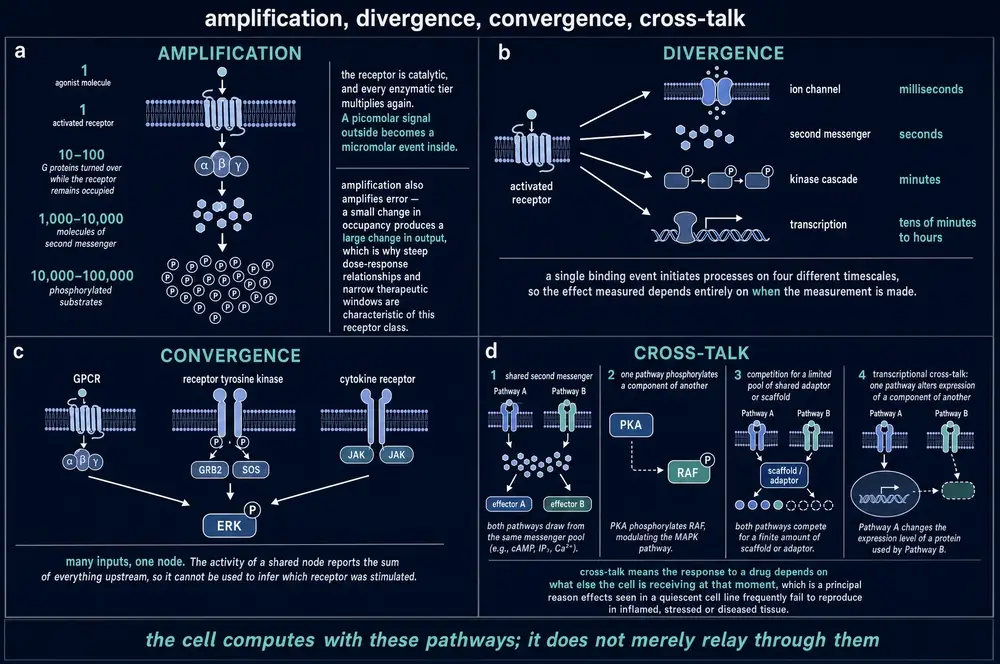
Signals also diverge and interfere. One receptor reaches several pathways; several receptors reach one pathway; and pathways interact through shared components. Cross-talk is not a vague notion — each edge has a mechanism, and naming the mechanism is what distinguishes a real interaction from a correlation. PKA phosphorylating a Raf isoform is a direct covalent edge. Calcium and DAG jointly required by conventional PKC isoforms is a coincidence-detection edge. Two receptors competing for a limited pool of G protein is a resource edge, and it produces interactions that look like pharmacological antagonism without any shared binding site. β-arrestin scaffolding ERK is an edge that belongs to the desensitisation machinery, which is where Part Four begins.
22Occupancy is not response
Part One established the principle historically. This section states it as a working rule, because it is the single most consequential fact in the document and it is routinely violated in practice.
For a full agonist in a tissue with substantial receptor reserve, the concentration producing half the maximal response can be one to two orders of magnitude below the concentration occupying half the receptors. The gap is not an error. It is the amplification described in section 21, expressed on a concentration axis. Three consequences follow, and each one is a mistake this document has seen made in the peptide literature:
- A maximal effect does not imply a saturated target. If a peptide produces its full effect at a concentration occupying a tenth of its receptors, then nine tenths of the receptor population is available for nothing to happen at, and raising the dose does not increase the wanted effect — it only recruits whatever else the compound binds. This is the mechanistic origin of a therapeutic ceiling coexisting with a rising adverse effect, and section 32 returns to it.
- Receptor loss is buffered until it is not. A tissue with tenfold reserve tolerates losing most of its receptors with no change in maximal response, and then fails abruptly. Downregulation during chronic exposure (section 26) is therefore invisible in the response until it crosses a threshold, which is one reason tolerance often appears suddenly rather than gradually.
- The efficacy needed to be a "full" agonist is a property of the tissue. A low-efficacy ligand is a full agonist where reserve is large and a partial agonist where it is small — the same molecule, two labels, neither wrong.
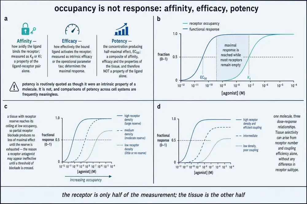
The reserve can be measured, by Furchgott's irreversible-antagonist method (Furchgott, 1959) or by fitting the operational model (Black & Leff, 1983), and it can now also be estimated from the ratio of binding affinity to functional potency for a full agonist (Buchwald, 2025). What it cannot be is assumed, and the most common way of assuming it is to work exclusively in an engineered cell line: raising receptor density raises reserve, which is why apparent pharmacology changes with expression level, and why a study of biased signalling found the bias itself disappearing when expression was varied (Li et al., 2023).
23Where non-linearity comes from
A concentration-response curve for a single ligand at a single site with response proportional to occupancy has a defined steepness: on a logarithmic axis it climbs from a tenth to nine tenths of maximum over about two orders of magnitude, a Hill slope of unity. Real curves are frequently steeper or shallower, and the four causes are distinguishable.
| Cause | Effect on slope | What distinguishes it |
|---|---|---|
| Positive cooperativity | steeper | Present in binding as well as in function; requires multiple interacting sites on one receptor or complex |
| Negative cooperativity | shallower | Also present in binding; insulin receptor is the classic peptide case (de Meyts et al., 1973) |
| Multiple receptor subtypes or states | shallower, sometimes biphasic | Binding is shallow or two-component; a selective antagonist resolves the components |
| Amplification with a saturating downstream step | steeper in function only | Binding slope is unity; the steepening appears only in the functional readout |
| Ligand depletion or non-equilibrium | artefactual, either direction | Slope changes with incubation time, cell number or assay volume |
| Ligand-induced dimerisation stoichiometry | bell-shaped | Response falls at high concentration; growth hormone receptor is the canonical case |
The last row is worth dwelling on because it is the clearest case in which more drug does less, for a reason that has nothing to do with toxicity or off-target binding. Where one ligand molecule must bridge two receptor chains, excess ligand saturates both chains separately and the productive complex cannot form. The concentration-response curve rises, peaks and falls. Any analysis that assumes monotonicity — including most curve-fitting software defaults — will misfit it, and a dose-ranging study that samples only the ascending limb will conclude the wrong thing about the plateau.
24Turning the signal off
Signalling does not stop when the ligand leaves. It is stopped, actively, by machinery that begins working within seconds of the signal starting, and that machinery is part of signalling rather than a separate process. The sequence was worked out on the β2-adrenergic receptor and generalises across the G protein-coupled family, peptide receptors included.
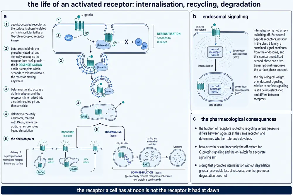
A G protein-coupled receptor kinase recognises the active conformation and phosphorylates the receptor's intracellular tail; the receptor kinase family was defined by Benovic and colleagues (Benovic et al., 1989). β-arrestin then binds the phosphorylated tail, which sterically blocks G protein coupling and terminates the signal — the protein was identified for exactly this function by Lohse and colleagues (Lohse et al., 1990). Because the kinase recognises the active state, only occupied receptors are silenced, which is what makes this homologous desensitisation. A second route, through PKA or PKC phosphorylating receptors regardless of occupancy, silences receptors that were never stimulated, and is heterologous.
The phosphorylation pattern is an instruction, not a switch. Different kinases place phosphates at different sites, arrestin adopts a different conformation depending on the pattern, and the downstream consequence differs. This "barcode" model has direct experimental support: at CXCR3, distinct phosphorylation barcodes were shown to direct biased chemokine signalling (Eiger et al., 2023), and biased ligands at the angiotensin II type 1 receptor promote distinct subcellular β-arrestin conformations rather than different amounts of recruitment (Chundi et al., 2025). Arrestin coupling has now been resolved structurally for a class B peptide receptor, the parathyroid hormone type 1 receptor (Zhai et al., 2026). And the picture continues to gain layers: β-arrestins have been reported to form biomolecular condensates that regulate receptor function (Anderson et al., 2026), a mechanism absent from every textbook account of desensitisation written before it.
Arrestin's second job is the one that complicated the field. Having terminated G protein signalling, it acts as a scaffold, assembling kinase complexes and initiating a second wave of signalling with a different time course and different outputs. Desensitisation and signal initiation are the same event seen from two sides, which is why the clean separation the biased- agonism programme hoped for (section 11) proved hard to obtain.
25Internalisation, recycling, degradation
Arrestin binding also recruits the endocytic machinery. The receptor is drawn into a clathrin-coated pit, pinched off by dynamin, and delivered to an early endosome — a route separable from arrestin's other functions, as Zhang and colleagues showed by dissociating dynamin-dependent internalisation from arrestin-dependent steps (Zhang et al., 1996). What happens next is a sorting decision, and it determines the timescale of everything the reader would call tolerance:
- Fast recycling back to the surface, dephosphorylated and resensitised, in minutes. Function is restored quickly and the cell is ready to respond again.
- Slow recycling through a perinuclear compartment, over tens of minutes to hours.
- Lysosomal degradation, usually following ubiquitination, which removes the receptor permanently. Recovery then requires new synthesis, which takes hours to days.
The decision is encoded in the receptor's own sequence and in the phosphorylation pattern from section 24. A dileucine motif in the calcium-sensing receptor was shown in 2025 to direct internalisation to spatially distinct endosomal populations (Wyatt et al., 2025), and the multiple roles of β-arrestins in this process have been reviewed comprehensively (Liu et al., 2025). Trafficking is also a design lever: altered intracellular trafficking has been used deliberately as a mechanism for prolonging the duration of receptor action (Kim et al., 2026). A 2025 preprint — not yet peer reviewed, and flagged as such here — reports that fast resensitisation depends on PI(4,5)P2-dependent sorting (Gulyas et al., 2025).
The finding that most changed the picture is that the endosome is not a terminus. Internalised receptors continue to signal, and for the parathyroid hormone receptor the endosomal phase generates a substantial and prolonged cyclic AMP signal under Gq/11-dependent control (White et al., 2020; Sutkeviciute & Vilardaga, 2020). Whether endocytosis prolongs or terminates a signal is receptor-specific, not general (Blythe et al., 2026), and for the GLP-1 receptor internalisation has been shown to control the spatiotemporal pattern of signalling produced by biased agonists (Fletcher et al., 2018). Internalisation is therefore not a synonym for switching off, and a paper that treats receptor loss from the surface as evidence of signal termination is making an assumption that has been falsified for several peptide receptors.
26Three words, three timescales
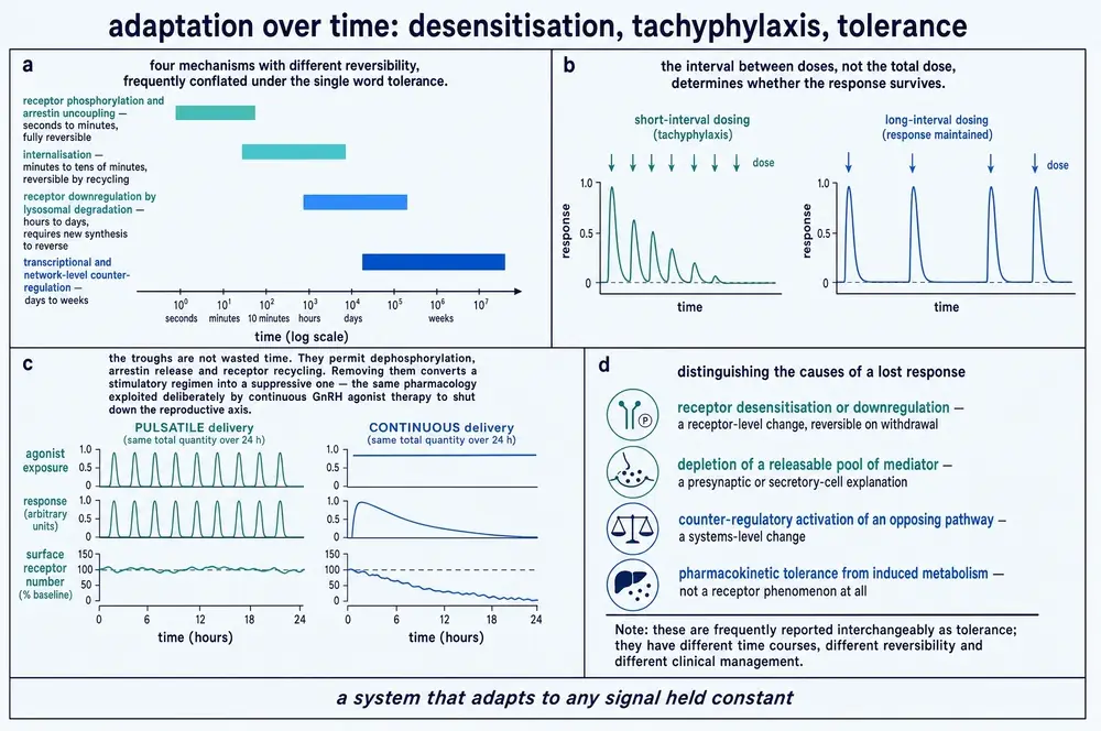
Desensitisation, downregulation, tolerance and tachyphylaxis are used interchangeably in a great deal of writing about peptides, and they name different things happening over different periods by different mechanisms. Keeping them apart is not fussiness: the distinction predicts how quickly an effect will fade, how quickly it will come back, and whether a dose interval can do anything about it.
| Term | Timescale | Mechanism | Reversal |
|---|---|---|---|
| Desensitisation | seconds to minutes | GRK phosphorylation and arrestin binding; receptor still present | Dephosphorylation on recycling; minutes |
| Tachyphylaxis | minutes to hours | Descriptive term for rapid loss of response on repeated dosing; usually desensitisation plus internalisation, sometimes depletion of a mediator | Requires an interval long enough for resensitisation |
| Internalisation | minutes | Receptor removed from the surface; may continue signalling from the endosome | Recycling; minutes to hours |
| Downregulation | hours to days | Net loss of receptor protein: lysosomal degradation exceeding synthesis, often with reduced transcription | New synthesis; hours to days |
| Tolerance | days and longer | Whole-system adaptation: the above, plus counter-regulation at other nodes and in other tissues (section 30) | Slow, and sometimes incomplete |
The clinically important point is that these do not simply add up. Receptor reserve buffers the early ones: a tissue with tenfold reserve can lose most of its surface receptors to internalisation with little change in maximal response, so desensitisation at the molecular level produces no observable tolerance until degradation has proceeded far enough to exhaust the buffer. A system-level demonstration comes from chronic peptide exposure in rodents, where tolerance in thalamic paraventricular nucleus neurons developed only after sustained treatment (Koita et al., 2025) — a result in rats, and reported as such.
The practical test a reader can apply to any report of tolerance is to ask which of these five the author has actually measured. A paper reporting that "response declined over two weeks of treatment" has measured the whole stack and identified none of it. A paper reporting reduced surface receptor number has measured internalisation and downregulation together and separated neither. Distinguishing them requires either a time course fine enough to resolve the mechanisms' different rates, or an intervention that acts on one and not the others — a washout long enough for recycling but too short for resynthesis, for instance, which separates the reversible arms from the degradative one. Where the literature has not done this, the honest reading of a tolerance claim is that something diminished and the mechanism is unassigned.
27Pulsatile versus continuous
This is the cleanest demonstration in endocrine pharmacology that exposure pattern is itself information, and it is the strongest single piece of evidence for this document's central claim. Two cases, both with opposite outcomes from the same molecule at the same total exposure.
Gonadotrophin-releasing hormone. Belchetz, Plant, Nakai, Keogh and Knobil, working in ovariectomised rhesus monkeys whose hypothalamic connections to the pituitary had been ablated, replaced the hormone by infusion. Delivered in hourly pulses it maintained gonadotrophin secretion. Delivered continuously at the same rate it suppressed it (Belchetz et al., 1978); the permissive role of pulsatile delivery in the primate cycle was set out subsequently (Knobil et al., 1980). The mechanism is receptor desensitisation and downregulation as described in the two previous sections: continuous occupancy drives the receptor down the trafficking pathway and it does not return. This is not a curiosity. It is the basis of an entire class of clinical intervention, in which a receptor agonist is used to switch a system off by never letting it recover — and it is worth noticing that the drug in that use is an agonist doing exactly what an agonist does.
Parathyroid hormone. The same molecule builds bone or destroys it depending on the pattern of exposure. Intermittent administration increased osteoblast number and bone formation markers in rats (Schmidt et al., 1995), while continuously elevated levels — as in hyperparathyroidism — produce net resorption. The human counterpart is the trial of once-daily parathyroid hormone (1–34) in postmenopausal women with osteoporosis, which reported increased bone mineral density and reduced fracture risk over a median of 21 months (Neer et al., 2001). Reported as a completed randomised trial in a defined population, at the schedule the trial used; no dose, route or schedule is recommended by this document. Modelling work has since attempted to reproduce the divergence from receptor kinetics alone, using a two-state receptor model driven by different dosing patterns (Martonová et al., 2023) — a computational result, labelled as such — and the cyclic AMP/PKA arm through which the anabolic signal runs has been reviewed (Martin, 2021).
28Tissue-specific expression and cell state
The last contribution the system makes is the most obvious and the most often left out of a mechanism diagram: the cell has to be listening, and what it hears depends on what it is.
Four variables, each independent of the ligand:
- Receptor presence. A tissue with no receptor has no response, and this is what makes hormonal signalling addressable at all — the message is broadcast in blood and read only where a reader exists.
- Receptor density. Which sets reserve, which sets the position and the shape of the dose-response curve, and which converts the same ligand from a partial to a full agonist between tissues.
- Transducer availability. A receptor that can couple to Gs and Gq does whichever the cell has more of. The same receptor, in two tissues, is functionally two receptors, and this is a mechanism rather than a metaphor.
- Prior state. A cell stimulated ten minutes ago has phosphorylated, partly internalised receptors and a different transducer pool from a resting one. The response to the second dose is not the response to the first. The extracellular milieu counts too: extracellular ATP has been shown to increase agonist potency and reduce response latency at class B G protein-coupled receptors (Zhu et al., 2025), so a tissue's local nucleotide environment changes the pharmacology of a peptide acting on it.
Accessory proteins add a fifth. Receptor activity-modifying proteins determine what a calcitonin receptor recognises; MRAP2 modifies the signalling and oligomerisation state of the melanocortin-4 receptor (Sohail et al., 2025); amylin receptor subunit interactions are themselves modulated by which agonist is bound (Gostynska et al., 2025). And genetic variation operates on all of it: the genetic architecture of an allosteric hormone receptor has been mapped at scale, showing how widely distributed the sequence determinants of receptor behaviour are (Stammnitz et al., 2026). Where a receptor system has been characterised to the standard this section demands — distribution, density, transducer coupling and species differences reported together — it is usually because an expert panel has done it: the IUPHAR review of apelin receptor pharmacology is a recent model of the form (Davenport et al., 2026).
29How a cellular response becomes a physiological one
Everything so far has happened inside one cell. A drug effect is not one cell's response; it is the summed, weighted response of a population of cells, propagated through tissue mechanics and organ architecture and then integrated by a body that has its own opinions. Each of those steps can amplify a cellular response, attenuate it, or reverse its sign.
Three things happen between the cell and the reader's clinical endpoint, and the document is precise about them because this is where mechanistic writing most often becomes wishful:
- Aggregation is weighted, not averaged. A tissue's response is dominated by whichever cells express the receptor most densely, not by the mean expression. A receptor present on ten per cent of cells at high density can carry the whole response, and a transcript-level measurement averaged over the tissue will understate it.
- Architecture converts a cellular change into a physiological one. Smooth muscle calcium becomes vascular tone becomes pressure only through geometry — vessel radius enters the resistance relation to the fourth power — so a modest cellular effect can produce a large haemodynamic one and a large cellular effect in the wrong vessels can produce none.
- Nothing in the body is measured in isolation. By the time an effect is observable it has already been sensed by whatever regulates the variable in question, and section 30 is about what that regulation does to it.
Where a mechanistic account can be followed all the way to a human physiological endpoint, this document says so. Where it cannot, it names the missing link rather than eliding it. That is not a hedge; the chain receptor → pathway → cell → tissue → organ → outcome is nowhere continuous in one experimental system, and a paper that appears to demonstrate it has almost always demonstrated two ends of it and assumed the middle. A review of how developments in receptor theory are changing the understanding of hypertension makes the point from the clinical side: the pharmacology that explains a blood-pressure response is not the pharmacology that was measured in the cell line (Watts et al., 2024).
30Feedback, and the body's answer to sustained pharmacology
Section 26 described adaptation inside the target cell. This section describes adaptation everywhere else, and it is the larger effect.
Endocrine systems are built as feedback loops. A hormone's output is sensed — sometimes by the gland that released it, sometimes by the hypothalamus and pituitary, sometimes by a distant tissue whose state is the regulated variable — and the sensing adjusts the input. Introduce a drug that mimics the hormone and the loop does exactly what it is built to do: it reduces endogenous production, and it may also change receptor number, transducer coupling, or the activity of an opposing system. The drug's effect on the regulated variable is therefore smaller than its effect on the receptor, and the difference grows with time.
The renin-angiotensin system is the clearest peptide example of a further refinement, which is that counter-regulation frequently runs through a second receptor for the same ligand rather than through a separate pathway. Angiotensin II acts at AT1 and at AT2, and the two oppose each other; the equilibrium between them, rather than the level of the peptide, is what determines the net effect (Colin et al., 2023). A ligand that engages both is self-limiting in a way that a receptor-selective one is not, and a selective antagonist changes the balance as well as the magnitude.
31Four ways a peptide drug causes harm
Adverse effects are usually sorted by severity or by organ. Sorting them by mechanism is more useful, because mechanism predicts what can be done about them. There are four causes, and only the fourth is what most readers mean by an off-target effect.
| Cause | What it is | What can address it |
|---|---|---|
| 1 · Intended pharmacology | The target receptor in the target tissue, doing exactly what it was meant to do, in a patient for whom that is unwelcome | Patient selection. Not a pharmacological problem |
| 2 · Exaggerated on-target effect | The same receptor in the same tissue, driven too far. Not a different mechanism — too much of the intended one | Dose; a partial agonist's ceiling; an allosteric modulator's saturability |
| 3 · On-target, off-tissue | The same receptor in a different organ, where its activation is unwanted. The commonest source of peptide-drug adverse effects | Tissue-restricted delivery, or biased signalling if the tissues differ in transducer coupling. Subtype selectivity helps only if the tissues differ in subtype |
| 4 · Off-target | Binding an unrelated receptor, generally at higher concentration | Selectivity ratio — and note that this is the only one of the four that a selectivity ratio addresses |
Two consequences are worth drawing out. The first is that raising selectivity does nothing at all for causes 1 to 3, which between them account for most of what goes wrong with peptide drugs. A perfectly selective agonist still produces cause 3 wherever its receptor is expressed, and receptor expression is not something a medicinal chemist controls. The second is that cause 2 is bounded by the ceiling discussed in section 22: if the therapeutic effect saturates at low occupancy and the adverse effect does not, then every increment of dose above the therapeutic plateau buys adverse effect alone. That is a mechanistic argument for dosing to the plateau, and it is the subject of the next section.
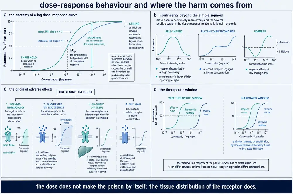
32The therapeutic window is a ratio of two curves
The therapeutic window is routinely described as a property of a molecule. It is not. It is the separation between two concentration-response curves — one for the wanted effect, one for the unwanted — and either curve can move without the molecule changing at all. Because the two curves are generated in different tissues with different receptor densities and different coupling, the window differs between individuals for reasons that have nothing to do with pharmacokinetics.
Four levers can widen it, and they are worth ranking by how much human evidence each actually has:
| Lever | Mechanism | Strength of human evidence |
|---|---|---|
| Exposure pattern | Intermittent exposure permits resensitisation and can invert the direction of a response entirely (section 27) | Strong. Demonstrated for parathyroid hormone in a randomised human trial (Neer et al., 2001) and exploited clinically in the opposite direction for GnRH |
| Subtype selectivity | Separates cause 4, and cause 3 where the tissues differ in subtype | Good for well-differentiated families; poor where paralogues share their binding surface (section 08) |
| Allosteric ceiling | A modulator's effect saturates when its own site fills, bounding cause 2 (section 10) | Mechanistically sound, thinly tested in humans for peptide receptors |
| Biased agonism | Separates wanted from unwanted where they run through different transducers | Weak, and the best-tested case was negative (section 11; Kliewer et al., 2019; Gillis et al., 2020a) |
A fifth lever has emerged from the trafficking work in section 25 and does not fit the classical scheme: altering where and for how long a receptor signals, rather than how strongly. Engineering prolonged duration through altered intracellular trafficking has been demonstrated (Kim et al., 2026), and preclinical work at the GLP-1 receptor reports that a cyclic-AMP-biased agonist maintained glycaemic control with reduced malaise and emesis in animal models (Baumer-Harrison et al., 2026) — a preclinical result, reported as such, and precisely the kind of claim section 11 says has a poor record of translating.
33Why the same molecule is not the same drug
The document's argument can now be stated in full, because every part of it has been earned.
A peptide arriving at a cell does one thing: it changes the probability distribution over that receptor's conformations. That is the entire contribution of the molecule, and it is a small contribution. Everything a reader would recognise as the drug's effect is added afterwards, by the receiving system, in a sequence of stages each of which can multiply or nullify what the previous stage delivered.
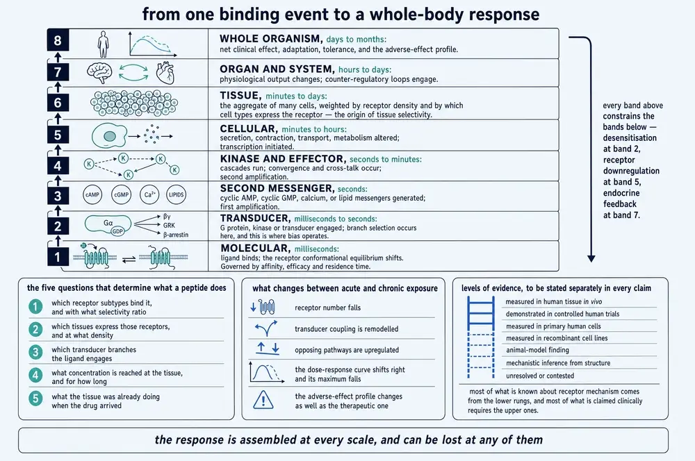
This is why potency tables do not transfer between preparations, why an effect established in a cell line so often fails in a tissue, why the same peptide is anabolic and catabolic on two schedules, and why tolerance appears suddenly rather than gradually. None of these is an anomaly. Each is a prediction of the account above, and taken together they are the reason a lock-and-key picture of peptide action is not merely incomplete but systematically misleading about which variable to change: it directs attention to the molecule when most of the available leverage is in the schedule, the tissue and the state.
34What this account does not explain
Five limits, stated plainly, because a document that has argued this hard for the importance of the system owes the reader an account of where its own evidence runs out.
The structures are photographs of ensembles. Every activation sequence in Part Three is assembled from static structures of stabilised states, frequently of different receptors, sometimes with engineered constructs, and the ordering between them is inferred rather than observed. The field's own dynamic methods — spectroscopy, single-molecule work, molecular dynamics — agree that the ensemble is broader than the structures suggest, and no current method resolves the transitions in a physiological membrane at physiological receptor density.
The pharmacology is not human, and mostly not tissue. Reserve, bias, coupling stoichiometry and trafficking itineraries are overwhelmingly measured in heterologous expression systems at receptor densities that do not occur in the body. Because reserve scales with density, this systematically inflates apparent potency and can convert a partial agonist into a full one. Every quantitative statement in this document about efficacy or bias inherits that limitation.
Human receptor occupancy is almost never measured. The quantity on which the whole occupancy-versus-response argument turns is, in humans, available only for the small number of systems with a suitable tracer. For peptide receptors it is largely unavailable, which means the reserve argument — correct in principle and demonstrated in tissue preparations — is applied to human pharmacology by extrapolation.
Biased agonism has an unresolved translation problem. The conformational basis is real and the structural evidence is improving. Whether a measured bias predicts a separated clinical profile is a different question, and the case tested most rigorously returned a negative answer for the endpoint that mattered (section 11).
And for several well-described peptide effects, the receptor is still not identified. A physiological action can be robust, reproducible and dose-dependent while its receptor remains unknown or contested. Where that is so, this document says the receptor is unidentified rather than assigning the effect to the nearest plausible candidate. The specific cases are set out in the disputed-mechanism appendix, which exists because collecting them in one place is what makes the pattern visible: the mechanistic confidence of a literature is not evenly distributed, and it is lowest exactly where the commercial interest is highest.
35Glossary
The quantitative vocabulary of receptor pharmacology is small, precise, and routinely used loosely. These definitions are the ones this document uses throughout, and they are placed here rather than at first use because a reader consulting a table needs them more than a reader following the argument.
| Term | Definition as used here |
|---|---|
| Affinity | How tightly a ligand binds, independent of what binding does. Reported as a dissociation constant. A property of the ligand-receptor pair alone. |
| Allosteric | Acting at a site topographically distinct from the orthosteric site, changing what happens at that site without competing for it. |
| Bias factor | The logarithmic ratio of transduction coefficients for two pathways, taken relative to a reference ligand. Meaningless without naming both the reference ligand and both pathways. |
| Constitutive activity | Receptor signalling in the absence of any ligand. What makes inverse agonism detectable. |
| Desensitisation | Loss of response over seconds to minutes with the receptor still present, by phosphorylation and arrestin binding. |
| Downregulation | Net loss of receptor protein over hours to days. Reversal requires new synthesis. |
| Efficacy | How strongly a bound ligand stabilises transducer-engaging conformations. Cannot be measured in a binding assay. |
| Emax | The maximal response a ligand produces in a given system. A system property as much as a ligand property. |
| EC50 | The concentration producing half the maximal response in that preparation. A composite of affinity, efficacy and amplification. |
| Hill slope | The steepness of a concentration-response curve. Unity for one ligand, one site, response proportional to occupancy; departures have four distinguishable causes (section 23). |
| Inverse agonist | A ligand that reduces signalling below the unliganded baseline. Requires constitutive activity to be demonstrable. |
| Kd | Equilibrium dissociation constant: the concentration at which half the receptors are occupied at equilibrium. Lower means higher affinity. |
| koff | Dissociation rate constant. Its reciprocal is the residence time, and it is what the equilibrium constant conceals. |
| Operational model | The Black-Leff framework separating a ligand property (affinity, efficacy) from a system property (the transducer ratio) with independent parameters. |
| Orthosteric | The site the endogenous ligand occupies. |
| Partial agonist | A ligand producing a sub-maximal response at full occupancy in that tissue. Not a fixed property of a molecule. |
| Potency | The concentration producing a given effect. A property of a molecule in a preparation, never of a molecule. |
| Probe dependence | The observation that an allosteric modulator's effect differs according to which orthosteric ligand it is modulating. |
| Receptor reserve | The excess of receptors over the number needed for a maximal response. A tissue property, measured as the displacement between occupancy and response curves. |
| Residence time | How long a ligand-receptor complex lasts. The reciprocal of the dissociation rate constant. |
| Tachyphylaxis | Rapid loss of response on repeated dosing. Descriptive; usually desensitisation plus internalisation. |
| Tolerance | Whole-system loss of response over days and longer, including counter-regulation in tissues the drug did not act on. |
| Transducer ratio (τ) | The amplification a system provides between receptor occupancy and response. The quantity that makes reserve possible. |
36Abbreviations
| Short | Expansion | Short | Expansion |
|---|---|---|---|
| AT1, AT2 | angiotensin II receptor types 1 and 2 | MAPK | mitogen-activated protein kinase |
| cAMP | cyclic adenosine monophosphate | MC4R | melanocortin-4 receptor |
| cGMP | cyclic guanosine monophosphate | MRAP2 | melanocortin-2 receptor accessory protein 2 |
| CGRP | calcitonin gene-related peptide | mTOR | mechanistic target of rapamycin |
| CREB | cAMP response element-binding protein | NPR | natriuretic peptide receptor |
| DAG | diacylglycerol | PAM / NAM | positive / negative allosteric modulator |
| ECD | extracellular domain | PDE | phosphodiesterase |
| EPAC | exchange protein directly activated by cAMP | PI3K | phosphoinositide 3-kinase |
| ERK | extracellular signal-regulated kinase | PIP2 | phosphatidylinositol 4,5-bisphosphate |
| FGFR1 | fibroblast growth factor receptor 1 | PKA / PKC / PKG | protein kinase A / C / G |
| GC-A, GC-B, GC-C | receptor guanylyl cyclases A, B and C | PTH | parathyroid hormone |
| GIP | glucose-dependent insulinotropic polypeptide | PTH1R | parathyroid hormone type 1 receptor |
| GLP-1, GLP-2 | glucagon-like peptide 1 and 2 | RAMP | receptor activity-modifying protein |
| GnRH | gonadotrophin-releasing hormone | RTK | receptor tyrosine kinase |
| GPCR | G protein-coupled receptor | RXFP | relaxin family peptide receptor |
| GRK | G protein-coupled receptor kinase | SH2 | Src homology 2 domain |
| IP3 | inositol 1,4,5-trisphosphate | SOCS | suppressor of cytokine signalling |
| JAK | Janus kinase | STAT | signal transducer and activator of transcription |
| KOR / MOR | κ- / μ-opioid receptor | TM1–TM7 | transmembrane helices 1 to 7 |
37Disputed and unresolved mechanisms
Six mechanistic questions in this document are open. They are collected here because §10.4 requires conflicting evidence to be presented as conflict, and because assembling them in one place makes visible a pattern that is invisible claim by claim.
Each row states the dispute rather than summarising a consensus, because in every one of these six there is no consensus to summarise. The right-hand column is this document's position, and it is a position rather than a survey: where the evidence permits a judgement it is made, and where it does not the row says so. Three of the six are questions about whether a mechanism demonstrated in a reduced system reaches human physiology, which is this document's recurring epistemic problem and the reason the closing plate's levels-of-evidence ladder is reproduced rather than paraphrased.
| Question | The dispute | Where this document stands |
|---|---|---|
| Does biased agonism translate? | Conformationally real and structurally supported; but phosphorylation-deficient μ-opioid receptor mice showed worse respiratory depression, and operational analysis attributes much reported bias to low intrinsic efficacy interacting with unequal amplification | Bias is real at the receptor; a reported bias is unreliable unless amplification, assay time and reserve were controlled; translation is unproven and the best-tested case was negative |
| Induced fit or conformational selection? | Long-standing, usually posed as an either/or | Both occur within one binding event at peptide receptors. The general question is not well posed and the document does not adjudicate it |
| How much of physiological signalling is endosomal? | Endosomal signalling is established for several class B receptors; its quantitative weight relative to surface signalling is not, and endocytosis prolongs some receptors' signals and terminates others' | Reported as established for the named receptors and unquantified in general. Surface disappearance is not termination |
| Does residence time predict efficacy? | Holds in some systems and not others; rebinding, partitioning and target turnover intervene between a purified measurement and a tissue | Not a general rule. Must be demonstrated case by case, and this document treats it as a hypothesis about function rather than a finding |
| Do peptides act at intracellular targets? | Claimed for several research peptides on the basis of an observed intracellular effect, usually without a demonstrated uptake route | Treated case by case, not as a class. A demonstrated membrane-crossing route is required before an intracellular mechanism is credited. Signalling from an internalised receptor is not intracellular target engagement and the two are routinely conflated |
| Which receptor mediates several well-described peptide effects? | Robust, reproducible, dose-dependent physiological actions whose receptor is unidentified or contested | Named as unidentified. A phospho-ERK or phospho-AKT signal in a cell line is not receptor identification (section 19), and this document does not assign an effect to the nearest plausible candidate |
38References
103 references, generated from verified NCBI records and sorted by first-author surname. House style §5: references in this series are never generated from recall. Every identifier below was resolved against PubMed at build time and the build refuses to run on an unresolved identifier.
PMID 42203872 · doi:10.1038/s41586-026-10539-y · PMC13345904
PMID 13229430
PMID 40172014 · doi:10.1021/acs.biochem.4c00812 · PMC12004451
PMID 41508705 · doi:10.1111/dom.70427 · PMC12981003
PMID 100883 · doi:10.1126/science.100883
PMID 2552582 · doi:10.1126/science.2552582
PMID 6095092 · doi:10.1038/312315a0
PMID 6141562 · doi:10.1098/rspb.1983.0093
PMID 41654131 · doi:10.1016/j.jbc.2026.111253 · PMC13049939
PMID 31244653 · doi:10.3389/fphar.2019.00605 · PMC6580154
PMID 40264679 · doi:10.3389/fphar.2025.1541872 · PMC12011844
PMID 41391570 · doi:10.1016/j.molmet.2025.102304 · PMC12808597
PMID 40410443 · doi:10.1038/s42003-025-08220-7 · PMC12102237
PMID 38047990 · doi:10.1186/s43556-023-00156-w · PMC10695916
PMID 40974303 · doi:10.1021/acs.biochem.4c00884 · PMC13267248
PMID 16993813 · doi:10.1113/jphysiol.1926.sp002314 · PMC1514867
PMID 16993814 · doi:10.1113/jphysiol.1926.sp002315 · PMC1514864
PMID 37513355 · doi:10.3390/molecules28145481 · PMC10383525
PMID 5167021
PMID 8197455 · doi:10.1126/science.8197455
PMID 41895070 · doi:10.1016/j.pharmr.2026.100130 · PMC13197934
PMID 6248546
PMID 4361269 · doi:10.1016/s0006-291x(73)80072-5
PMID 3010132 · doi:10.1038/321075a0
PMID 18086673 · doi:10.1074/jbc.M708118200
PMID 31670461 · doi:10.1111/febs.15115
PMID 37030291 · doi:10.1016/j.chembiol.2023.03.006 · PMC10147449
PMID 41475549 · doi:10.1016/j.jbc.2025.111117 · PMC12857286
PMID 37081311 · doi:10.1038/s41589-023-01321-6 · PMC10530065
PMID 30195733 · doi:10.1016/j.bcp.2018.09.003
PMID 13667424
PMID 35523760 · doi:10.1111/bph.15866 · PMC9541843
PMID 32665252 · doi:10.1124/mol.119.119214
PMID 33097283 · doi:10.1016/j.tips.2020.09.009
PMID 40828907 · doi:10.1126/scisignal.adt8127 · PMC12410446
PMID 40378294 · doi:10.1021/acs.biochem.5c00022 · PMC12138978
PMID 40568163 · doi:10.1101/2025.03.28.645988 · PMC12190482
PMID 31727777 · doi:10.1083/jcb.201907210 · PMC7039211
PMID 42303626 · doi:10.1038/s41392-026-02729-x · PMC13272830
PMID 38568193 · doi:10.7554/eLife.88144 · PMC10990489
PMID 38856028 · doi:10.1042/BST20230358 · PMC11346446
PMID 23681003 · doi:10.1038/nrd3954-c2
PMID 42159093 · doi:10.1021/jacs.6c02192 · PMC13244458
PMID 30664663 · doi:10.1038/s41467-018-08162-1 · PMC6341117
PMID 6766566 · doi:10.1126/science.6766566
PMID 37286611 · doi:10.1038/s41586-023-06169-3 · PMC10307627
PMID 40456612 · doi:10.1523/ENEURO.0249-24.2025 · PMC12177709
PMID 40489055 · doi:10.1007/s43440-025-00748-z · PMC12241192
PMID 16992931 · doi:10.1113/jphysiol.1908.sp001271 · PMC1533580
PMID 37305033 · doi:10.3389/fendo.2023.1179079 · PMC10248450
PMID 36961836 · doi:10.1371/journal.pone.0283477 · PMC10038289
PMID 30209400 · doi:10.1038/s41586-018-0535-y · PMC6166790
PMID 39981925 · doi:10.1002/pro.70072 · PMC11843475
PMID 41381542 · doi:10.1038/s41467-025-67156-y · PMC12799616
PMID 2163110 · doi:10.1126/science.2163110
PMID 38894878 · doi:10.1093/pnasnexus/pgae225 · PMC11185183
PMID 37629047 · doi:10.3390/ijms241612866 · PMC10454066
PMID 35126319 · doi:10.3389/fendo.2021.833221 · PMC8807523
PMID 36996072 · doi:10.1371/journal.pone.0283544 · PMC10062658
PMID 39660453 · doi:10.1161/HYPERTENSIONAHA.124.21709 · PMC11732264
PMID 38029963 · doi:10.1016/j.jbc.2023.105505 · PMC7615481
PMID 11346808 · doi:10.1056/NEJM200105103441904
PMID 6935665 · doi:10.1073/pnas.77.11.6516 · PMC350316
PMID 42213608 · doi:10.1159/000552360 · PMC13399965
PMID 38961287 · doi:10.1038/s41586-024-07587-7 · PMC11998091
PMID 37694948 · doi:10.1021/acs.jcim.3c00926 · PMC11791896
PMID 37844653 · doi:10.1016/j.phrs.2023.106961 · PMC10713092
PMID 21772288 · doi:10.1038/nature10361 · PMC3184188
PMID 4993962
PMID 39978339 · doi:10.1016/j.molcel.2025.01.024 · PMC7617694
PMID 8095262
PMID 1326293 · doi:10.1016/0896-6273(92)90177-f
PMID 7588250 · doi:10.1210/endo.136.11.7588250
PMID 42139356 · doi:10.1126/sciadv.aeb7558 · PMC13178572
PMID 37047169 · doi:10.3390/ijms24076187 · PMC10094638
PMID 18621717 · doi:10.1073/pnas.0804246105 · PMC2481318
PMID 2901039 · doi:10.1038/334708a0
PMID 40998819 · doi:10.1038/s41467-025-63988-w · PMC12462500
PMID 7984244 · doi:10.1038/372478a0
PMID 29298813 · doi:10.1124/mol.117.111062 · PMC5820538
PMID 41922352 · doi:10.1038/s41467-026-70341-2 · PMC13216263
PMID 13383117 · doi:10.1111/j.1476-5381.1956.tb00006.x · PMC1510558
PMID 6605482 · doi:10.1038/306067a0
PMID 13549488
PMID 32571882 · doi:10.1074/jbc.REV120.009348 · PMC7450137
PMID 41570980 · doi:10.1016/j.molmet.2026.102321 · PMC12925471
PMID 39775170 · doi:10.1038/s41589-024-01812-0 · PMC12202187
PMID 31436533 · doi:10.7554/eLife.48630 · PMC6721835
PMID 40280422 · doi:10.1016/j.jbc.2025.108530 · PMC12145835
PMID 41556500 · doi:10.1002/pro.70455 · PMC12817300
PMID 17644195 · doi:10.1016/j.tips.2007.06.006
PMID 42362532 · doi:10.1038/s41392-026-02759-5 · PMC13309543
PMID 38150382 · doi:10.1093/ajh/hpad121 · PMC10941088
PMID 39667424 · doi:10.1016/j.ejphar.2024.177192 · PMC12984539
PMID 32184323 · doi:10.1073/pnas.1918158117 · PMC7132270
PMID 39730334 · doi:10.1038/s41467-024-55381-w · PMC11680790
PMID 39151001 · doi:10.1126/sciadv.adn7771 · PMC11328905
PMID 40510119 · doi:10.1016/j.isci.2025.112651 · PMC12159506
PMID 42034616 · doi:10.1038/s41467-026-72448-y · PMC13319199
PMID 42082465 · doi:10.1038/s41467-026-72633-z · PMC13347004
PMID 8702465 · doi:10.1074/jbc.271.31.18302
PMID 28538729 · doi:10.1038/nature22394 · PMC5587415
PMID 40378650 · doi:10.1016/j.molpha.2025.100040 · PMC12264551
39How this document was assembled
Two literature layers, merged by PMCID and never summed, plus a foundational layer verified one paper at a time.
| Stage | What it did | Result |
|---|---|---|
| 01 | Scanned every project-05 JATS asset; scored each on fourteen concept families; applied the two-part identity test | 10,204 assets scanned |
| 02 | Fifteen MeSH-built axis queries against the indexed record, screening in front of the fetch | 85,905 indexed records on the surface; 3,082 retrieved |
| 02b / 02c / 02d | Foundational papers located by targeted query, printed in full and confirmed by reading, in three passes | 51 works verified |
| 03 | Fetched PMC full text; applied the substantive-use screen on the same ruler | 1,652 full texts with readable bodies |
| 05 | Keyed merge on PMCID; corpus inventory | 975 documents read, ~18,048 printed-page equivalents; 2,134 retained overall |
| 06 / 06a | Assembly with all build gates; figure numbering generated, not hand-maintained | one continuous Figure series |
| 07 / 07c | Both editions rendered; running head and folio stamped | light and dark |
| 08 | Commissioned plates verified, encoded and interleaved | fourteen admitted, one withheld |
| 11 / 11b / 11c / 12 | Margin audit, page-density gate, figure-placement search, contrast audit | reported in the compliance audit filed with the project |
The surface and the corpus are different numbers, and the difference is the point. 10,204 local assets were scanned and 85,905 external records were on the query surface; 975 documents were read. Reporting the surface as coverage would be a claim this document cannot support. Thirty-one documents appeared in both layers and are counted once.
Two rejection causes are reported separately, because they mean opposite things. Five hundred and eighty-nine documents engaged four or more concept families — they are genuinely about receptor signalling — and carried too few peptide-ligand markers to be about peptide receptor action. They are the small-molecule GPCR and kinase literature, and rejecting them says the query was wide. A far larger number engaged the mechanism barely or not at all, and rejecting those says the concept ruler is working. Pooling the two into a single "rejected" figure would credit the identity gate with work the query did badly.
The figures. Twenty-three, in one numbered series: nine authored for this document as inline vector charts, and fourteen commissioned plates. No third-party figure has been reproduced, adapted or redrawn. Fifteen further authored figures were drafted and retired when the plates arrived, under a single criterion — where an authored figure and a plate carried the same idea, the one carrying a verified number was kept and the other discarded. Where a figure shows a shape rather than a measurement, its caption says so in place and its axes are left unnumbered, because a reader takes the picture and not the note.
One plate was withheld. Its quantification panel printed a bias-factor expression whose reference term was wrong in a subscript, with the effect that the correction it exists to apply cancels and the expression reduces to the raw uncorrected ratio — the quantity the same panel's own note says must not be used. Quantification was that panel's entire payload and a caption cannot repair a formula a reader will read. The two authored figures covering the same ground were kept instead. A second plate was admitted with its caption naming a discrepancy: its inverse-agonist trace is drawn falling below zero on an axis whose zero is marked, and receptor activity cannot fall below zero. The full record of every printed value checked, verified, qualified or found wanting is in the plate register filed with the project.
40Evidence handling
Every mechanistic finding in this document is labelled by the system that produced it, in the sentence that reports it. This is house style §10.2, and it carries more weight here than in any compound monograph in the series for a structural reason: the mechanistic literature of receptor pharmacology is overwhelmingly non-human and largely not primary tissue. Activation sequences come from purified protein and heterologous expression; trafficking itineraries from transfected cell lines carrying tagged receptors at non-physiological density; reserve and bias from tissue preparations and engineered assays. Human data exist for outcomes and, in a few tracer-accessible systems, for occupancy.
Four separations are maintained throughout and are worth restating because collapsing any of them is the commonest failure in writing about peptides. Binding is not function — a dissociation constant is a dissociation constant. Pathway activation is not clinical effect — a phosphorylation is a phosphorylation, and because MAPK and PI3K-AKT are reached from every receptor architecture in this document, an observed phosphorylation is weak evidence about which receptor was engaged. A biomarker change is not patient benefit. And a modelled interaction is not a measured one — docking poses, homology-derived contacts, molecular-dynamics trajectories and structure predictions are labelled as modelled wherever they appear, including one cytokine-receptor assembly used here that is an AlphaFold prediction rather than an experimental structure.
Where accounts conflict, both are given with their designs and the reason one does or does not supersede the other, and the six live disputes are collected in section 37 rather than resolved by authorial preference. Where a claim rests on a preprint that has not been peer reviewed, the caption or the sentence says so.
The corpus is open-access biased. Both literature layers are built from PubMed Central, so work in journals depositing no open-access full text is reachable only through its abstract. That bias falls hardest on the oldest material: several founding papers of receptor theory are indexed with no abstract at all and are cited for the claim the indexed record supports and nothing further. No statement in this document should be read as implying that the closed literature was searched.
Finally, a limit that is specific to this subject. Receptor density in a transfected cell is not receptor density in a tissue, and a very large fraction of the quantitative pharmacology available anywhere was measured in the former. Because receptor reserve scales with density, that inflates apparent potency and can convert a partial agonist into a full one. This is treated throughout as a named confound rather than as a caveat, and it is the single largest reason the numbers in this literature travel badly.
Continue exploring