
Peptide Pharmacokinetics and Delivery Moving fragile molecules through the human body
A peptide is a message the body is built to erase; a peptide medicine is a message that must persist. Everything a clinician measures — half-life, clearance, volume, bioavailability, the shape of the concentration–time curve — is the readout of that argument. This monograph follows one molecule from the point of a needle or the back of the throat to the receptor and the kidney, and explains why the route, the timing and the duration of exposure are not delivery details but the drug itself.
Section 01The argument with evolution
Consider glucagon-like peptide-1, a hormone your gut releases within minutes of a meal. It tells the pancreas to secrete insulin, tells the stomach to empty more slowly, and tells the brain that you are full. Then, almost as quickly as it appeared, it is gone. The enzyme dipeptidyl peptidase-4 clips two amino acids from its front end, a second enzyme called neutral endopeptidase cuts it elsewhere, and what the kidney does not filter away is finished within a few minutes of its release.14 This is not a design flaw. A signal that cannot be switched off is useless; the body builds its peptide messengers to be destroyed quickly and locally precisely so that the message can stop.
That single fact governs everything in this document. Evolution optimised peptides to be erased. Pharmacology asks the opposite of them: persist long enough, in the right place, to be useful as a medicine. A therapeutic peptide is therefore a molecule caught in a permanent argument with the two machines the body uses to erase its own signals — a population of ubiquitous, fast, high-capacity enzymes that cut the bonds between amino acids, and a kidney whose filter passes almost anything much smaller than the blood protein albumin. The whole of pharmacokinetics, for this class of drug, is the study of how a given molecule, sent by a given route, resists those two machines without losing the potency and selectivity that made it worth using.
The class is large and growing. “Therapeutic peptide” conventionally means an ordered chain of roughly two to fifty amino acids — a molecular weight between about 500 and 5000 daltons — and more than eighty such drugs have reached the market.14 They sit in a deliberate gap: larger and more fragile than the small molecules a chemist can make swallowable, smaller and simpler than the antibodies that survive in the blood for weeks. That in-between size is the source of both their promise and their difficulty, and it is why their disposition looks like neither of the neighbouring classes.
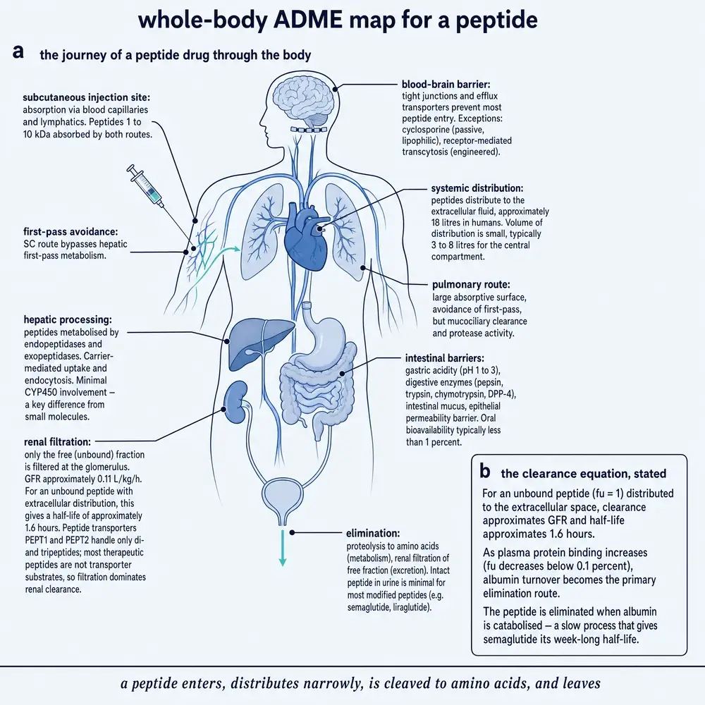
Figure 1 traces the journey the rest of this monograph dissects: a molecule is introduced, must survive or fail at a barrier, reaches the circulation or a local target, distributes through the body water, produces an effect that depends on how its concentration rises and falls, and is finally degraded and cleared. Each of those stages is a place where the two erasing machines act, and each is a place where a delivery technology can intervene.
It is worth naming the machines precisely, because the rest of the argument depends on them. The first is proteolysis: the blood, the gut, the liver, the kidney and the surface of nearly every cell carry peptidases that hydrolyse amide bonds. They are abundant and fast, which is why an intact peptide dropped into the small-intestinal lumen has a half-life measured in minutes, not hours.13 The second is glomerular filtration: the kidney's filter is a sieve with an effective cutoff near the size of albumin, so a small peptide passes into the urine almost as freely as water does, unless something holds it back.14 Neither machine was built to attack drugs. Both attack drugs superbly, because a peptide drug looks, to an enzyme or a glomerulus, exactly like the endogenous signal it was designed to erase.
Three ideas run through every Part that follows, and it is fair to state them at the outset. First, binding is the lever: because a peptide lives in the body water and its volume of distribution barely varies, its half-life is tuned almost entirely by how fast it is cleared, and clearance is dominated by the fraction that is free rather than bound to a plasma protein. Second, the route is chosen by the molecule, not by the marketing: whether a peptide can be given orally, nasally, by lung, or only by a depot injection is decided by its own half-life, potency and therapeutic window, not by which route would be most convenient to sell. Third, every value has a species and a setting or it has no meaning: a half-life is a number about a molecule in a particular animal or person under particular conditions, and this document prints both.
Section 02Clearance, and the discovery that the body has a rate
The word “clearance” hides a nineteenth-century insight that is easy to take for granted: the body does not remove a drug in fixed amounts but at a rate proportional to how much is present. Clearance is defined as the volume of blood or plasma that is completely cleared of a substance per unit time — litres per hour, not milligrams. It is the single most important parameter in pharmacokinetics because, together with the dose, it sets the average exposure a tissue sees.
For most small-molecule drugs, clearance happens in the liver and is often perfusion-limited: the liver can only clear what the blood delivers to it, so clearance cannot exceed the rate at which blood flows through the organ, and it certainly cannot exceed cardiac output. Peptides break this intuition. Because the enzymes that destroy them are distributed throughout the blood and the well-perfused organs rather than concentrated behind a single organ's blood supply, peptide metabolic clearance is not perfusion-limited and can in fact exceed cardiac output — a value that would be physically impossible for a liver-cleared small molecule.14 The molecule is being destroyed wherever it goes, not only where it is delivered. This is the first sign that peptide disposition needs its own rules.
Section 03The glomerulus and the size threshold
The kidney's contribution is more geometric. Each glomerulus is a tuft of capillaries wrapped in a filtration barrier whose pores pass small molecules freely and hold back large ones, with an effective cutoff in the region of 30 kilodaltons. A therapeutic peptide, at 0.5 to 5 kilodaltons, is far below that threshold; to the glomerulus it is essentially a small solute, and it is filtered into the urine at a rate set by how much of it is free in the plasma and by the glomerular filtration rate itself — a physiological constant of roughly 0.11 litres per kilogram per hour in an adult.14
A useful approximation follows: renal clearance of a small peptide is close to its free fraction multiplied by the filtration rate. The reason this simple form holds is that linear peptides are not substrates for the intestinal and renal di- and tri-peptide transporters PEPT1 and PEPT2, which evolved to reclaim the two- and three-residue fragments of digested protein. A therapeutic peptide is usually too long to be reabsorbed by that route, so tubular handling is minor and filtration dominates.14 The consequence is stark: a fully unbound peptide distributed only into the extracellular water would have a half-life of roughly 1.6 hours from renal filtration alone, before any enzyme touches it.14 Left to itself, a peptide is a short-lived drug.
Section 04Albumin, the reversible depot
The escape from that fate was discovered in the study of plasma protein binding. Albumin, the most abundant protein in blood, carries fatty acids, hormones and drugs on a set of reversible binding sites. A molecule bound to albumin is, for the moment, too large to be filtered by the glomerulus and partly shielded from the enzymes; only the free fraction is cleared. Albumin therefore behaves as a circulating depot, holding a reservoir of drug in a protected, inactive form and releasing it as the free concentration falls.
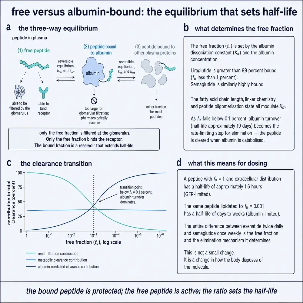
Figure 2 shows the arithmetic of that shielding. If ninety-nine percent of a peptide is bound at any instant, only the remaining one percent is available to the glomerulus and the peptidases, and the effective clearance drops by roughly the same factor. This is the mechanism modern long-acting peptides exploit: attach a fatty-acid chain that inserts into albumin's lipid-binding pocket, and the molecule spends most of its time protected. Liraglutide, acylated with a sixteen-carbon fatty acid, is more than ninety-nine percent bound to plasma protein and carries a half-life near thirteen hours after subcutaneous injection.14 Semaglutide, with an eighteen-carbon diacid linker and enzyme-resistant substitutions, binds albumin strongly enough to reach a half-life of about a week; its physicochemistry and enzyme-resistant design have been characterised in detail.131425
But the depot has a ceiling, and it is set by albumin itself. Albumin is synthesised at about ten and a half grams a day, is cleared at three to four milligrams per kilogram per day, and turns over with a circulating half-life of roughly twenty-one days. A peptide can only hide behind albumin for as long as that albumin persists; when the carrier is degraded, so is its passenger. Reversible albumin binding can therefore extend a peptide's half-life only up to a physiological limit of about sixteen to twenty-three days — no chemistry that relies on albumin can beat the turnover of the protein it hides behind.14 The lever is powerful, but it is not infinite.
Section 05The compartment and the curve
The last idea Part One needs is the concentration–time curve, the basic data object of pharmacokinetics. When a drug is given, its plasma concentration rises to a peak (the maximum concentration, Cmax, reached at a time Tmax), then falls; the total exposure over time is the area under that curve (the AUC), and the lowest point before the next dose is the trough. The rate of the terminal fall defines the half-life. Every one of these quantities is a description of the same curve, read at a different place.
Given intravenously, a peptide shows a two-phase curve: a fast initial drop as the molecule distributes from the blood into the well-perfused kidney and liver, followed by a slower decline as it exchanges with the more poorly perfused tissues and is cleared.14 The shape of the curve is not fixed by the molecule alone, however; it is set jointly by the molecule and the way it is delivered.
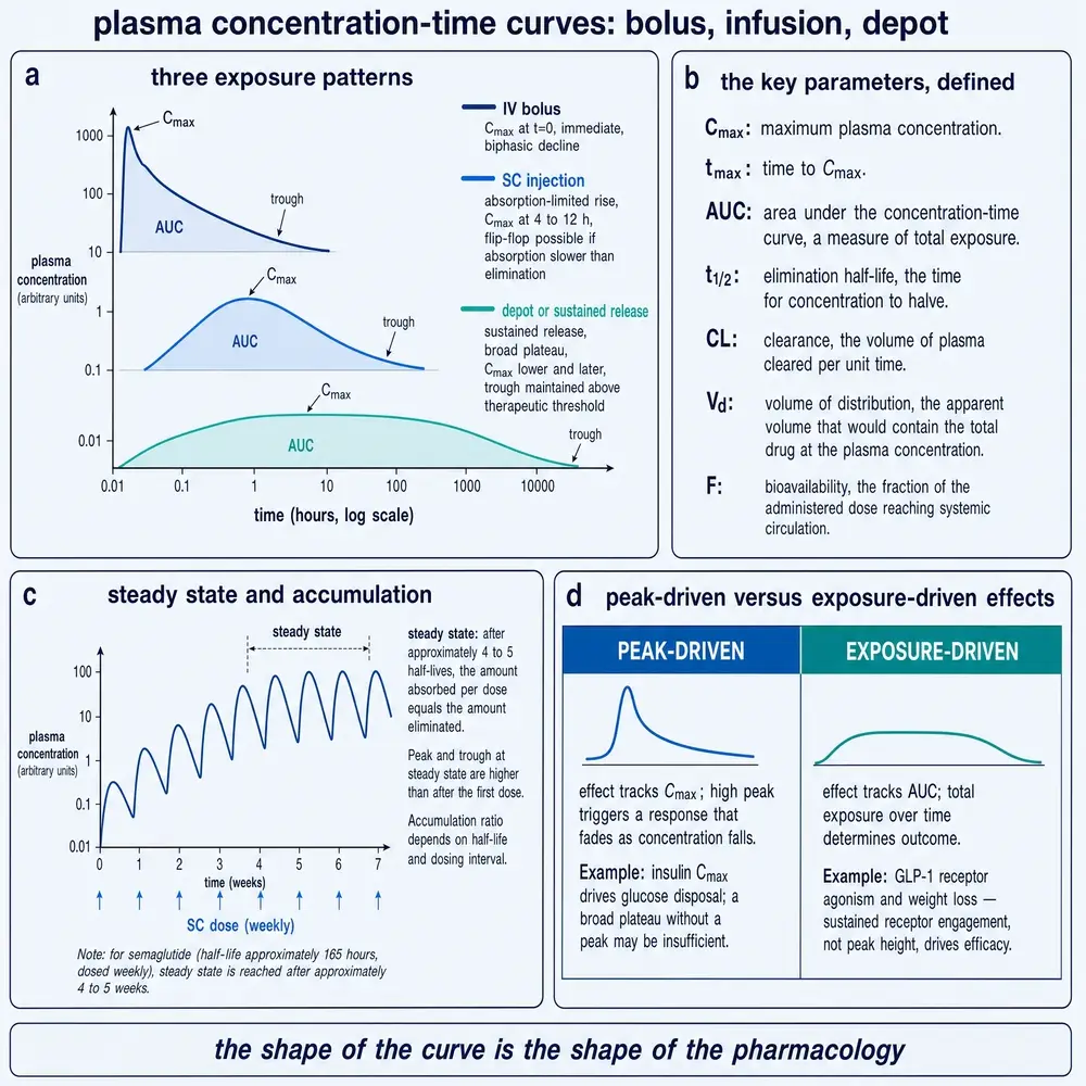
Figure 3 contrasts three delivery patterns for the same total dose: an intravenous bolus, which produces a tall early peak and a rapid fall; a steady infusion, which climbs to a plateau; and a subcutaneous depot, which releases slowly and produces a low, broad hump. These are three different drugs in every way that a tissue can perceive, built from one molecule. Part Five returns to this figure to show that the difference is not cosmetic — that the same dose on a different clock can select for a completely different biological effect.
Section 06Where a peptide goes
Once a peptide reaches the blood, the next question is where it can travel. The answer, for most peptides, is: not far into the cells. A peptide is large and water-loving, and cell membranes are thin sheets of fat; a molecule that cannot cross a membrane cannot enter a cell, so it is confined largely to the water outside the cells — the plasma and the interstitial fluid between cells, together the extracellular fluid, about eighteen litres in an adult.14
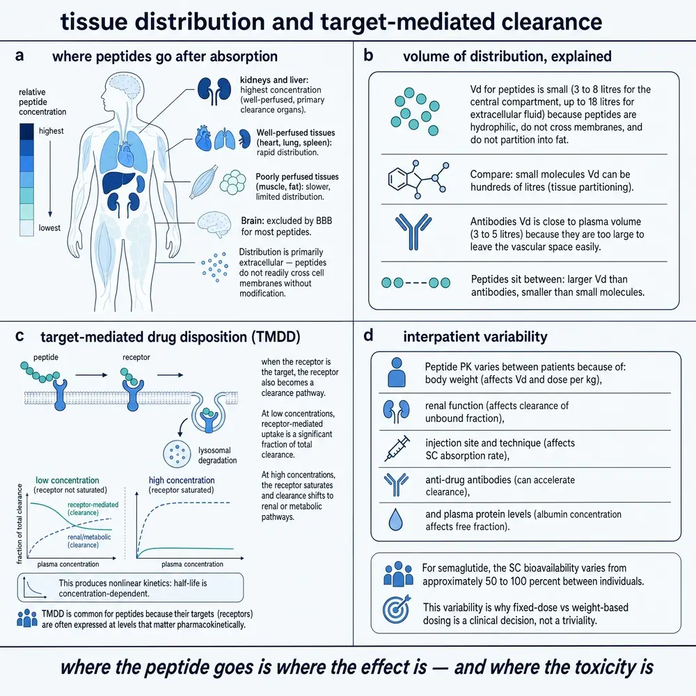
This confinement is measured by the volume of distribution, a parameter with a slightly counterintuitive definition. It is not a real anatomical volume but the apparent volume that would be needed to hold all the drug in the body at the concentration measured in the plasma. For a molecule locked out of cells, that apparent volume is close to the real extracellular volume, and it is strikingly consistent across peptides: roughly 0.10 to 0.26 litres per kilogram of body weight, far narrower than the range seen for small molecules, many of which partition into fat or bind inside tissues and show apparent volumes many times the size of the body.14 Figure 4 shows the picture: a peptide fills the body water and lines the interstitial space, but is largely shut out of the intracellular compartment and the fat — and, when receptor-mediated uptake matters, the same panel set shows how target saturation bends clearance.
That narrow volume is the quiet hero of the half-life story. Because a peptide's volume of distribution barely moves from molecule to molecule, it cannot be used as a design lever; a chemist cannot meaningfully extend a half-life by making the drug distribute more widely. The only lever left is clearance, and clearance, as Part One established, is dominated by the free fraction. This is why binding, not distribution, is the master control for this class — the reverse of the small-molecule intuition, where volume often does much of the work.
Two transport routes deserve a note because they matter for larger and lipidated peptides. After a subcutaneous injection, a small peptide is absorbed mostly through the blood capillaries, but a large or albumin-bound one is taken up preferentially by the lymphatic system, which drains the interstitial space and returns slowly to the blood. In lymph-cannulated sheep given a PEGylated insulin, less than two percent of the dose was recovered in lymph, and the molecule's terminal phase reflected the slow absorption rather than elimination.8 The lymphatic detour is one reason large peptides absorb slowly and incompletely, and it is a route delivery engineers now try to exploit deliberately.
Section 07Proteolysis in detail
Proteolysis is not one process but a layered one, and where a peptide is cut determines what it becomes. In the blood, dipeptidyl peptidase-4 trims specific N-terminal sequences — the very cut that ends native GLP-1 — while neutral endopeptidase and a family of aminopeptidases attack elsewhere along the chain.14 In the tissues, membrane-bound peptidases on the surface of endothelial and epithelial cells finish the work. The practical lesson for design is that a single vulnerable bond can dominate a molecule's fate: substituting one amino acid to block the DPP-4 cut, as semaglutide does, removes a major clearance route at a stroke.14
Proteolysis also raises the question of metabolites. When an enzyme cuts a peptide, the fragments are usually inactive — the message is destroyed because its shape is destroyed. Occasionally a fragment retains or changes activity, and then the metabolite must be tracked as a drug in its own right. The default assumption for a linear peptide is that its fragments are inactive and are themselves rapidly degraded and filtered, but this is an assumption to be tested per molecule, not a law.14
Section 08Renal and hepatic processing
For most peptides the kidney is the dominant organ of elimination, and it eliminates by filtration followed by destruction, not by excretion of the intact drug. A filtered peptide is largely reabsorbed into the cells lining the proximal tubule and hydrolysed there; very little intact peptide appears in the urine. This is why measuring a peptide in the urine understates renal clearance so badly, and why renal clearance is estimated from the free fraction and the filtration rate rather than from urinary recovery.14
The liver plays a smaller part for most peptides than it does for small molecules, because peptides are not generally substrates for the cytochrome P450 enzymes that dominate small-molecule metabolism. Hepatic uptake and biliary handling exist but are usually minor routes. A more specialised elimination pathway is receptor-mediated clearance: when a peptide binds its target receptor, the receptor–peptide complex can be pulled into the cell and degraded, which removes the drug from circulation. For most peptides this is a minor contribution, but for a few it is large enough to bend the entire concentration–time curve, which is the subject of Section 10.14
Section 09The half-life equation, assembled
The pieces now assemble into the equation that governs the class. Half-life is proportional to the volume of distribution divided by the clearance:
t½ = ln2 × Vd / CL
Read this equation with the two facts Part Two established. The volume of distribution is nearly fixed, because a peptide lives in the body water. So the half-life is set almost entirely by clearance — and clearance falls steeply as plasma-protein binding rises, because only the free drug is filtered and hydrolysed. Binding is therefore the lever that tunes half-life, exactly the relationship a small-molecule pharmacologist would not expect.14
Figure 5 places real peptides on a ladder spanning almost four orders of magnitude of half-life, with the species and setting printed on each rung. Oxytocin lasts one to six minutes; glucagon, minutes; vasopressin analogues, ten to thirty-five minutes; the rapid insulin analogues, one to a few hours; liraglutide, about thirteen hours; and semaglutide, about a week — roughly 168 hours.1314 Every step up that ladder was bought by increasing the fraction of time the molecule spends bound or protected, and the top of the ladder runs into the albumin ceiling of Section 04: no albumin-dependent chemistry pushes the half-life much past three weeks.14
Section 10Nonlinear kinetics and target-mediated disposition
The tidy proportionality of the half-life equation assumes that clearance is a constant — that doubling the dose doubles the exposure. For a class of peptides this fails, because one of their clearance routes is the target itself. When a peptide is eliminated substantially by binding to its receptor and being internalised, the number of receptors is finite. At low doses the receptors are plentiful relative to the drug, and clearance by that route is fast; at high doses the receptors saturate, that clearance route stalls, and the drug lingers far longer than a low-dose experiment would predict.14
Figure 4, panel (c), shows the resulting nonlinearity: exposure rises faster than dose, and the apparent half-life lengthens as the dose climbs. This behaviour is called target-mediated drug disposition, and it means that for such molecules a single half-life number is a fiction — the value depends on the dose. The disposition of these peptides has to be assessed with the receptor in mind: where it is expressed, how tightly the peptide binds it, and how fast the complex is internalised.14 It is a reminder that for peptides, pharmacology and pharmacokinetics are not separate subjects; the effect and its clearance can be the same event.
Section 11Reading a person: variability, impairment, body size, age
Every parameter so far has been a population average, but a drug is given to a person, and people differ. The most consequential differences for peptides are in the organs of clearance. Because renal filtration is the dominant elimination route for most peptides, impaired kidney function raises exposure directly. In a study of somapacitan, a long-acting growth-hormone derivative given once weekly, exposure was considerably higher in subjects with severe renal impairment and those on haemodialysis, and significantly higher in moderate hepatic impairment, than in matched subjects with normal organ function.2 A dose that is ordinary for one kidney can be an overdose for another.
Design can blunt this dependence. When clearance is shifted away from the kidney — by attaching a large PEG chain that makes the molecule too big to filter, or by binding it to albumin — renal impairment matters less. A PEGylated insulin showed clearance essentially unchanged by surgical removal of five-sixths of the kidney mass in rats, whereas the unmodified insulin was cleared more than three times more slowly in the same nephrectomised animals.8 Protecting a peptide from the kidney also protects its dosing from the patient's kidney.
Body size is handled through allometric scaling, one of the oldest quantitative regularities in biology. Across species, physiological rates scale with body weight raised to a power near three-quarters — Kleiber's law — and peptides obey it well. In a set of nine peptides scaled across mouse, rat, dog, monkey and human, clearance scaled with an exponent near 0.72 and volume with an exponent near 0.98, both with high correlation.14 Figure 6 shows these lines. The practical payoff is that preclinical peptide pharmacokinetics predicts human disposition reasonably well — better than for many small molecules — because the underlying machines, filtration and proteolysis, are conserved across mammals. What scaling does not license is extrapolation of a target-mediated or immune response, which depends on species-specific biology and can differ sharply between the animal and the person.
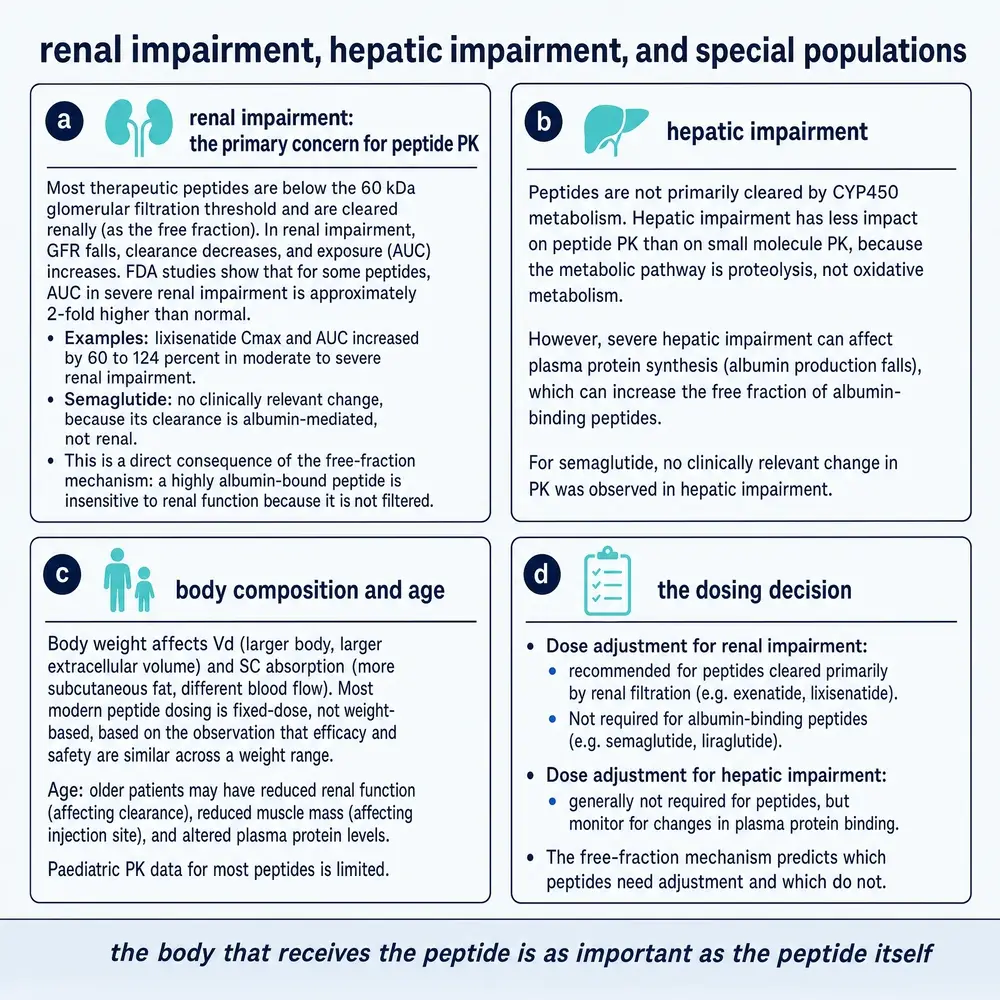
Figure 7 gathers the special-population consequences of the free-fraction mechanism. Age enters through the same organs: the newborn and the elderly kidney filter more slowly, and a peptide cleared by filtration will accumulate accordingly. Every number in this Part, in short, is a number about a body, and the body has to be specified.
Section 12The gut: acid, enzymes, mucus, epithelium
People have tried to feed insulin to patients since the year it was discovered, and for more than a century the gut has won almost every time. The reason is not one obstacle but a series of them, each sufficient on its own to defeat a naked peptide. Systemic-acting peptides taken by mouth typically reach the blood with an absolute bioavailability below one percent; oral semaglutide, the most successful case, delivers roughly half a percent to one percent of the swallowed dose, and effectively nothing when taken with food.13 To appreciate how a molecule can be so thoroughly erased between the mouth and the blood, it helps to walk down the tract.
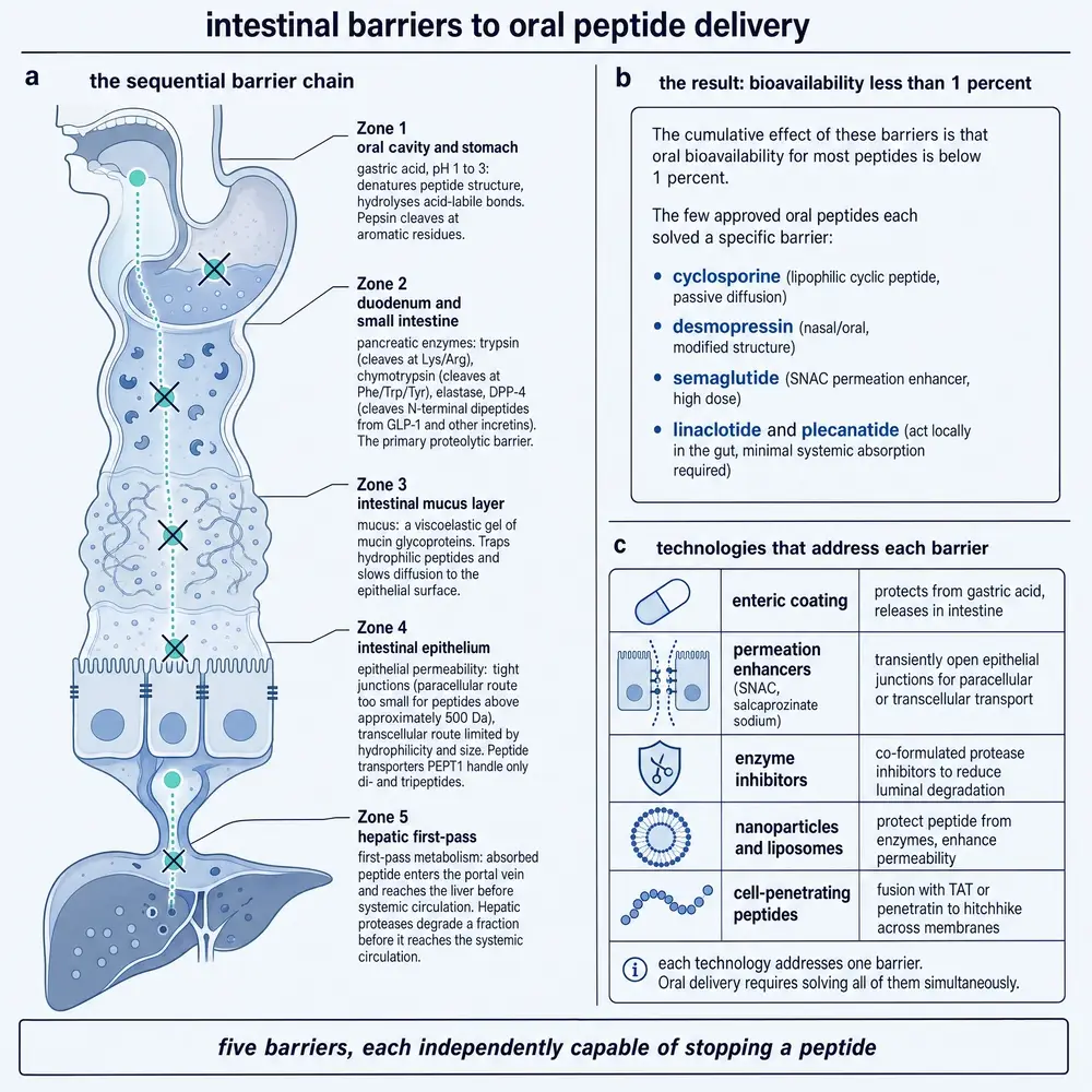
Figure 8 lays out the descent. The stomach is first: an acidic bath in which the enzyme pepsin begins cleaving the peptide. Those that survive reach the small intestine, where the pancreas pours in trypsin, chymotrypsin and elastase, a battery of enzymes evolved specifically to reduce dietary protein to fragments; a therapeutic peptide is, to them, simply lunch. What is left runs into the brush border of the intestinal wall, studded with further peptidases, and then the cytoplasm of the absorptive cells, which carries still more. Intestinal half-lives for intact peptides are measured in minutes.1318
Suppose a molecule survives the enzymes. It now meets the permeability barrier, and this one is arguably worse, because it cannot be blocked by chemistry. The intestinal lining is a sheet of cells sealed to one another by tight junctions, and the gaps those junctions leave open are far smaller than a peptide. Nor can most peptides slip through the cells themselves, being too large and too water-loving to cross the lipid membrane. Insulin's measured permeability across the intestinal epithelium is orders of magnitude below the threshold that would give a useful dose.13 A layer of mucus over the epithelium adds a final diffusion barrier and a last population of enzymes. The gut, in other words, is not accidentally hostile to peptides; it is a purpose-built protein-destruction and exclusion system, and it treats a peptide drug exactly as it treats a piece of steak.
Section 13First pass and the portal detour
Even a peptide that crosses the intestinal wall is not yet home. Blood leaving the gut does not go straight to the body; it is collected into the portal vein and routed first through the liver. This is the first-pass effect, and for many orally absorbed molecules it removes a large fraction before they ever reach the general circulation. For peptides the portal detour compounds the problem: the same molecule that survived the lumen and the epithelium now passes through an organ rich in uptake and degradation, so the fraction that reaches a distant receptor is smaller still. Any honest accounting of oral bioavailability is an accounting of survival across all of these serial filters multiplied together, which is why the surviving fraction is so often below one percent.13
Section 14Skin, nose, lung
If the gut is impassable, the body offers other surfaces, and each is a different compromise between accessibility and defence. The skin is the most convenient and the least permeable: its outer layer, the stratum corneum, is a brick-and-mortar wall of dead cells and lipid that passes essentially nothing above about 500 daltons by simple diffusion — well below the size of any peptide.13 Passive transdermal delivery of a peptide is therefore a non-starter; the skin becomes useful only when a device physically breaches the stratum corneum, which is the subject of the microneedle section in Part Four.
The nose and the lung are more generous, because their job is exchange rather than exclusion. The nasal mucosa is thin and well supplied with blood, and nasal peptide bioavailabilities of about thirty to forty percent are achievable; nasal glucagon, absorbed fast enough to rescue a person from hypoglycaemia, is the proof of concept, and the same mucosa offers a debated nose-to-brain route for peptides aimed at the central nervous system.13 The lung is the most generous of all: its alveolar surface is enormous, on the order of a hundred square metres, and the barrier between air and blood is only a fraction of a micron thick, which is why inhaled insulin reaches bioavailabilities around twenty to twenty-five percent.13 Yet both surfaces defend themselves by clearance rather than by a wall. The nose sweeps its mucus toward the throat within minutes; the lung carries ciliary escalators and a standing population of macrophages that engulf foreign particles. A peptide delivered to either surface is racing a clearance mechanism, and the formulation's job is to be absorbed before it is swept away.
Section 15The blood–brain barrier and the hard tissues
Some targets sit behind walls the body defends with particular determination. The blood–brain barrier is a specialised endothelium whose cells are sealed by tight junctions and backed by transporters that actively pump many molecules back out; it exists to keep the brain's chemical environment stable, and it keeps peptides out with the same efficiency. Delivering a peptide to the brain therefore usually means either bypassing the barrier physically — the intrathecal route, injecting into the cerebrospinal fluid — or attaching the peptide to a shuttle that the barrier's own transporters will carry across, a strategy Part Four returns to.13 The interest is not academic: neuropeptides are candidate treatments for ischemic brain injury, and their delivery across this barrier is the central obstacle to using them.27
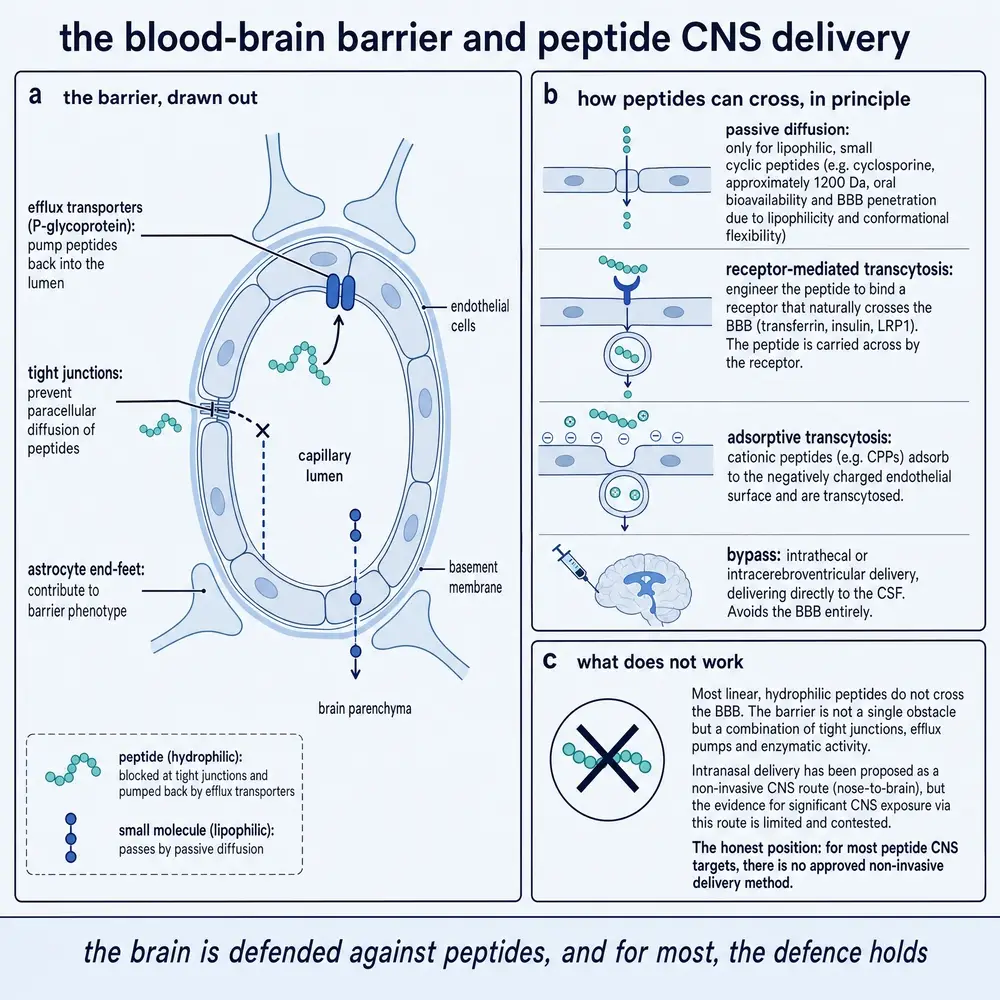
Figure 9 is the structural companion to that argument.
Solid tissues raise a different kind of wall. A tumour builds a dense, disorganised extracellular matrix and a leaky, high-pressure vasculature that together resist the penetration of anything large; fibrotic tissue lays down collagen that does the same. These are not membranes to be crossed but thickets to be pushed through, and a peptide's ability to reach a target buried inside them depends on diffusing through a matrix engineered, in effect, to stop it. The extracellular matrix is thus both a barrier to distribution and, for delivery engineers, a feature to be exploited — a place where a locally-activated or matrix-degrading system can be made to release its cargo.
Section 16The cell membrane and the endosome
The last barrier is the one every peptide with an intracellular target must face: the cell membrane itself. Most peptide drugs act on receptors displayed on the outside of cells precisely because the inside is so hard to reach. A peptide that must act within a cell has to cross the lipid membrane, and the usual way in — endocytosis, in which the membrane folds inward and swallows a parcel of fluid — delivers the peptide not into the cell's working interior but into an endosome, a membrane-bound bubble that typically matures into a degradative compartment. The peptide is inside the cell and still trapped, a problem known as endosomal sequestration.
Escaping the endosome before it destroys its contents is one of the central unsolved problems of intracellular delivery. Cell-penetrating peptides — short, often arginine-rich sequences — can drag a cargo across the membrane and improve endosomal escape, and newer chemistries push this further: fluorinated oligoarginines have been reported to act as “supra-enhancers” of intracellular and transepithelial delivery, precisely by improving the escape step.17 But efficiency remains low, and the fraction of an internalised dose that reaches its intracellular target is often a small one. For now, the cell interior remains the frontier where peptide delivery is least solved.
Section 17The accumulation gate
Before surveying the technologies, it is worth stating the rule that decides which of them can possibly work for a given molecule, because it eliminates most combinations before any chemistry is attempted. When a drug is dosed repeatedly, it accumulates only if a meaningful amount of the previous dose remains when the next arrives. The accumulation factor makes this quantitative:
R = 1 / (1 − e−kτ), k = 0.693 / t½
where τ is the dosing interval and k the elimination rate constant. Read the two extremes. If the half-life is much shorter than the interval between doses, the exponential term collapses, R approaches one, and each dose is gone before the next arrives — no accumulation, no build-up of exposure. If the half-life is long relative to the interval, R climbs: once-daily semaglutide, with its week-long half-life, accumulates roughly ten- to fifteen-fold before reaching a steady state after four or five half-lives.13
Figure 10 plots R against half-life for a fixed interval, and the shape of that curve is the single most important piece of negative-selection logic in peptide delivery. A molecule with a half-life of minutes cannot reach a useful steady state on any practical oral schedule, no matter how clever the formulation, because it is erased between doses. This is why the century of oral insulin failures was not merely a formulation problem: insulin's short half-life places it on the flat part of the curve, where no amount of protection or absorption enhancement can build sustained exposure from intermittent doses.13 Formulation cannot fix a short half-life; it can only help a molecule that already has a workable one. The gate is applied first, and it decides what the rest of Part Four is even allowed to attempt.
Section 18Molecular protection: enteric coats, enzyme inhibitors, permeation enhancers
The first family of technologies tries to get a peptide across the gut alive. Enteric coatings are the oldest idea: a pH-sensitive shell that stays closed in the acidic stomach and dissolves in the more neutral intestine, sparing the peptide the stomach's pepsin. Enzyme inhibitors go further, co-formulating agents that transiently blunt the intestinal proteases so the peptide has a few more minutes to be absorbed. Neither, on its own, solves the permeability barrier.
The workhorses of modern oral peptides are permeation enhancers, and two dominate the marketed and late-stage products. Sodium caprate, a ten-carbon fatty acid usually written C10, transiently fluidises the epithelial membrane and loosens the barrier; it is the enhancer in the oral small-molecule candidate MK-0616 at a dose of 180 milligrams, and is used in the range of roughly 180 to 500 milligrams.13 Salcaprozate sodium, known as SNAC, works by a different mechanism: at about 300 milligrams it buffers the local pH to protect the peptide from pepsin and facilitates its transcellular passage, and it is the enhancer that makes oral semaglutide possible.13 The mechanistic claim that such enhancers genuinely raise intestinal permeability has been measured directly for peptide drugs in controlled permeability studies.5 The cost, always, is that an enhancer that opens the barrier to the drug opens it to other things as well, and that the fraction absorbed remains small and variable — which returns the molecule to the accumulation gate of Section 17.
Section 19Half-life by chemistry: lipidation, PEGylation, fusion, albumin binding
The second family does not fight a barrier at all; it re-engineers the molecule so the two erasing machines act on it more slowly. These are the chemistries behind the half-life ladder of Section 09, now read as engineering rather than as observation. Lipidation — attaching a fatty-acid chain — buys reversible albumin binding, which shields the drug and lengthens its half-life up to the albumin ceiling; it is how liraglutide reaches thirteen hours and semaglutide reaches a week.14 The interaction is physical and tunable: acylating glucagon-like peptide-2 changes how the molecule inserts into lipid membranes and how fast it is degraded in vitro, showing that the chain length and chemistry are design variables, not a single fixed trick.21
PEGylation attaches a chain of polyethylene glycol, which does something subtler than binding: it enlarges the molecule's hydrodynamic size until it is too big for the glomerulus to filter. A twenty-kilodalton PEG on insulin lispro raised its effective size roughly fourfold and shifted its clearance from renal to non-renal — clearance became insensitive to loss of kidney mass — while its subcutaneous absorption became largely lymphatic and its terminal phase came to reflect that slow absorption rather than elimination.8 Fusion is a third route: joining the peptide to albumin or to an antibody Fc domain borrows the carrier's long residence, and GLP-1 fusion chimeras act as potent, long-acting agonists on exactly this principle.24 Every one of these fixes carries a cost — larger molecules absorb more slowly and less completely, added chemistry can provoke an immune response, and a longer half-life means a mistake takes longer to clear — which is why the choice of chemistry is always a negotiation, never a free lunch.
Section 20Particles and matrices: nanoparticles, liposomes, hydrogels, microspheres
The third family packages the peptide rather than modifying it. Nanoparticles and liposomes wrap the drug in a protective shell intended to shield it from enzymes and, in the oral case, to ferry it across the epithelium; hydrogels and microspheres embed it in a matrix that releases it slowly over days or weeks. Figure 11 sorts these and the device technologies of the next section into the problem-classes they address.
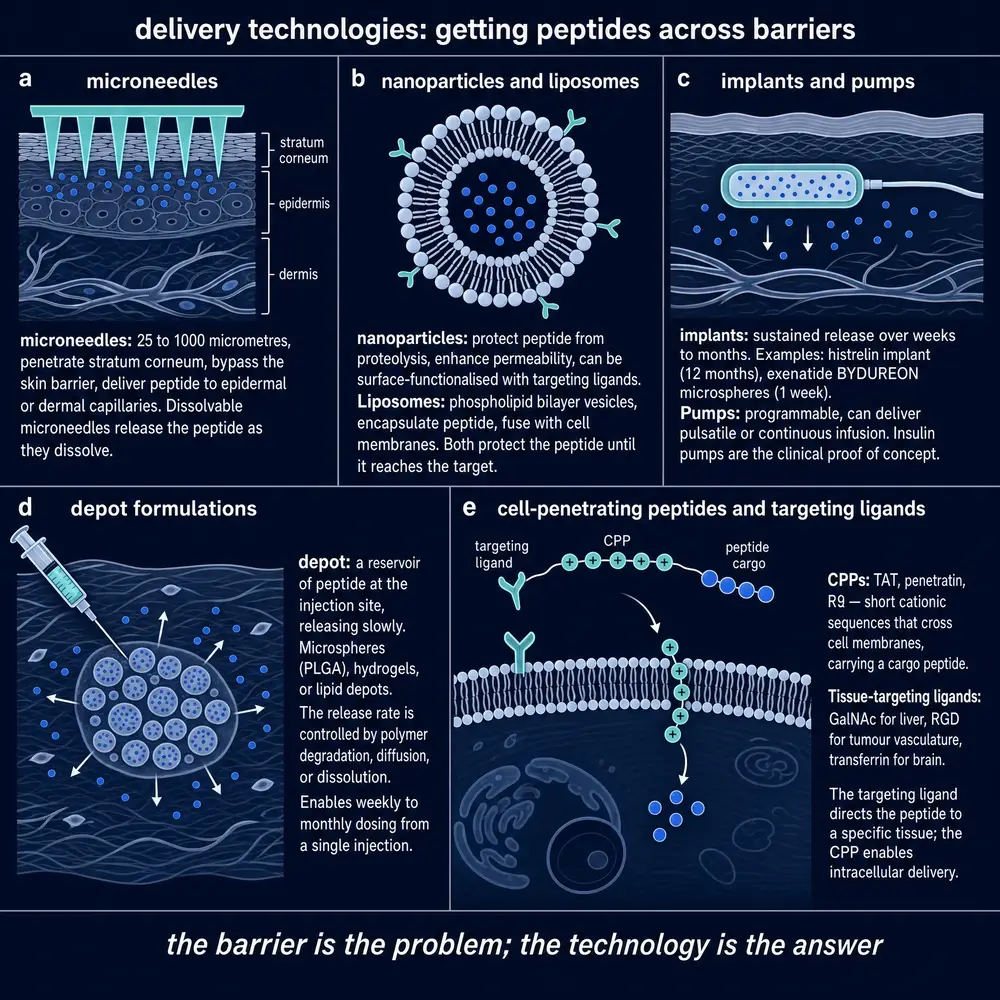
The honest record of oral nanocarriers is a cautionary one. Many improved absorption impressively in rodents and then failed to translate to humans; the harsher and more variable human gastrointestinal environment, together with the difficulty of manufacturing such particles reproducibly, negated the benefit seen in animals.13 This is one of the clearest examples in the field of a result that is real in one species and absent in another, and it is why this document treats a rodent nanoparticle success as species-limited evidence rather than as a promise. The matrix depots, by contrast, have a solid clinical record by the parenteral route: peptides can be remote-loaded into poly(lactic-co-glycolic acid) microspheres from aqueous solution, and triptorelin acetate microspheres deliver the drug over weeks in patients.626 In-situ forming systems extend the same idea — a liquid injected under the skin that self-assembles into a liquid-crystalline or gel depot — and smart hydrogels aim to make the release respond to a local trigger rather than run on a fixed clock.2219 The pattern is consistent: packaging works most reliably when it is used to slow release from a depot, and least reliably when it is asked to force a peptide across the gut.
Section 21Devices: microneedles, pumps, implants, swallowed injection
Where chemistry and packaging reach their limits, hardware takes over. Microneedles are arrays a few hundred microns tall that pierce the stratum corneum without reaching the pain nerves, converting the impassable skin into a minimally invasive intradermal or subcutaneous route; a rocket-shaped microneedle patch has been shown to deliver a hormone as a sustained depot in an animal model.3 Pumps and implants take the logic of the depot to its conclusion, holding a reservoir of drug and metering it out at a controlled rate for weeks to months, which turns an intermittent injection into a continuous infusion and lets the exposure pattern be designed rather than accepted.
The most striking devices attack the oral problem by refusing to solve it chemically at all. The self-orienting millimeter-scale applicator and the ingestible intestinal-microneedle capsule are, in effect, swallowed injections: they carry a solid dose of peptide to the stomach or intestinal wall and inject it mechanically past the epithelium, bypassing absorption entirely. Because they deliver the whole dose through the wall, their performance is governed by device reliability and the safety of repeatedly perforating tissue, not by bioavailability — they are combination products, judged as devices as much as drugs.13 Meanwhile the more conventional oral programs continue to inch forward: small-intestine-targeted long-acting oral insulin formulations, engineered to release where absorption is best, are an active line of work.20
Section 22Targeting and conditional activation
The most sophisticated delivery does not just move a peptide into the body; it sends the peptide to a particular place and keeps it inert until it arrives. Peptide–drug conjugates and homing ligands exploit the peptide's own selectivity, using a short targeting sequence to concentrate a payload on cells that display a particular receptor. A radiolabelled gastrin-releasing peptide receptor antagonist, for instance, uses the peptide purely as a delivery address, carrying a radionuclide to receptor-bearing tumour cells for imaging or therapy.9 Shuttle strategies apply the same idea to the blood–brain barrier, borrowing the barrier's own transporters to carry a cargo across.
Conditional activation adds a second layer of control: the system is built to release or activate its cargo only in response to a local cue. Self-immolative conjugates that are triggered by inflammation have been reported to enable oral peptide delivery, staying intact through the gut and unmasking the active drug only where the target tissue's chemistry sets them off.4 Programmable and stimulus-responsive hydrogels pursue the same goal from the materials side, releasing on a schedule or a trigger rather than by simple diffusion.19 These systems are early, but they point at the direction of the field: from moving a molecule into the body, toward controlling exactly where and when it becomes active.
Section 23Route triage
All of the foregoing can be compressed into a single decision. Given a molecule, which route is even feasible? The answer follows from the three threads of this monograph: a route is feasible only if the molecule's half-life, potency and therapeutic window match what the route can deliver, and only if the molecule's effect can tolerate the variability the route imposes.
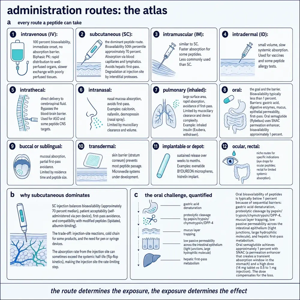
Figure 12 assembles the atlas, pairing each route with the reported bioavailabilities established in Part Three: subcutaneous injection, the workhorse, with a median bioavailability near seventy percent across therapeutic peptides;14 pulmonary at roughly twenty to twenty-five percent; nasal at thirty to forty; buccal and sublingual offering partial first-pass avoidance but only for small doses, as approved sublingual desmopressin shows; and oral, at well under one percent, feasible only for an unusually potent molecule with a long half-life and an area-driven effect.13 The comparison between once-weekly subcutaneous semaglutide and daily oral semaglutide is the field's clearest single lesson: the same molecule is far more pharmacokinetically predictable as a depot injection than as a pill, and the pill is possible at all only because the molecule already sits on the favourable side of the accumulation gate.13 The most heavily engineered delivery systems now in development — the oral GLP-1 formulations and the small-molecule-versus-injectable comparisons of the current literature116 — are all, in the end, attempts to move a molecule from the impossible side of that gate to the merely difficult side.
Section 24Bolus, infusion, sustained, pulsatile
The final Part returns to the concentration–time curve of Section 05 and makes the claim that the outline promised: the same total dose, delivered on a different clock, is a different drug. A bolus produces a tall, brief peak; a steady infusion holds a plateau; a depot releases a low, broad exposure; and a pulsatile schedule delivers the drug in repeated spikes separated by troughs. These are four temporal patterns, and a receptor does not respond to the total amount of drug alone — it responds to the pattern.
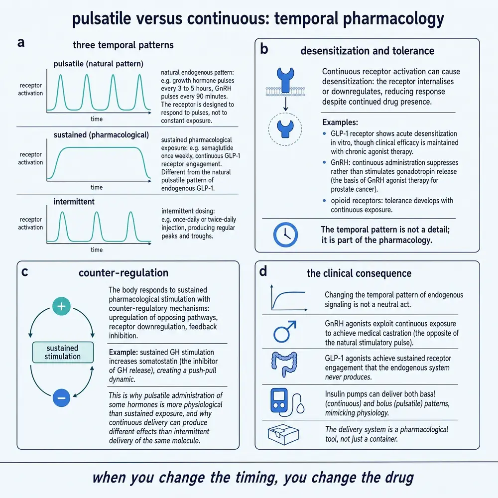
Figure 13 contrasts the patterns that matter most for signalling: continuous exposure and pulsatile exposure. The distinction is not a subtlety. Many of the body's own peptide hormones are secreted in pulses — growth hormone, insulin, the gonadotropin-releasing hormone that governs reproduction — and their target tissues are tuned to that rhythm. A drug that delivers the same molecule as a flat, continuous exposure is sending a message the tissue was never built to receive, and the response can differ from the pulsatile case even when the total exposure is identical.
Section 25Peak-driven versus exposure-driven effects
The cleanest demonstration that pattern, not amount, can drive the effect comes from a controlled experiment in rats. When insulin was infused as a fixed total dose — half a unit per kilogram over two hours — but broken into an increasing number of pulses, the integrated pharmacodynamic effect grew with the number of pulses, plateauing above about four pulses near the value produced by a continuous two-hour infusion, and it did so without raising the plasma insulin concentration. The temporal pattern of delivery, not the exposure, changed the biological effect, a result the investigators reproduced with indirect-response models of receptor binding and glucose-transporter translocation.11 The same amount of the same drug did more work when it arrived in the right rhythm.
This is why peptides split into two pharmacodynamic kinds, and why the split governs which delivery routes they can tolerate. Some effects are peak-driven: insulin covering a meal, glucagon rescuing a hypoglycaemic patient, oxytocin, the short parathyroid-hormone fragments — each needs a sharp rise at the right moment, and each is easily ruined by a formulation that blunts the peak or delivers it at the wrong time. Other effects are exposure-driven, depending on the area under the curve over days: semaglutide's control of appetite and glucose is of this kind. The practical consequence is that exposure-driven peptides tolerate the variability of an absorption-limited route such as oral delivery, because a fluctuating input still integrates to a similar area, whereas peak-driven peptides do not, because the peak is the therapy and a variable peak is a variable drug.13 It is no accident that the peptide which crossed into oral dosing, semaglutide, is an area drug; a peak drug could not have made the same crossing.
Section 26Desensitisation, tolerance, counter-regulation
Overwriting the body's natural rhythm has a cost beyond a blunted effect: the receptor can adapt. A receptor exposed to a continuous, unrelenting signal often responds by desensitising — withdrawing from the cell surface or uncoupling from its downstream machinery — so that the same concentration produces a smaller effect over time. This is the molecular basis of tolerance, and it is one reason continuous exposure is not always superior to pulsatile exposure even when it is easier to achieve. The body's pulsatile secretion of many hormones appears to exist partly to prevent this adaptation, giving the receptor a trough in which to reset between signals.
Beyond the single receptor, the body defends its set points with counter-regulation: push one signal continuously in one direction, and opposing systems activate to push back. A delivery pattern that ignores these feedback loops can find its effect eroded not because the drug stopped working but because the body mounted a response against it. Amylin, whose mode of action and clinical development are the subject of current review, is one of the hormones whose therapeutic use has to be understood in the context of this counter-regulatory web rather than as a simple dose-response.23
Section 27When timing is the therapy
The corollary of everything in this Part is that the delivery schedule is not a convenience wrapped around the drug; for many peptides it is the drug. Intermittent dosing that mimics a natural pulse can produce an effect that continuous dosing of the same molecule cannot, and dosing timed to the body's circadian rhythm can align a signal with the physiology built to receive it. The choice among bolus, infusion, depot and pulse is therefore a pharmacodynamic decision made upstream of any formulation, and it is made per molecule. Characterising it demands careful pharmacokinetic comparison across formulations and subjects — the kind of single-dose, head-to-head studies that distinguish one delivery system from another715 and the cross-species work, such as exenatide's characterisation in non-human primates, that connects an animal model to a human expectation.10 The literature on GLP-1 receptor agonists, now the most studied peptide class in medicine, is in large part a literature about exactly this question of how to shape exposure over time.12
This monograph describes published research on how peptides move through the body and how delivery systems change that movement. It weighs observed and simulated values, labels species and study type, and prefers recent evidence except where a preponderance of older evidence contradicts it. It does not recommend the human use of any compound and specifies no dose, route or schedule for any person. Every quantitative value is drawn from a cited source or is a labelled pedagogical calculation; none is invented.
Section 28References
The list below is numbered and sorted by first-author surname. Every entry was resolved from the source record's own metadata — author, title, journal, year, volume, issue, pages and identifiers — and never from recall. In-text citations are the superscript numbers throughout the document.
- Abbasi M, Sun K, Huggins KW, Heath B, DeLoit H, McGinness L, et al.. Advances in GLP-1 receptor agonists delivery systems for obesity and diabetes. Acta Pharmaceutica Sinica. B. 2026;16(5):2682.
PMID 42180536 · doi:10.1016/j.apsb.2026.01.035 · PMC13198345 - Bentz Damholt B, Dombernowsky SL, Dahl Bendtsen M, Bisgaard C, Højby Rasmussen M. Effect of Kidney or Hepatic Impairment on the Pharmacokinetics and Pharmacodynamics of Somapacitan: Two Open-Label, Parallel-Group Trials. Clinical Pharmacokinetics. 2021;60(8):1015-1027.
PMID 33754315 · doi:10.1007/s40262-021-00990-7 · PMC8332591 - Chen Y, Chen K, Yu K, Pang S, Yang H. Rocket-Like Microneedle Patch for Sustained Hormone Therapy in Prostate Cancer. ACS Biomaterials Science & Engineering. 2025;11(8):4830.
PMID 40676997 · doi:10.1021/acsbiomaterials.5c00127 · PMC12344640 - Cheng J, Wu P, Li C, Han Y, Sun M, Dou Y, et al.. Inflammation-triggered self-immolative conjugates enable oral peptide delivery by overcoming gastrointestinal barriers. Science Advances. 2026;12(3):eaea2989.
PMID 41533788 · doi:10.1126/sciadv.aea2989 · PMC12802832 - Dahlgren D, Olander T, Sjöblom M, Hedeland M, Lennernäs H. Effect of paracellular permeation enhancers on intestinal permeability of two peptide drugs, enalaprilat and hexarelin, in rats. Acta Pharmaceutica Sinica. B. 2021;11(6):1667.
PMID 34221875 · doi:10.1016/j.apsb.2020.12.019 · PMC8245904 - Giles MB, Hong JKY, Liu Y, Tang J, Li T, Beig A, et al.. Efficient aqueous remote loading of peptides in poly(lactic-co-glycolic acid). Nature Communications. 2022;13:3282.
PMID 35676271 · doi:10.1038/s41467-022-30813-7 · PMC9177552 - Hu X, Zhang Q, Zheng Y, Zhai Y, Xu N, Zhao Q, et al.. A single-dose, randomized, open-labeled, parallel-group study comparing the pharmacokinetics, pharmacodynamics and safety of leuprolide acetate microspheres 3.75 mg and Enantone® 3.75 mg in healthy male subjects. Frontiers in Pharmacology. 2022;13:946505.
PMID 36059939 · doi:10.3389/fphar.2022.946505 · PMC9437282 - Knadler MP, Nguyen T, Campanale K, De Veer MJ, Beals JM, Li S, et al.. Addition of 20-kDa PEG to Insulin Lispro Alters Absorption and Decreases Clearance in Animals. Pharmaceutical Research. 2016;33(12):2920-2929.
PMID 27528391 · doi:10.1007/s11095-016-2014-1 · PMC5093203 - Kumar N, Bilinska A, Menéndez E, Läppchen T, Kanellopoulos P, Orlova A, et al.. Preclinical Evaluation of a 177Lu-Labeled Gastrin-Releasing Peptide Receptor Antagonist and Prostate Cancer Treatment with Monotherapy and in Combination with Everolimus. ACS Pharmacology & Translational Science. 2025;9(1):59-68.
PMID 41536273 · doi:10.1021/acsptsci.5c00491 · PMC12797171 - Li Y, Vaughan KL, Tweedie D, Jung J, Kim HK, Choi H, et al.. Pharmacokinetics of Exenatide in nonhuman primates following its administration in the form of sustained-release PT320 and Bydureon. Scientific Reports. 2019;9:17208.
PMID 31748513 · doi:10.1038/s41598-019-53356-2 · PMC6868133 - Miyazaki M, Hayata M, Samukawa N, Iwanaga K, Nagai J. Pharmacokinetic–pharmacodynamic modelling of the hypoglycaemic effect of pulsatile administration of human insulin in rats. Scientific Reports. 2020;10:18876.
PMID 33139788 · doi:10.1038/s41598-020-76007-3 · PMC7608663 - New RRC, Bogus M, Travers GN, Hahn U, Vaiceliunaite A, Burnet M, et al.. Glucagon-like peptide-1 receptor agonists for treatment of diabetes and obesity: advantage of oral delivery. Frontiers in Drug Delivery. 2024;4:1456654.
PMID 40836975 · doi:10.3389/fddev.2024.1456654 · PMC12363265 - Niazi SK. Oral delivery of peptides and proteins: pharmacokinetic boundaries, negative selection, and route triage. Frontiers in Drug Delivery. 2026;6:1776167.
PMID 41939723 · doi:10.3389/fddev.2026.1776167 · PMC13047097 - Nordell P, Jansson-Löfmark R, Gennemark P. Systemic Pharmacokinetic Principles of Therapeutic Peptides. Clinical Pharmacokinetics. 2026;65(4):571-582.
PMID 41661442 · doi:10.1007/s40262-025-01615-z · PMC13038672 - Pan Q, Wang X, Li W, Chen X, Zhuang Y, Zhou Q, et al.. Pharmacokinetics, pharmacodynamics, and safety of prandial oral insulin (N11005) in healthy subjects. Frontiers in Endocrinology. 2023;14:1172327.
PMID 37564986 · doi:10.3389/fendo.2023.1172327 · PMC10411801 - Patel D, Muacevic A, Adler JR. Small-Molecule Oral Versus Injectable Glucagon-Like Peptide-1 (GLP-1) Receptor Agonists: Comparative Efficacy, Safety, and Future Clinical Perspectives. Cureus. 2026;18(4):e107202.
PMID 42153087 · doi:10.7759/cureus.107202 · PMC13180026 - Rong G, Fan Q, Chen K, Cheng Y, Hu J. Fluorous oligoarginines as supra-enhancers for intracellular and transdermal peptide delivery. Bioactive Materials. 2025;59:305.
PMID 41551769 · doi:10.1016/j.bioactmat.2025.12.027 · PMC12804165 - Shaji J, Patole V. Protein and Peptide Drug Delivery: Oral Approaches. Indian Journal of Pharmaceutical Sciences. 2008;70(3):269.
PMID 20046732 · doi:10.4103/0250-474X.42967 · PMC2792531 - Shi G, Yang Z, Lan S, Chen R, Wu Z, Li L, et al.. Versatile smart hydrogels for spatiotemporal drug delivery to orchestrate diabetic bone regeneration. Bioactive Materials. 2025;55:754.
PMID 41216378 · doi:10.1016/j.bioactmat.2025.09.049 · PMC12596967 - Thamizhchelvan AM, Li Y, Padelford J, Yang C, Yang C, He P, et al.. Small Intestine-Targeted Long-Acting Oral Insulin Formulation Based on Engineered Milk Protein Nanoparticles. ACS Applied Bio Materials. 2025;9(2):1083.
PMID 41442621 · doi:10.1021/acsabm.5c02114 · PMC12820955 - Trier S, Linderoth L, Bjerregaard S, Andresen TL, Rahbek UL, Castanho MARB. Acylation of Glucagon-Like Peptide-2: Interaction with Lipid Membranes and In Vitro Intestinal Permeability. PLoS ONE. 2014;9(10):e109939.
PMID 25295731 · doi:10.1371/journal.pone.0109939 · PMC4190408 - Vitek M, Zvonar Pobirk A, Roškar R, Matjaž MG. Exploiting the potential of in situ forming liquid crystals: development and in vitro performance of long-acting depots for peptide drug thymosin alpha 1 subcutaneous administration. Drug Delivery. 2025;32(1):2460708.
PMID 40066714 · doi:10.1080/10717544.2025.2460708 · PMC11899226 - Volčanšek Š, Koceva A, Jensterle M, Janež A, Muzurović E. Amylin: From Mode of Action to Future Clinical Potential in Diabetes and Obesity. Diabetes Therapy. 2025;16(6):1207.
PMID 40332747 · doi:10.1007/s13300-025-01733-8 · PMC12085449 - Wang Q, Chen K, Liu R, Zhao F, Gupta S, Zhang N, et al.. Novel GLP-1 Fusion Chimera as Potent Long Acting GLP-1 Receptor Agonist. PLoS ONE. 2010;5(9):e12734.
PMID 20856794 · doi:10.1371/journal.pone.0012734 · PMC2939854 - Wasilewska B, Petruczynik A. Semaglutide - properties, action and chromatographic analysis. Journal of Diabetes and Metabolic Disorders. 2025;24(2):197.
PMID 40937273 · doi:10.1007/s40200-025-01711-8 · PMC12420565 - Wu G, Zhou F, Wang H, Liu K, Yu D, Fan L, et al.. Effectiveness, pharmacokinetics, and safety of triptorelin acetate microspheres in patients with locally advanced and metastatic prostate cancer. Therapeutic Advances in Medical Oncology. 2024;16:17588359241307818.
PMID 39734709 · doi:10.1177/17588359241307818 · PMC11672368 - Yu J, Shen M, He T. The neuropeptide in ischemic brain injury: insights, challenges, and horizon of targeted interventions. Journal of Translational Medicine. 2026;24:366.
PMID 41664200 · doi:10.1186/s12967-026-07801-2 · PMC12983573
Section 29How this document was assembled
The evidence base was built by an automated pipeline over a shared full-text library. 10,204 open-access and peptide-sciences full texts were scanned; a two-armed identity gate retained a document only if it was genuinely about a peptide and genuinely engaged pharmacokinetics or delivery, which removed the large class of papers that merely dosed a peptide without reasoning about its disposition. 500 documents passed as on-topic, of which 85 scored highly enough on the coverage families to be read in full, totalling roughly 9,670 page-equivalents of text. From that reading corpus, the 27 sources cited here were selected as the load-bearing evidence for specific claims. The scan counts, tier definitions and every rejection class by name are recorded in the project's corpus report; the scoring configuration is versioned alongside it.
Section 30Evidence handling
Three rules governed how evidence was weighed. First, study type is labelled at the point of use: a value from a human study, an animal study naming the species, an in-vitro assay, or a model is identified as such, because a half-life from a sheep and a half-life from a person are not interchangeable. Second, observed values are distinguished from simulated ones, and any illustrative calculation is labelled hypothetical (Appendix C). Third, recency is preferred but not blindly: a 2025–2026 value is weighted above an older one unless a preponderance of evidence contradicts it. Two contradictions were handled explicitly. The mechanistic volume-of-distribution equation overpredicts the observed volume in about half of cases, so it is presented as a bounded estimate rather than a point prediction. And the impressive oral-nanoparticle results seen in rodents are contradicted by the human record, so they are reported as species-limited rather than as a general promise. No numerical value in this document was invented, and no human use, dose, route or schedule is recommended anywhere in it.
Appendix AGlossary
Absorption. The passage of a drug from its administration site into the systemic circulation.
Accumulation factor (R). The factor by which drug builds up with repeated dosing; R = 1/(1−e−kτ).
Albumin. The most abundant blood protein; a reversible carrier that acts as a circulating depot for lipidated peptides.
Area under the curve (AUC). The integral of concentration over time; the measure of total exposure.
Bioavailability. The fraction of an administered dose that reaches the systemic circulation intact.
Clearance (CL). The volume of plasma cleared of drug per unit time; the primary determinant of average exposure.
DPP-4 (dipeptidyl peptidase-4). A protease that trims the N-terminus of GLP-1 and related peptides, a major clearance route.
Extracellular fluid. The body water outside cells — plasma plus interstitial fluid — where most peptides distribute.
First-pass effect. Loss of an orally absorbed drug during its initial passage through the liver via the portal vein.
Flip-flop kinetics. The situation in which absorption is slower than elimination, so the terminal phase reflects absorption.
Glomerular filtration. The kidney's size-selective filtration of plasma; the dominant elimination route for small peptides.
Half-life (t½). The time for concentration to fall by half; t½ = ln2·Vd/CL.
Lipidation. Attachment of a fatty-acid chain to enable reversible albumin binding and extend half-life.
Permeation enhancer. A co-formulated agent that transiently increases epithelial permeability (e.g. C10, SNAC).
PEGylation. Attachment of polyethylene glycol to enlarge a molecule beyond the glomerular filtration threshold.
Target-mediated drug disposition (TMDD). Nonlinear kinetics arising when binding to a saturable target is a significant clearance route.
Volume of distribution (Vd). The apparent volume relating total body drug to plasma concentration; near-fixed for peptides.
Appendix BAbbreviations
ADME absorption, distribution, metabolism, elimination · AUC area under the curve · BBB blood–brain barrier · C10 sodium caprate · CL clearance · Cmax maximum concentration · DPP-4 dipeptidyl peptidase-4 · GFR glomerular filtration rate · GLP-1 glucagon-like peptide-1 · GLP-2 glucagon-like peptide-2 · IV intravenous · NEP neutral endopeptidase · PEG polyethylene glycol · PEPT1/PEPT2 di/tri-peptide transporters 1 and 2 · PLGA poly(lactic-co-glycolic acid) · PK pharmacokinetics · PD pharmacodynamics · SC subcutaneous · SNAC salcaprozate sodium · TMDD target-mediated drug disposition · t½ half-life · Vd volume of distribution.
Appendix CQuantitative-methods appendix
Every equation used in this monograph is listed here with its variables, assumptions, cited model and validated domain. Observed values are cited in the text; the worked example below is explicitly hypothetical and is included only to make the arithmetic reproducible.
C.1 Renal clearance of a small peptide
CLr = fu × GFR
where CLr is renal clearance, fu the unbound fraction in plasma (0–1, dimensionless), and GFR the glomerular filtration rate (≈ 0.11 L/kg/h in an adult). Assumptions: the peptide is small enough to be freely filtered, is not reabsorbed by PEPT1/PEPT2, and undergoes negligible tubular secretion. Domain: linear peptides below the glomerular cutoff; it does not apply to PEGylated or fusion molecules enlarged past the filtration threshold. Source: PMC13038672.14
C.2 Half-life from volume and clearance
t½ = ln2 × Vd / CL
where Vd is the volume of distribution (L/kg) and CL total clearance (L/kg/h). Assumptions: one-compartment approximation for the terminal phase; Vd constant with dose. Domain: linear kinetics; it fails under the target-mediated disposition of Section 10, where CL is dose-dependent. Source: PMC13038672.14
C.3 Accumulation factor
R = 1 / (1 − e−kτ), k = 0.693 / t½
where R is the accumulation factor (dimensionless), τ the dosing interval (h) and k the elimination rate constant (h−1). Assumptions: linear kinetics, complete and reproducible dosing, constant interval. Domain: repeated dosing at fixed intervals; steady state is reached in roughly four to five half-lives. Source: PMC13047097.13
C.4 Allometric scaling
Y = a × Wb
where Y is a pharmacokinetic parameter, W body weight (kg), a a coefficient and b the allometric exponent. For the nine-peptide dataset, the clearance exponent was ≈ 0.72 (range 0.58–0.88) and the volume exponent ≈ 0.98, consistent with Kleiber's 0.75. Assumptions: conserved elimination mechanisms across species. Domain: filtration- and proteolysis-driven disposition; it does not license extrapolation of species-specific target or immune effects. Source: PMC13038672.14
C.5 Worked example (hypothetical)
To show the binding lever numerically: assume a peptide confined to the extracellular water with Vd = 0.20 L/kg and cleared only by filtration at GFR = 0.11 L/kg/h. If the peptide is fully unbound (fu = 1), then CLr = 1 × 0.11 = 0.11 L/kg/h and t½ = 0.693 × 0.20 / 0.11 ≈ 1.3 h — consistent with the ≈ 1.6 h reported for a fully unbound extracellular peptide, the small difference reflecting the assumed volume.14 Now bind it so that only one percent is free (fu = 0.01): CLr falls to 0.01 × 0.11 = 0.0011 L/kg/h, and t½ rises to 0.693 × 0.20 / 0.0011 ≈ 126 h — from about an hour to about five days, from a single design change in binding, holding volume fixed. This is the arithmetic behind the half-life ladder of Section 09; it is illustrative and not a prediction for any specific compound.
Appendix DModel and assumption register
The document invokes five models, each with a bounded domain. The well-stirred clearance model underlies the statement that peptide clearance is not perfusion-limited; its assumption is instantaneous mixing, and it is used only qualitatively here. The free-fraction filtration model (C.1) assumes free filtration and no transporter handling. The one-compartment half-life model (C.2) assumes a constant volume and linear clearance, and is explicitly voided by target-mediated disposition. The accumulation model (C.3) assumes linear, reproducible, fixed-interval dosing. The allometric model (C.4) assumes conserved mechanisms across mammals. The one model whose known failure mode is flagged in the text is the mechanistic volume-of-distribution estimate, which overpredicts observed volume in about half of samples and is therefore used as a bounded estimate only.14
Appendix EUnresolved-questions register
Three questions are left open by the current evidence and are flagged rather than papered over. First, endosomal escape remains inefficient; no delivery system reliably places a peptide into the working interior of a cell, and the reported supra-enhancer chemistries are early.17 Second, the oral-to-human translation gap for nanocarriers is unexplained mechanistically; the failure is documented but not fully attributed, leaving open whether manufacturing, the gastrointestinal environment, or both dominate.13 Third, nose-to-brain delivery is claimed for several peptides but the quantitative fraction reaching the central nervous system by this route, as opposed to systemic re-entry, is not firmly established.13
Continue exploring