
Peptides as Biological Messengers The chemical language of the human body
You are a colony of roughly thirty trillion cells, and almost none of them can see each other. They coordinate anyway — growth, hunger, sleep, defence, birth, repair — and a great deal of that coordination is written in short chains of amino acids. This monograph is about that writing system: what the words are made of, how a cell composes one, how it travels, how it is read, and how it is silenced. Therapeutic peptides work because medicine learned to write in a language the body was already speaking. Understanding the language comes first.
01The message in the blood
In January 1902, in a laboratory at University College London, two physiologists cut every nerve they could find running to a dog's pancreas. This was not vandalism. It was the decisive step in an argument.
Everyone agreed on the phenomenon. Acid arrives in the upper intestine from the stomach, and the pancreas answers by pouring out juice that neutralises it and digests the meal. The question was how the intestine told the pancreas to start. The answer everyone expected was nerves, because nerves were the only communication system anatomy had found. Ivan Pavlov's laboratory had built a reputation on exactly this kind of reflex.
William Bayliss and Ernest Starling cut the nerves and the reflex should have died. They introduced acid into a loop of jejunum whose nerve supply had been destroyed, leaving it connected to the rest of the animal only by blood vessels. The pancreas responded anyway.
Then they did the experiment that settled it. They scraped the lining from a length of intestine, ground it up with sand and dilute acid, filtered the result, and injected that filtrate into a vein. The pancreas responded to an extract of gut — delivered nowhere near the gut — as though the meal itself had arrived (Bayliss & Starling, 1902). Something in the intestinal lining, released into the blood, was carrying an instruction. They named it secretin.
Three years later Starling gave the Croonian Lectures at the Royal College of Physicians and proposed a name for the whole class of such substances, built from a Greek verb meaning to arouse or set in motion: hormone (Starling, 1905, not indexed in PubMed). The word arrived before almost any of the chemistry did. For the next fifty years, physiologists would be able to name what these messengers did long before anyone could say what they were.
That gap — between an effect you can measure and a molecule you can hold — is the shape of this entire story. It closes four times in the twentieth century, and each closing required a different kind of instrument.
02What a peptide is, and what it is not
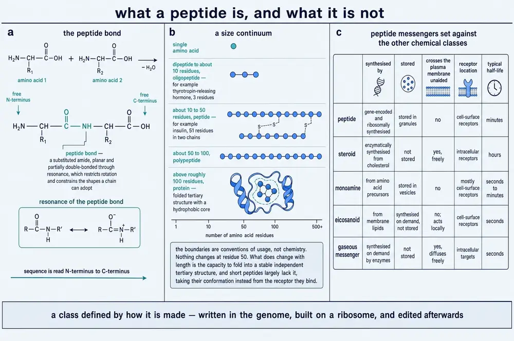
Start with the alphabet. Twenty amino acids do the work in human proteins. Each has a common backbone and a distinguishing side chain: some are electrically charged, some repel water, some are small enough to let a chain turn a tight corner, one contains sulphur that can bond to another sulphur and staple two distant points together.
Join two amino acids and you make a peptide bond: the acid group of one condenses with the amine group of the next, water leaves, and what remains is a link far more stable than the chemistry of the cell would casually break. Chains have direction. One end retains a free amine group, the other a free acid group, and by universal convention a sequence is written and synthesised from the amine end to the acid end. Direction matters as much as it does in writing: the same letters in the opposite order are a different word, and in almost every case a biologically inert one.
Where does a peptide stop and a protein begin? There is no natural boundary. The convention in most of the literature places the transition somewhere around fifty residues, and like most conventions it is approximately useful and occasionally absurd: insulin, at fifty-one residues across two chains, is called a protein by some authors and a peptide hormone by nearly all of them. What actually changes with length is not category but behaviour. Short chains are floppy and often adopt a definite shape only once they are gripped by something else. Long chains fold into stable structures with pockets and clefts, and can therefore do chemistry — catalysis, transport, mechanical work. Peptides do not usually do chemistry. They carry meaning.
It is worth asking why a body would use them at all, because it has other options. Steroid hormones are small, greasy, and pass straight through cell membranes to act on receptors inside the cell — which makes them powerful and slow, and hard to stop once released. Amine transmitters are tiny, fast and made in one or two enzymatic steps. Nitric oxide is a gas with a lifetime measured in seconds and a range measured in cell diameters. Lipid mediators are made on demand from the membrane itself.
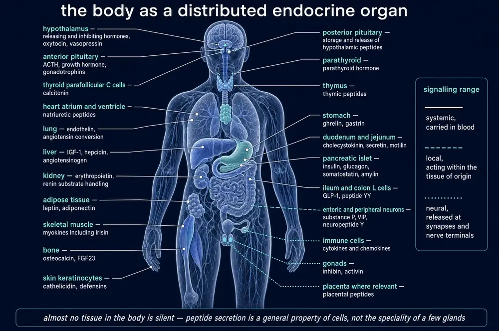
Peptides occupy a particular niche in that landscape, and the niche explains most of what follows in this document. A chain of ten to forty residues is large enough to be unambiguous — the number of possible sequences is astronomically greater than the number of messages a body needs to send, so a receptor can be built that recognises one and essentially nothing else. It is small enough to be manufactured quickly from a gene, without the folding problems of a large protein. And it is destroyed easily, by enzymes that are everywhere, which sounds like a weakness and is in fact the single most important thing about it. A signal that cannot be switched off is not a signal; it is a condition. Section 25 returns to this at length.
03Learning to read a sequence
By 1950 the hormone concept was secure and the chemistry was not. Insulin had been in clinical use since 1922, when Frederick Banting, Charles Best, James Collip and their colleagues in Toronto published the results of injecting a pancreatic extract into patients with diabetes (Banting et al., 1922). It worked, spectacularly, and nobody knew what it was. The prevailing assumption was that a protein was not a definite object at all but a statistical one — a population of similar molecules with amino acids in roughly, but not exactly, the same arrangement.
Frederick Sanger spent a decade demonstrating otherwise. Working with insulin, he developed reagents that would label the free amine end of a chain, broke the molecule into fragments, identified the fragments, and reassembled the order by overlap — the same logic later used to sequence genomes, executed by hand. The B chain came first (Sanger & Tuppy, 1951), then the A chain (Sanger & Thompson, 1953), and finally the disulphide bridges that hold the two together (Ryle et al., 1955).
The result was more important than the molecule. Insulin had one sequence. Every insulin molecule in every ox was the same object, specified exactly. A protein was a defined chemical entity, and therefore — in principle — something that could be written down, compared across species, and eventually made.
Vincent du Vigneaud made it. Working on the two hormones of the posterior pituitary, he established the sequence of oxytocin as a nine-residue chain closed into a ring by a disulphide bond, with a three-residue tail (du Vigneaud et al., 1953a) — and then built the same molecule in glassware and showed that the synthetic material contracted a uterus exactly as the natural extract did (du Vigneaud et al., 1953b, not indexed in PubMed). It is difficult to overstate what this settled. The activity was not in some undetectable contaminant, not in a property of living tissue that chemistry could not reach. The sequence was the message. Nothing else needed to be present.
And immediately next to oxytocin sat the proof of how much a sequence means.
Making peptides remained an ordeal until Bruce Merrifield proposed attaching the growing chain to an insoluble bead. Every reagent could then be washed away rather than separated, and the chain could be extended, washed, extended, washed, indefinitely. Solid-phase peptide synthesis turned an achievement into a procedure and eventually into a machine (Merrifield, 1963, not indexed in PubMed; described by its author in Merrifield, 1985). Almost every synthetic peptide discussed anywhere in this series exists because of it.
04Learning to measure a whisper
A second obstacle was subtler. Even with sequences known and synthesis solved, physiologists could not watch hormones. Bioassays — inject an extract, measure a uterus contracting or a blood glucose falling — were the standard, and they were crude, slow, and blind to anything present in genuinely small amounts.
Rosalyn Yalow and Solomon Berson, working at a Veterans Administration hospital in the Bronx, arrived at the answer sideways. They had been studying what happened to injected insulin and found that patients previously treated with animal insulin carried antibodies against it. The immunological finding was itself contentious. What they saw in it was an instrument.
If an antibody binds insulin, then radioactively labelled insulin and unlabelled insulin will compete for it. Add a plasma sample containing an unknown quantity of the patient's own insulin and it displaces the labelled material in proportion to how much is there. Measure the radioactivity and you have measured the hormone (Yalow & Berson, 1960). The conceptual move is the elegant part: stop measuring what a hormone does, and start measuring what binds it.
Radioimmunoassay reached concentrations that had previously been unreachable, and in doing so it changed the questions physiology could ask. Almost everything in Parts Three and Four of this document — that hormones are released in pulses, that pulse frequency carries information, that concentrations follow the clock and the meal, that feedback loops have measurable gain — is invisible without an assay sensitive enough to follow a hormone minute by minute in a living animal. The instrument did not merely confirm the picture. It produced it.
05The hypothalamic decade
By the 1960s a specific and embarrassing gap remained. The pituitary was understood to command the thyroid, the adrenal cortex and the gonads. Something was understood to command the pituitary, and the evidence pointed at the hypothalamus, connected to it by a small private circulation. But nobody could produce the commanding substances, and a good many physiologists doubted they existed.
The problem was quantity. Hypothalamic releasing factors are present in vanishing amounts, and there was no way to enrich them. Roger Guillemin and Andrew Schally — who had worked together briefly and then spent two decades as rivals — both concluded that the only route was brute force: process hypothalami from slaughterhouse animals in industrial quantities and chase the activity through fraction after fraction.
The first target fell in 1969. Thyrotropin-releasing factor turned out to be three residues long, chemically modified at both ends in ways that had frustrated every earlier attempt to characterise it (Burgus et al., 1970). Two years later Schally's group isolated the factor that releases luteinising hormone from porcine hypothalamus (Schally et al., 1971), a decapeptide now called GnRH, which returns in Section 19 as the centre of this document's argument.
Then, in 1973, Guillemin's group found something they had not been looking for. While pursuing a factor that would release growth hormone, they isolated a fourteen-residue peptide that powerfully inhibited it, and named it somatostatin (Brazeau et al., 1973). Its sequence was settled the same year by mass spectrometry (Ling et al., 1973).
Somatostatin mattered out of proportion to its size, because it changed the shape of the model. Until then the hypothalamus looked like a command centre issuing orders to start. Somatostatin showed that it also issues orders to stop, and that the pituitary's output is the arithmetic of the two. That is not a chain of command. It is a control system — and control systems have properties that command chains do not: set points, gain, oscillation, instability. Part Four is largely an examination of those properties.
06Nothing is made in its final form
In 1967 Donald Steiner was studying insulin production in a human islet-cell tumour and found something that should not have been there: a single-chain molecule, larger than insulin, carrying insulin's two chains joined by an extra stretch of sequence. It was not a degradation product. It was the starting material (Steiner & Oyer, 1967; Steiner et al., 1967).
The finding generalised almost immediately, and it is now the rule rather than the exception. A peptide hormone is not transcribed and translated as itself. It is made as a longer precursor and cut down — often more than once, often by more than one enzyme, and often in different ways in different cells.
Why would a cell manufacture something in order to destroy most of it? There are at least three good answers, and this Part is organised around them.
The first is folding. Insulin's two chains must be joined by disulphide bonds in exactly the right pairing. Held together in one chain by the connecting segment, the correct pairing is geometrically favoured; as two separate chains in solution it is a lottery. The extra sequence is a jig.
The second is safety. A precursor is inactive. A cell can accumulate a large quantity of a potent signal in a form that cannot act, and convert it only at the moment and place of its choosing.
The third is economy of information, and it turns out to be the most consequential. One precursor can yield several different products, and which products a cell obtains depends on which cutting enzymes it happens to own. That is where the specificity of an entire endocrine tissue can live.
07The address written on the front
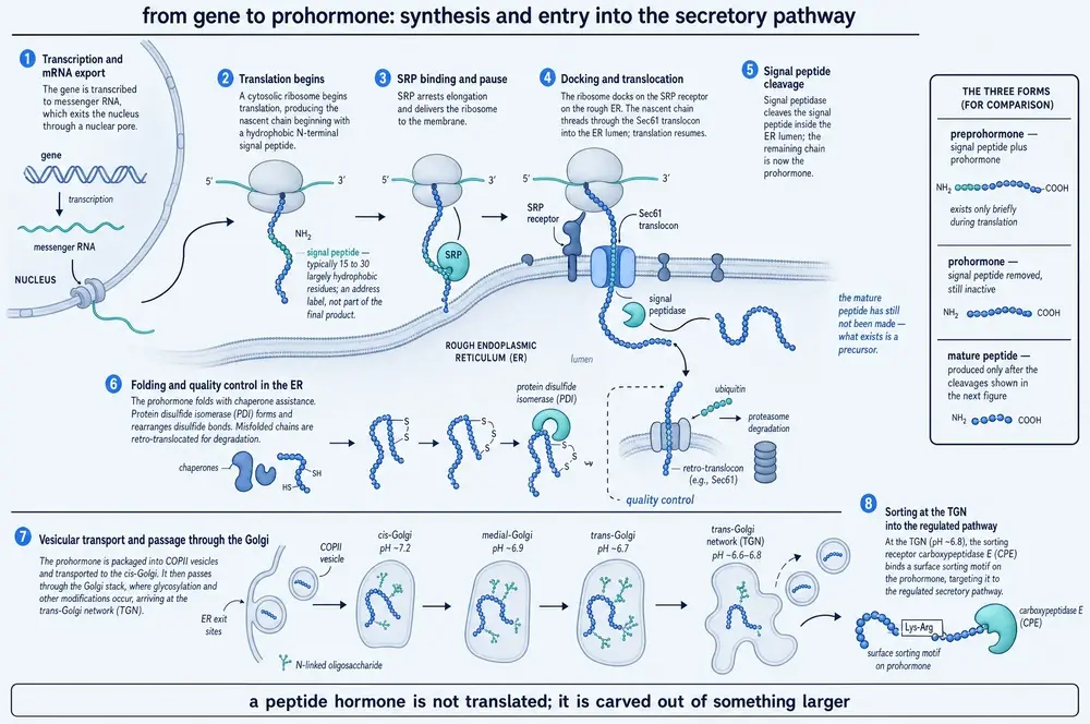
A peptide destined for secretion has a problem before it has a sequence: it is being built by a ribosome in the cytoplasm, and it needs to end up outside the cell. Something has to route it.
The solution, proposed by Günter Blobel and tested through the 1970s, is a stretch of about twenty residues at the front of the chain. As it emerges from the ribosome it is recognised by the signal recognition particle, which halts translation and delivers the whole assembly to a channel in the endoplasmic reticulum membrane. Translation resumes with the growing chain threading directly into the lumen, and an enzyme on the far side snips the signal off (Blobel & Dobberstein, 1975). The particle doing the recognising turned out to be built around an RNA (Walter & Blobel, 1982), with a dedicated receptor waiting in the membrane (Gilmore et al., 1982).
The elegance is that the address is read while the message is still being written, and then removed, so the finished product carries no trace of how it was delivered.
What makes a signal peptide a signal peptide is startlingly crude. Across signal peptides generally, the composition runs to roughly 37 per cent leucine and 0.4 per cent lysine — a bias of about ninety-fold in a twenty-five residue stretch (Shah et al., 2026). That compositional skew is what "hydrophobic core" means in practice. And evolution guards it: of 1,008 recoding RNA-editing sites catalogued in the fruit fly, exactly two fall inside a signal peptide, and both are the same substitution (Duan et al., 2025). One fly protein is twenty-one amino acids long, of which eighteen are signal peptide — eighty-six per cent of the protein is address label.
The cell also polices these sequences aggressively, in a way discovered only recently. Deleting two leucines from a model signal peptide dropped the reporter's messenger RNA to 0.29 of normal but its protein to 0.11 — the transcript was destroyed and the survivors were translated less. The surveillance factor turned out to be ZAP, a protein known for antiviral defence, which does this by binding the very RNA inside the signal recognition particle. Remove ZAP and overexpress a signal peptide alone, with no cargo attached, and a cellular stress-response gene rises about fiftyfold (Shah et al., 2026, in human cell lines). A twenty-residue address label, loose in the cell, is toxic.
The rule has exceptions that are worth stating because they show how the rule was tested. Keratin-19 carries no signal peptide and nevertheless enters the endoplasmic reticulum by the classical route, using its N-terminal head domain instead — and the control that makes this credible is that of sixty-three proteins found enriched in the same compartment, sixty-two did have a signal peptide or a transmembrane domain (Moresco et al., 2025). One exception among sixty-three is an exception. Sixty-three among sixty-three would have been an artefact.
08The scissors and the finishing shop
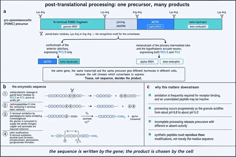
Inside the Golgi and the maturing secretory granule, the prohormone is cut. The enzymes that do it are a family of proprotein convertases — PC1/3, PC2, furin and others — which recognise pairs or runs of the basic amino acids lysine and arginine. Cutting there leaves basic residues hanging off the new end, and a carboxypeptidase trims them. Many peptides are then finished by an enzyme that converts the C-terminal carboxyl group to an amide, which protects the end and is frequently required for receptor binding.
Here is the key idea of this Part, and it is best shown twice.
Proglucagon is one gene and one precursor. In the intestinal L-cell, PC1/3 cuts it into GLP-1 and GLP-2. In the pancreatic alpha cell, PC2 cuts the identical precursor into glucagon — a hormone with the opposite effect on blood sugar to the one the intestine makes from the same starting material (Han et al., 2026; Irwin, 2021). That a single proglucagon gene carries glucagon and two related peptides was established by cloning in 1983 (Bell et al., 1983). The information distinguishing "raise blood glucose" from "lower blood glucose" is not in the gene. It is in which enzyme the cell expresses.
The regulation goes deeper than that. A single RNA-binding protein, HuD, binds the untranslated regions of both the precursor's messenger RNA and PC1/3's messenger RNA, so one regulator sets the supply of the substrate and of the scissors at once (Han et al., 2026, in mouse and cell models).
Pro-opiomelanocortin makes the same point across more products. Cloning its complementary DNA in 1979 showed a single precursor encoding ACTH and β-lipotropin together (Nakanishi et al., 1979) — the first direct demonstration that one gene carries several hormones. One precursor yields ACTH in the corticotroph of the anterior pituitary and, where different convertases are present, α-MSH and β-endorphin. The first cut is at a specific bond, KR164, and testing six candidate enzymes in cells lacking a regulated secretory pathway produced a genuine surprise: furin performed it, PC7 barely, and PC1/3 — the textbook enzyme for this step — scored zero. The authors state that their result challenges the prevailing account (Coppola et al., 2022, in cell lines and mice).
Processing is also where a cell decides how much. Activin-A's precursor carries a run of five arginines. Mass spectrometry shows furin cutting after the fifth and a different protease, kallikrein-8, cutting after the first, second or third — and the two are separated by acidity, since kallikrein-8 requires prior acidification, which disables furin. Mutating the fifth arginine to alanine silenced the pathway entirely in a tumour model: no mature product, no growth advantage, no cachexia, circulating Activin-A below detection where the wild type reached about 35 ng/mL. Deleting the same arginine outright restored everything, which shows the block was conformational rather than arithmetic (Bulliard et al., 2025, in mouse models and in vitro).
And a cut is not always sufficient. Myostatin is released from its propeptide by furin — but the propeptide stays wrapped around it, and only a second cut, by a different protease, sets it free. Mice engineered with a cleavage-resistant propeptide carry more than tenfold higher circulating myostatin and have muscle weights approaching those of an animal with no myostatin at all (Lee, 2023). Abundance is irrelevant if the lock is not picked.
09What happens when the scissors fail
The strongest evidence that a step is load-bearing is what happens when it breaks, and human genetics supplies the cleanest examples.
Mutations in the human PC1/3 gene produce obesity together with a complex endocrine disorder, and the mechanism is exactly what the model predicts: prohormones accumulate and their products do not appear (Jackson et al., 1997). The fuller phenotype, described later, includes severe small-intestinal dysfunction — because the gut is one of the tissues that depends on this enzyme (Jackson et al., 2003). In the mouse, a mutation in carboxypeptidase E — the trimming enzyme, one step further on — produces hyperproinsulinaemia and obesity (Naggert et al., 1995).
A more recent case shows how small the failure can be. In prepro-orexin, substituting one lysine for arginine converts the cleavage site from GKR to GRR. The convertases still work, but much less well: about fiftyfold worse for PCSK1 and about eleven-hundred-fold worse for PCSK2. Carriers had an odds ratio of 5.36 for idiopathic hypersomnia across 598 cases and 9,826 controls. The detail that makes this case instructive is that the single carrier whose cerebrospinal fluid was measured read 227 pg/mL — comfortably normal — which the authors attribute to the standard assay largely measuring degraded fragments. The cleavage defect itself was demonstrated in vitro; the study does not show impaired processing in a living carrier, and the normal assay reading is consistent with, rather than proof of, that interpretation (Miyagawa et al., 2022).
The same study contains a lesson about instruments rather than biology. The prepro-orexin coding sequence is 73 per cent GC-rich, which exome sequencing under-reads: 36.9 per cent of variant carriers had near-zero exome coverage at that position against none by whole-genome sequencing. The variant is also East-Asian specific — 0.34 per cent in Han Chinese, 0.013 per cent in non-Finnish Europeans, absent in the African and Finnish samples. It was hiding in the wrong population, read by the wrong instrument.
10Stored under pressure
A cell that has manufactured a peptide now faces a timing problem. Making one takes minutes to hours. Physiology frequently needs one in under a second.
The answer is a warehouse. Peptides destined for controlled release are packaged at the trans-Golgi network into dense-core secretory granules, which mature by acidifying and concentrating their contents — processing continues inside them, which is why the convertases of Section 08 have acidic pH optima. The granules then wait, sometimes for a long time, until a signal arrives.
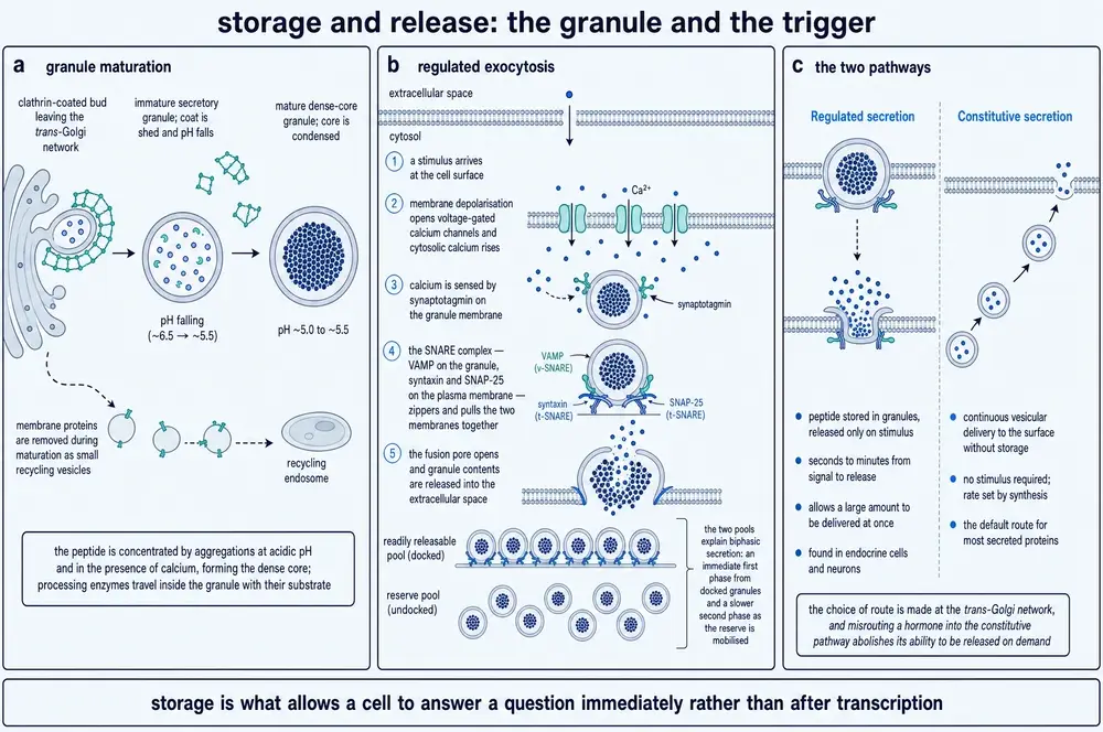
This is the regulated secretory pathway, and it exists alongside a constitutive one in which vesicles carry their contents to the membrane and release them continuously, without waiting. Which route a protein takes is a sorting decision made in the Golgi. The distinction matters more than it sounds: a peptide released constitutively cannot be pulsatile, cannot be released in a burst, and cannot answer a stimulus in milliseconds. Everything in Section 19 about timing as information presupposes a regulated pathway.
11Release
The trigger is calcium. A stimulus — depolarisation, a receptor signal, a rise in glucose — raises calcium in the cytoplasm; calcium is detected by sensor proteins on the granule; and the granule membrane fuses with the cell membrane, spilling its contents outside. The machinery is shared with neurotransmitter release, which is why the same protein names recur in endocrinology and neuroscience.
Two features of this arrangement matter for the rest of the document.
The first is that release is frugal. In mouse central neurons, only about one to six per cent of the dense-core vesicle pool is released even under robust stimulation (Baginska et al., 2024; Abramian et al., 2024). The warehouse is not emptied; a small fraction is dispatched and the rest is held.
That restraint is actively enforced rather than merely a limit on the machinery. A pancreatic beta cell carries on the order of a hundred and twelve thousand copies of syntaxin-2, a SNARE protein that inhibits fusion, at roughly thirty-two copies per docking site — about ten times the number of fusion complexes a granule actually needs (Kang et al., 2022, in human and rodent beta cells). Deleting a single brake protein can triple output from an unchanged pool (Abramian et al., 2024, in mouse neurons). Storage capacity is almost never the limiting term. Permission is. That is the correct way to think about every regulated secretory cell in this document: it is not struggling to produce enough, it is holding back nearly all of what it has.
The second is that the store is nonetheless exhaustible, and the consequences are visible in people. In septic shock the posterior pituitary's vasopressin stores are depleted within about two hours — observable as loss of the normal bright signal on T1-weighted MRI (Lajoye et al., 2025). A system that stores its messages can run out of them, and Section 22 returns to why peptide stores in particular are slow to replace.
12Five ranges of voice
A cell that has released a peptide has not yet communicated anything. What happens next depends almost entirely on where the message goes, and the categories physiology uses to describe that are categories of distance and route rather than of chemistry.
Endocrine signalling releases the peptide into the bloodstream, which carries it everywhere. Paracrine signalling releases it into the fluid between cells, where it acts on neighbours. Autocrine signalling acts on the cell that released it. Neurocrine signalling releases a peptide from a neuron, either into a synapse or into the surrounding tissue, or — in the case of the hypothalamus — directly into a specialised blood supply. Juxtacrine signalling requires the two cells to touch. And intracrine signalling never leaves the cell at all: the peptide acts on targets inside its own cytoplasm or nucleus.
The crucial point, and the one most easily lost, is that these are not properties of the molecule. The same peptide can and often does use more than one route. Somatostatin acts as a paracrine brake inside the pancreatic islet and as a circulating hormone released from the gut; islet delta cells are reported to supply only about five per cent of the somatostatin in the circulation, which means the somatostatin governing insulin and glucagon output essentially never enters the blood (Hartig & Cox, 2020, a review of islet paracrine biology).
How local is local? Less is settled here than the textbooks suggest. A computational analysis of gene-expression correlation between cells in rat pituitary tissue found correlation strongest below about twenty-four micrometres — roughly two cell diameters — and faded by about thirty-five (Momiji et al., 2019). But the same analysis separated contact-dependent from diffusion-dependent signalling by how steeply correlation fell with distance, found the contact-dependent fingerprint between nine and a hundred and forty times steeper, and concluded on that basis that juxtacrine signalling dominated in that tissue, with no strong evidence of paracrine signalling at all. The distance is therefore a contact-signalling correlation length, not a paracrine range, and it is quoted here as such.
If mode is not a property of the molecule, what decides it? Partly the chemistry of how tightly the peptide sticks to the tissue around it — and this has been demonstrated by changing it. Swapping three amino acids in the region of fibroblast growth factor 1 that binds heparan sulphate (Lys127Asp, Lys128Gln, Lys133Val) released the protein from the extracellular matrix and let it behave as a circulating hormone rather than a local one (Zhao et al., 2019, in engineered constructs tested in cells and mice). Whether a growth factor is paracrine or endocrine turned out to be an editable chemical property, not a category it belongs to.
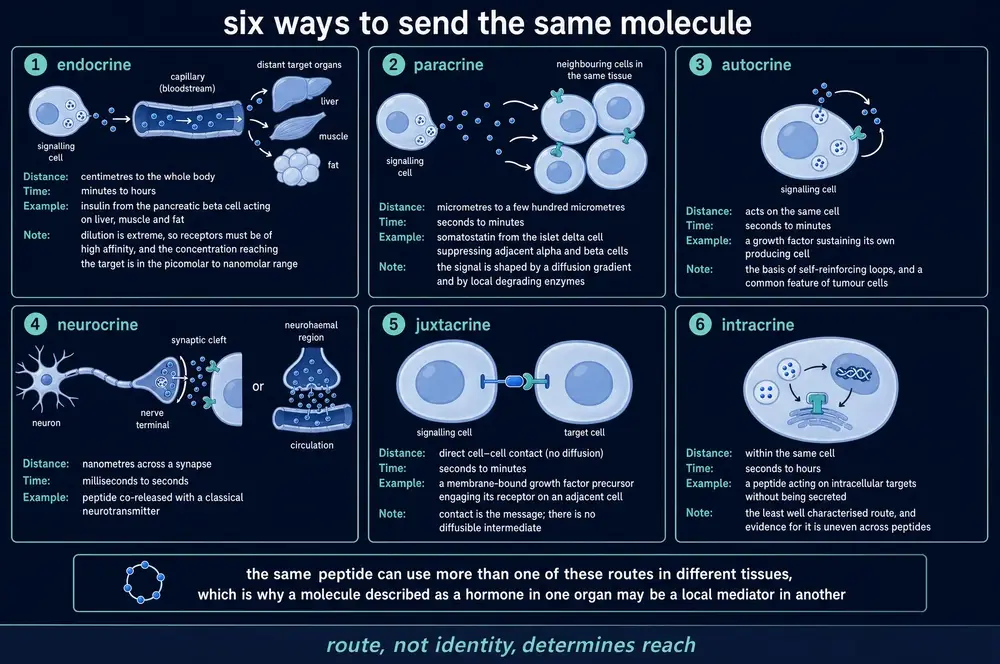
Partly, too, the arithmetic of production and capture: whether a signal stays local depends on how much of it is caught by nearby receptors before it escapes, and on how fast it is degraded (Segers & De Keulenaer, 2021). And in the intracrine case, on trafficking alone. Parathyroid hormone-related protein carries a nuclear localisation sequence in residues 67–94; in vascular smooth muscle cells the same protein inhibits proliferation when it acts through the cell-surface receptor PTH1R and increases it when routed to the nucleus (Edwards & Johnson, 2021, reviewing cell-culture work). One molecule, two destinations, opposite outcomes.
13Dilution as a design constraint
Consider what the bloodstream actually does to a message. An adult carries roughly five litres of it. A cell releasing a peptide into that volume has performed an act of extraordinary dilution, and the resulting concentration — often picomolar, as Section 04 established — is the price of reaching everywhere.
The alternative is not to release into blood at all. A peptide delivered into the narrow space between two cells occupies a volume smaller by many orders of magnitude, and the concentration there can be enormously higher. Within a pancreatic islet, a single insulin granule is reported to raise the insulin concentration in the fluid between neighbouring cells to more than a hundred times the circulating level (Hartig & Cox, 2020, a review citing earlier primary work; the figure is a ratio and no absolute concentration is given). The neighbouring beta cell and the rest of the body are reading two different signals from the same act of secretion.
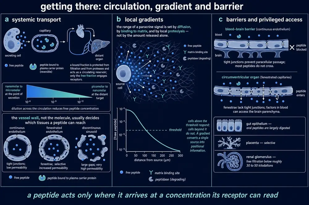
Half-life does the same work as geometry. A peptide destroyed within seconds of release cannot be anything but local, whatever the anatomy allows. C-type natriuretic peptide is cleared from blood so rapidly that it is classified as a local signal by default, while its close structural relatives ANP and BNP survive long enough to reach the kidney and act as hormones. Among the gut peptides, GLP-1 survives one to two minutes in human blood, GIP and GLP-2 about seven minutes each (Morrow et al., 2021; Segers & De Keulenaer, 2021). Section 24 takes up what destroys them and why the body bothers.
14Recognition
A message arriving at a cell is inert unless something there is built to read it. For most peptide hormones that something belongs to a family called class B1 G-protein-coupled receptors: a protein threaded seven times through the membrane, with a substantial extracellular domain of roughly a hundred and twenty residues sitting above it.
The standard account of how these receptors capture their ligand is a two-step, two-domain mechanism. The extracellular domain first grips the back half of the peptide, which tethers it and dramatically raises the local concentration of its front half; the front half then inserts into the pocket formed by the membrane-spanning helices and triggers the conformational change that activates the receptor (Austin & Tomas, 2026, a review). It is an elegant solution to a real problem: catching a floppy chain out of a dilute solution is far easier in two stages than in one.
What specificity means at this scale can be seen most sharply in what happens when a single atom is removed. In the orexin-1 receptor, mutating the tyrosine at position 6.48 to alanine cost a hundred and sixty-four-fold of orexin-A's potency. Mutating the same tyrosine to phenylalanine — which removes only the hydroxyl group and keeps the aromatic ring — cost 1.6-fold (Heifetz et al., 2013, in cell-based assays with computational modelling). The receptor cares about the ring. It barely notices the chemistry hanging off it.
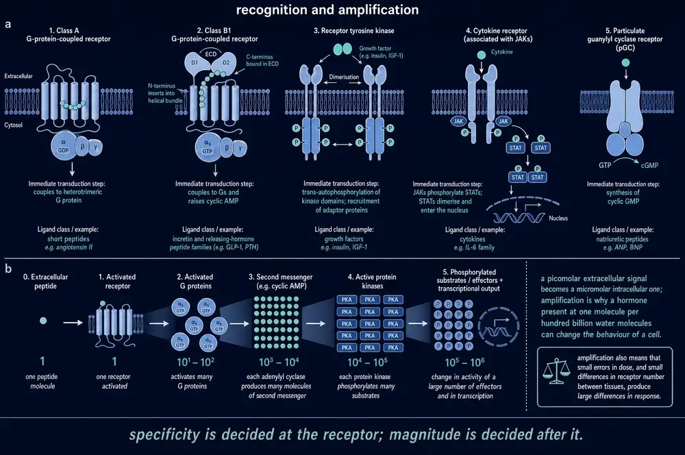
And specificity is not fixed by the receptor gene alone. The calcitonin-receptor-like receptor is inert to CGRP on its own. Paired with an accessory protein called RAMP1 it becomes a CGRP receptor; paired with RAMP2 or RAMP3 the identical receptor protein becomes an adrenomedullin receptor, with roughly fiftyfold lower potency for CGRP (Zhu et al., 2025, in transfected cells; the modulating role of the accessory proteins is reviewed by Shao et al., 2022). One gene, three receptors, decided by what the cell chooses to build alongside it.
15Amplification
Here is the question the reader has been carrying since Section 04. A hormone present at a few picomolar — perhaps a few thousand molecules in the fluid around a cell — produces a response involving the movement of millions of ions and the phosphorylation of millions of protein molecules. How?
The answer, in outline, is that each step of the receiving apparatus is catalytic. One activated receptor does not activate one G protein and stop; it activates G proteins repeatedly for as long as it stays in its active conformation. Each activated G protein switches on an enzyme — adenylyl cyclase, for the receptors under discussion — and each of those enzymes manufactures second-messenger molecules continuously while it remains active. Cyclic AMP then binds protein kinase A, which phosphorylates targets catalytically in turn. A cascade of catalysts multiplies at every stage, and the product of four multiplications is a very large number.
Amplification is also more spatially organised than the funnel suggests. Cyclic AMP signalling at the GLP-1 receptor has been described as confined to nanodomains roughly sixty nanometres across, which keep the signal local at low hormone concentrations and merge into a single undifferentiated pool under high agonist exposure (Austin & Tomas, 2026, reviewing imaging work). If that account holds, spatial precision is a property of the system at physiological concentrations that pharmacological concentrations destroy — a point Part Five returns to.
Nor is the cascade instantaneous. At several class B peptide receptors, a low agonist concentration produces a lag of ten to thirty minutes before signalling reaches full rate; adding extracellular ATP abolishes the delay and raises potency up to fiftyfold. The authors excluded purinergic receptors, ATPase and ectokinase activity, accessory-protein involvement, changes in intracellular ATP, receptor oligomerisation and internalisation, and concluded that they do not know what the delay is (Zhu et al., 2025, in transfected cells). It is worth recording an unexplained result of this size in a system this well studied.
16The same word in different rooms
Everything so far leads to a conclusion that sounds deflationary and is in fact the central practical fact of the field: the question "what does peptide X do" has no answer. A peptide does what the receiving tissue is equipped to do with it.
The equipment varies in at least four ways. Which receptor subtypes a cell expresses; which G proteins those receptors couple to in that cell; what accessory proteins are present, as the RAMP example showed; and what state the cell is in when the message arrives.
The most striking demonstration in this document's evidence base is an experiment where the readout was three cell types away from the receptor being blocked. Blocking the somatostatin receptor SSTR2 raised cyclic AMP in mouse beta cells — which express no SSTR2 at all. Pull the islet apart into single cells and the effect vanishes entirely, because the signal had been travelling through alpha cells (Hart et al., 2026, in mouse islets). The drug acted on one cell; the measurement changed in another; and the connection existed only while the tissue was intact.
Receptor behaviour after binding varies too. A receptor may signal from the cell surface, or be internalised and continue signalling from inside endosomes, or be internalised and silenced. Which of these happens shapes the duration and character of the response — and the rules are actively contested. One recent study reports endosomal cyclic AMP signalling that does not require β-arrestin, against a well-established consensus that it does (Blythe & von Zastrow, 2024). Another concludes that the benefit of G-protein-biased agonism at the GLP-1 receptor comes from slowing internalisation rather than from reducing β-arrestin recruitment, while conceding that this conflicts with established endosomal-signalling work and that no head-to-head human trial of oppositely biased agonists exists (Tran et al., 2026). Both are recent, both are careful, and they are not yet reconciled.
17The gut, which never stops talking
The intestine is not usually thought of as an organ of communication. It should be. Scattered through its lining is a population of cells that taste what you have eaten and report it — to the pancreas, to the brain, to the appetite centres of the hypothalamus — in peptides.
These enteroendocrine cells are rare. They make up roughly one per cent of the intestinal epithelium (Nwako & McCauley, 2024, reviewing human and mouse work). What that one per cent is worth can be read from what happens without it: mice in which the population is deleted genetically die within one to two weeks of birth from diarrhoea and fat malabsorption, and a child carrying a mutation in NEUROG3, the transcription factor that specifies these cells, passed more than eighty per cent of ingested protein straight into the stool.
It is worth pausing on the setting. The intestinal epithelium replaces itself roughly every five days, across a surface amplified sixty- to one-hundred-and-twenty-fold by coiling, crypt-villus folds and microvilli, and this continuously rebuilt apparatus extracts ninety to ninety-eight per cent of what is eaten. The endocrine cells embedded in it are being replaced on the same schedule.
The old textbook picture of these cells — a lettered alphabet of types, each making one hormone — has not survived. More than seventy per cent of enteroendocrine cells express several hormones at once, in combinations that change as the cell ages and migrates up the villus; one sequencing study found over fourteen hundred genes changing across a cell's short life, including a hundred and seventy-two transcriptional regulators (Nwako & McCauley, 2024). A cell can change which hormone it makes during the journey. There is no stable census of types to write down, and this document does not offer one.
Some of these cells do something stranger still. A subset grows a long cytoplasmic process — a neuropod — and forms an actual synapse with a nerve fibre, transmitting the detection of sugar to the brain in milliseconds. That is not endocrine signalling at all. It is the gut using the nervous system's own channel, and it means the gut–brain axis has at least two routes operating on timescales three orders of magnitude apart.
The classical demonstration that the gut reports to the pancreas is the incretin effect — quantified in people across a range of glucose loads in 1986 (Nauck et al., 1986) — introduced in Section 01's successor question: swallow glucose and the insulin response is much larger than it is when the identical glucose is delivered into a vein at a matched blood concentration. The difference is attributed to peptides released by the gut in response to food arriving.
Two findings from the gut peptides illustrate points made in earlier Parts. The first: GLP-1(9-36), long dismissed as the spent breakdown product of GLP-1, suppresses glucagon almost as strongly as the intact hormone — half-maximal at 4 pmol/l against 2.5 pmol/l — but through a different, inhibitory G-protein route (Gandasi et al., 2024). What degradation produces is not necessarily nothing. Section 24 returns to this.
The second: hunger has an antagonist nobody expected. LEAP2, a forty-residue liver peptide isolated in 2003 and characterised then as antibacterial, turns out to block the ghrelin receptor. It circulates at nanomolar concentrations while ghrelin sits at picomolar, it falls with starvation and rises in obesity — mirroring ghrelin in both directions (Valdés-Calero et al., 2026, a review). A molecule filed under host defence for fifteen years was in fact half of an appetite switch.
18The axes, and the arithmetic of feedback
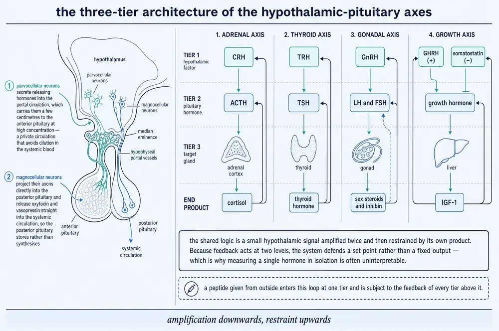
Section 05 ended with somatostatin turning a chain of command into a control system. The general architecture is worth stating plainly, because Parts Four and Five both depend on it.
A hypothalamic peptide instructs the pituitary. A pituitary hormone instructs a target gland. The target gland's product feeds back to inhibit both levels above it — the long loop — and the pituitary hormone feeds back on the hypothalamus — the short loop. The system therefore has a set point, a gain, and a delay, and like any such system it can be driven into oscillation or into silence.
What such a loop regulates is less obvious than it looks. It is easy to assume the controlled variable is a concentration. Often it is not. Feedback failure can present as a disturbance of level or as a disturbance of pattern, and these are different diseases: purely ectopic ACTH secretion produces persistently high, non-cyclical cortisol with loss of circadian rhythm, whereas tumours co-secreting ACTH and CRH produce cyclical, higher-amplitude output. In functional hypothalamic amenorrhoea the LH pulse pattern regresses toward an early-pubertal form, and prolactin pulse frequency and amplitude move in opposite directions.
19Frequency is information
This is the centre of the document's argument, and it rests on an experiment that is nearly fifty years old and has never needed repeating.
By the mid-1970s GnRH was available synthetically and the obvious clinical application was infertility: give the missing hormone, restart the axis. In monkeys whose hypothalamic connection had been destroyed, giving GnRH did restart it — and then, on continued infusion, the axis shut down completely. Not attenuated. Off.
Belchetz, Plant, Nakai, Keogh and Knobil then delivered the identical total dose of the identical peptide as an hourly pulse rather than as a continuous infusion, and the axis recovered fully (Belchetz et al., 1978, in rhesus monkeys). Same molecule, same amount, opposite result. The information was in the timing.
Why does continuous delivery silence the axis? The mechanism appears to be partly arithmetic and partly molecular. Luteinising hormone circulates with a half-life somewhere between thirteen and forty minutes (Goodman et al., 2022, reviewing human and rat data). Drive GnRH faster than that and each pulse begins before the last has cleared, so the troughs fill in and the signal flattens toward the continuous case. Drive it faster still and the receiving cascade desensitises. Output describes an inverted U: more stimulus, less hormone. This is why continuous GnRH analogues are used clinically to switch the reproductive axis off — the shutdown that looked like a failure in 1978 became a therapeutic mechanism.
What does a pulse actually look like? Sampling the pituitary portal blood of conscious ewes at thirty-second intervals resolves an abrupt rise over about two minutes, a plateau of four to five, and an abrupt fall over three, with the level between pulses essentially undetectable (Goodman et al., 2022). It is close to a square wave.
Getting that measurement was not straightforward. Told that simultaneous portal and peripheral sampling in a conscious animal was impractical, Iain Clarke went home and looked up neurosurgeons in the Melbourne telephone directory. The device the field eventually standardised on — known simply as "the gadget" — was two blunt needles glued together with dental acrylic and a cup cut from a microcentrifuge tube (Moenter & Evans, 2022). In the definitive natural-cycle experiment, eleven of twelve ewes showed a clear GnRH surge; the twelfth was explained at autopsy, where the lesioning stylet had struck the sphenoid bone instead of the portal vessels.
The pre-ovulatory surge turns out not to be simply faster pulses. At thirty-second sampling — fast enough to resolve individual pulses easily at other times — no discrete pulses can be found during the surge at all, and the surge continues for hours after the LH surge it triggered has ended (Moenter & Evans, 2022). It appears to be a different mode of secretion. This is genuinely unsettled: Knobil's monkeys produced LH surges under unchanging hourly GnRH replacement (Knobil et al., 1980), which argues that no change in pattern is required, and the sheep portal evidence argues that one occurs. Both observations stand; they have not been reconciled.
One further finding deserves recording because nobody can explain it. Removing the gonads shifts a male mouse's pulse generator from roughly one pulse every hundred and seventy minutes to one every fifteen. The same operation in a female monkey leaves the frequency at about one an hour, exactly where it started (Goodman et al., 2022).
If the pattern is the message, one prediction follows: a system that varies its output should vary the frequency and leave the pulse itself alone. Oxytocin neurons during lactation supply the cleanest test in this document's evidence base. Across five independent manipulations — suckling deprivation, stage of lactation, pup age, mouse strain, and weaning — pulse amplitude and waveform never changed, while frequency changed every time; the intervals between pulses within a burst are statistically memoryless (Yaguchi et al., 2023, in mice). The generator is behaving like a device that transmits by rate and holds its symbol constant.
The reproductive axis gives the same answer from the opposite direction. Knocking down the progesterone receptor specifically in the arcuate nucleus of mice raised LH pulse frequency significantly, while pulse amplitude, basal LH, mean LH and total output were all unchanged (Glendining et al., 2026). An intervention that moved one parameter and left four others alone is about as clean a dissociation as this kind of physiology offers, and it identifies which parameter the system is actually regulating.
Rhythm on a longer timescale is built the same way, and by a surprisingly indirect route. The suprachiasmatic nucleus — the body's master clock — has essentially no direct connection to the CRH neurons that drive the stress axis. The signal is relayed through two further nuclei, and destroying the first relay abolishes 97.7 per cent of the corticosterone rhythm in constant darkness. Deleting that relay's GABA transporter does nothing, so the molecule actually carrying the timing has not been identified (Ramirez-Plascencia et al., 2026, in mice).
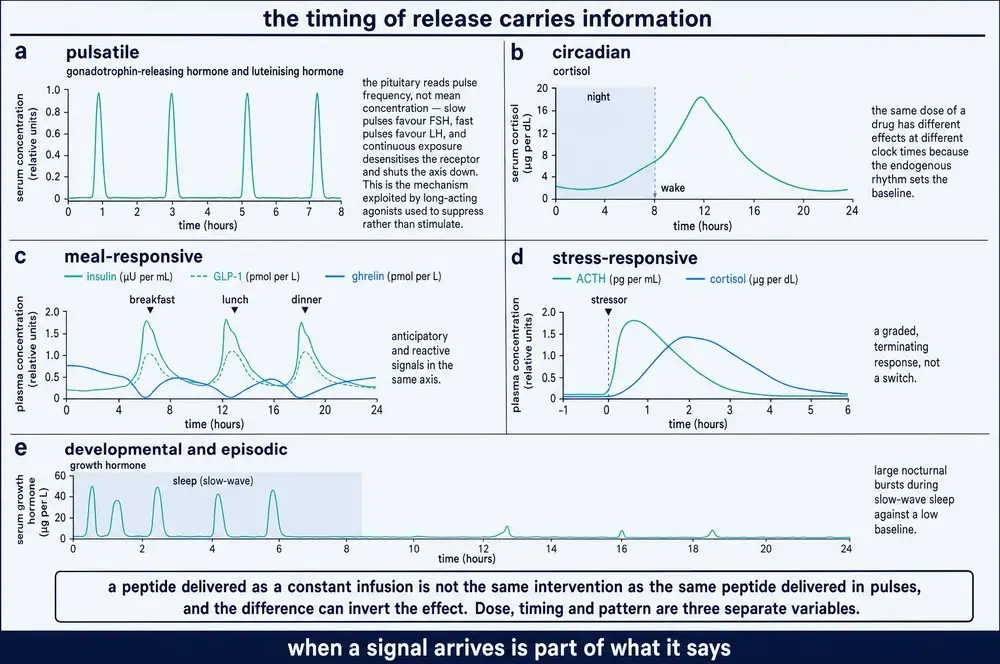
What the rhythm is worth can be measured by removing it. Human pancreatic beta cells transplanted into mice and then kept under constant light dropped from 92 per cent of grafts responding to glucose to 20 per cent (Sen et al., 2025). These were stem-cell-derived beta-like cells rather than primary human islets, which bounds how far the result can be carried; what it does show is that removing the timing, and nothing else, removed most of the response.
20The heart, which nobody expected to be a gland
In 1981 Adolfo de Bold and colleagues injected an extract of atrial muscle into rats and watched them produce a rapid and dramatic flow of sodium-rich urine (de Bold et al., 1981). The heart, an organ understood for three centuries as a pump, was secreting a hormone that told the kidney what to do.
The natriuretic peptides are worth this document's attention less for their clinical importance than for what they demonstrate: that a mechanical organ can be a chemical sensor. Stretch of the atrial wall — the direct physical consequence of too much circulating volume — is the stimulus. The response reduces the volume. It is a feedback loop in which the sensor is a muscle.
The three natriuretic peptides are close structural relatives of different lengths — ANP twenty-eight residues, BNP thirty-two, CNP twenty-two — each carrying a disulphide-bonded ring (Malsawmzuali et al., 2026). Their most visible legacy is not physiological but diagnostic: a hormone the heart releases in proportion to how stretched it is turned out to be measurable in blood, and thresholds derived from it now sit in cardiology guidelines. A messenger became an instrument.
The renin–angiotensin system makes the complementary point, and it connects Part Four back to Part Two. It is not a hormone but a cascade of cuts. The liver releases a large precursor, angiotensinogen. Renin — itself activated from a precursor by furin in the kidney, the same enzyme family met in Section 08 — cuts it to the ten-residue angiotensin I. Angiotensin-converting enzyme, sitting on the surface of vascular endothelial cells, removes two more residues to give the eight-residue angiotensin II, which is the potent one. And the cascade does not stop there: further enzymes convert angiotensin II onward to angiotensin III, angiotensin IV, angiotensin-(1–7) and alamandine, each acting at its own receptor, and several of them opposing the actions of angiotensin II (Unal et al., 2026).
Proteolysis here is not destruction. It is composition — the same principle Section 08 described inside the secretory granule, running instead in the open circulation, with each successive cut producing a molecule that means something different.
One clinical observation illustrates how a peptide system's state governs the response to an intervention. In a trial of seventy-two treated hypertensive patients, of whom forty-eight cut dietary sodium and twenty-four continued their usual diet, those in the intervention arm whose aldosterone response was intact dropped about nine millimetres of mercury of systolic pressure; those whose aldosterone barely moved dropped about three, despite the same reduction in sodium (Duus et al., 2026). The intervention was identical. The signalling state was not. It should be noted that this comparison was defined after the data were seen, splits the intervention arm into groups of thirty-two and sixteen, and is described by its own authors as observational.
21Defence, written in peptides
A quite different branch of peptide signalling does not signal at all in the ordinary sense: it acts directly on the intruder. The class came into focus in 1987, when Michael Zasloff isolated antimicrobial peptides from the skin of a frog he had been using for an unrelated purpose (Zasloff, 1987). Antimicrobial or host-defence peptides are typically short, positively charged, and shaped so that one face is greasy and the other charged. That architecture lets them distinguish a microbial membrane, whose outer surface carries a strong negative charge, from a human cell membrane, which does not and which is stiffened with cholesterol.
The human cathelicidin LL-37 shows both the selectivity and its limits. At its half-maximal concentration of 5.3 ± 0.3 µM it lysed fewer than ten per cent of uninfected human red cells but 18.3 ± 2.0 per cent of malaria-infected ones, rising to 34.9 ± 1.5 per cent at ten times that concentration; the selectivity was traced to phosphatidylserine appearing on the infected cell's outer surface together with cholesterol depletion (He et al., 2026, in human red cells in vitro). Against a hundred and two clinical Campylobacter jejuni isolates its minimum inhibitory concentration fell between 5.5 and 88 µg/mL for eighty-eight of them, the remaining fourteen requiring more than 88 µg/mL (Li et al., 2026a). And it kills Candida at 0.8 to 8 µM while being cytotoxic to human cells at 1 to 10 µM (Svensson & Nilsson, 2025) — a therapeutic window that overlaps, which the source states openly.
These molecules have a second life. Many are also immune signals — recruiting cells, modulating inflammation, acting on receptors rather than on membranes. A peptide filed under "antibiotic" is frequently also a messenger, which is one more instance of the pattern this document keeps meeting: the category belongs to the observer, not to the molecule.
22The nervous system's slow channel
Neurons signal with small molecules across synapses in under a millisecond. They also signal with peptides, and almost everything about that second channel is different.
The decisive difference is manufacturing. A classical transmitter is synthesised locally in the terminal, packaged into small vesicles, released, recovered and repackaged — a loop that can run indefinitely at the terminal itself. Peptides cannot do this. They require transcription and translation, which happen only in the cell body, and the large dense-core vesicles that carry them are not recycled at all: replenishment requires new synthesis in the soma and motor-driven transport back down the axon (Merighi et al., 2026, reviewing largely rodent work). A peptide released at a terminal a metre from the cell body must be replaced from a metre away.
That resupply line has been timed. Dense-core vesicles travel along the axon at close to one micrometre per second in both directions, spending some ninety-four per cent of their time moving rather than paused (Lim et al., 2017, in fruit-fly motor axons). One micrometre per second is roughly three and a half millimetres an hour. For a nerve running the length of a human leg, that is an arithmetic worth pausing on — and it is the physical reason a peptide signal cannot be topped up on demand the way a classical transmitter can.
That constraint has consequences. Release is frugal — in mouse central neurons only about six per cent of the vesicle pool is released even on robust stimulation (Baginska et al., 2024) — and stores are exhaustible. The clearest human illustration is the posterior pituitary in septic shock, where vasopressin stores are depleted within about two hours, visible as loss of the normal bright signal on T1-weighted MRI (Lajoye et al., 2025).
Release is also gated by firing pattern, and this is the most elegant part of the arrangement. Below about five hertz a hippocampal interneuron releases GABA only. At ten to twenty hertz and above it additionally recruits neuropeptide Y from large dense-core vesicles sitting a hundred to three hundred nanometres back from the active zone. Antibody microprobe studies in the spinal dorsal horn show substance P appearing only during high-intensity bursts while glutamate appears at every stimulus (Merighi et al., 2026). The peptide is not a parallel message; it is what the neuron adds when the news is urgent.
Having been released, a neuropeptide does not stay in the cleft. It diffuses into the surrounding tissue and acts on receptors that may be some distance from any synapse — volume transmission, as against the "wired" transmission of a synapse. In rat lumbar spinal cord, oxytocin-containing vesicles have been imaged at axonal varicosities that form no synapse at all with the neurons they influence (Oti et al., 2021), and oxytocin receptor density is high in hippocampus and ventromedial hypothalamus, regions receiving minimal oxytocin projection (Liu et al., 2021).
How far a neuropeptide actually travels was for a long time inferred rather than measured. It has now been measured, at least once. Releasing a caged opioid peptide by a flash of light in mouse striatal slices and watching a fluorescent sensor report its arrival gives an apparent diffusion coefficient of 1.4 ± 0.4 µm² per second — fast enough for the peptide to reach receptors more than a hundred micrometres from the release point within several seconds (Dong et al., 2024, in mouse brain tissue). For comparison, a synapse is a fraction of a micrometre across. This peptide is acting at a range some hundreds of synaptic widths from where it was released.
23Redundancy, and why single knockouts disappoint
A reader who has followed the argument this far might expect that deleting a peptide would produce a clear, interpretable deficit. Usually it does not, and the reasons are instructive.
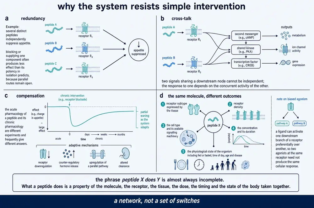
Redundancy is built into the architecture at the level of the gene. A single precursor in the nematode C. elegans encodes seven copies of the same peptide; three copies restore normal swimming and two restore normal crawling, so the remaining copies are spare capacity (Golinelli et al., 2026 — a bioRxiv preprint, not peer-reviewed). The same multi-copy design appears in mammalian proenkephalin and prodynorphin. Redundancy is also built in at the level of the ligand set: the same organism encodes forty insulin-like peptides converging on a single receptor, some agonist and some antagonist, and no single deletion reproduces the receptor mutant (Leptich et al., 2026). Replacing one of them with a paralogue at the same genomic locus rescued a learning defect — the address in the genome mattered more than the peptide's own sequence.
Those are worm experiments and are reported here as worm experiments. What they establish is that the design principle exists and is ancient, not that human physiology works identically.
In mammals, redundancy shows up as knockouts that under-deliver and as phenotypes that turn out to belong to a network rather than to a gene. Deleting myostatin produces the famous double-muscled cattle — and also denser bones, less fat, better insulin sensitivity, difficult births, smaller litters and delayed egg-laying, because myostatin shares up to ninety per cent of its mature sequence with GDF11 and signals through the same receptors (Chen et al., 2026). Removing it does not release one brake; it rebalances a network.
And the physiological state of the animal can abolish a genetic result entirely. Mice lacking a particular signalling kinase in fat were protected from diet-induced metabolic disease at normal room temperature and at six degrees — and the protection vanished when they were housed at thermoneutrality (Han et al., 2021). The "genetic" effect was a thermogenesis effect wearing a genetic costume.
The same lesson holds for interventions in people. A three-hour GLP-1 infusion reduced glomerular filtration by about six per cent in obese, insulin-resistant men and did nothing measurable in healthy men — same peptide, same dose, same duration, different body (Ryan & Acosta, 2015, reviewing human studies). This is the practical form of Section 16's conclusion: what a peptide does depends on the receiver, and physiological state is part of the receiver.
24A message that cannot be stopped is not a message
Every previous Part has been about getting a signal made, sent and heard. This one begins with the opposite requirement, which turns out to be equally engineered: the signal has to end.
Four mechanisms do the work, and they operate on different timescales and in different places.
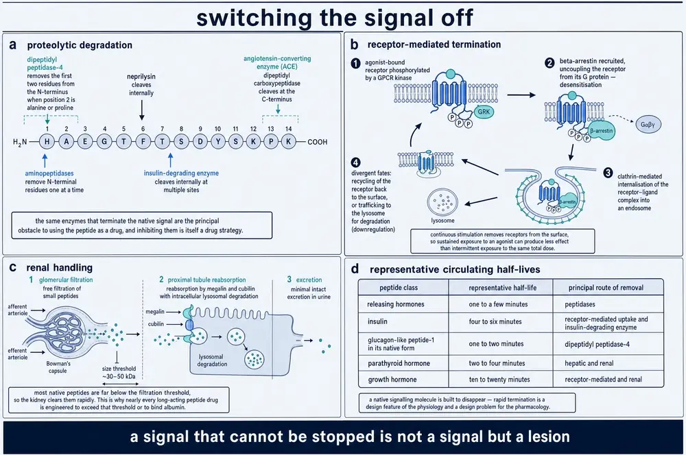
Proteolytic degradation is the fastest and the most specific. The best-characterised example is dipeptidyl peptidase 4, an enzyme that removes exactly two amino acids from a peptide's front end when the second residue is alanine or proline. In GLP-1 that residue is alanine, so GLP-1 is a substrate — and because an intact N-terminus is required for GLP-1 to bind its receptor, this single clip both degrades the peptide and terminates the signal in one event (Tschöp et al., 2023). It is worth appreciating how economical that is. The enzyme does not need to destroy the molecule. It needs to remove two residues.
The identification came in 1993, when Mentlein, Gallwitz and Schmidt showed that DPP-4 hydrolyses both GIP and GLP-1(7–36)amide (Mentlein et al., 1993). Degradation is essentially complete on a single pass through some vascular beds: perfusing isolated mouse hearts with intact GLP-1 and analysing the effluent by mass spectrometry recovered almost entirely the cleaved product (Li et al., 2017, reviewing mouse ex vivo work).
And the enzyme is not dedicated to one hormone. Its substrates include GIP, substance P, B-type natriuretic peptide and the chemokine CXCL12, whose N-terminal removal abolishes its receptor affinity outright (Li et al., 2017; Li et al., 2026b). A single termination enzyme sits across several signalling systems at once, which is the first hint of why interfering with it has consequences beyond the intended one.
Renal clearance works by a different principle: size. Peptides are small enough to pass the glomerular filter, and the kidney is also where DPP-4 is most abundant, localising to the glomeruli and to the brush border of the proximal tubule, shown in mouse kidney (Li et al., 2026b) — so filtration and degradation are co-located.
Receptor-mediated uptake terminates a signal by removing the receptor along with the ligand: the complex is internalised, and whether the receptor then continues signalling from inside the cell, is recycled, or is destroyed, sets how long the response persists. Section 16 recorded that the rules here are actively contested. It is also not universal — the mammalian GnRH receptor lacks the C-terminal tail that internalisation requires and cannot internalise at all, so its signal is terminated by the ligand disappearing and by nothing else (Kayo et al., 2025). For a system whose whole message is carried in pulse timing, that is a striking piece of design: the receptor has been stripped of the one mechanism that would blur a pulse.
Two of these routes are frequently confused, and the natriuretic peptides separate them cleanly. Cutting one of these peptides outside its disulphide-bonded ring leaves its activity intact; cutting inside the ring abolishes it (Selezneva et al., 2025, in vitro). Proteolysis is not the same thing as termination. Where the enzyme cuts decides which of the two has happened.
And dilution is a mechanism in its own right, particularly for paracrine signals: a peptide that diffuses away from a narrow interstitial space into a larger volume has been switched off by geometry alone, without any enzyme acting.
25Why a short life is a feature
Native GLP-1 survives one to two minutes in human blood. From an engineering standpoint that looks like a catastrophic design flaw, and for most of the history of peptide pharmacology it was treated as one.
It is not a flaw. It is the point.
Consider what Section 19 established. The reproductive axis reads GnRH pulse frequency, and it can only do that if each pulse ends cleanly before the next begins. Luteinising hormone's half-life of roughly thirteen to forty minutes is what sets the ceiling on how fast the axis can be driven before the troughs fill in and the message degrades into a continuous one. The degradation rate is not an obstacle to the signalling system. It is one of the system's parameters.
The same logic runs through everything earlier in this document. A gut that reports on each meal must stop reporting between meals. A stress response that persists after the stressor is a disease rather than a response. A paracrine signal that did not decay within a few cell diameters would not be paracrine at all — it would be an endocrine signal with a small source. Precision in time and precision in space are both purchased with the same currency: destruction.
One caution against reading destruction as annihilation. Section 17 recorded that GLP-1(9–36) — what DPP-4 leaves behind, and the predominant circulating form of the peptide — suppresses glucagon nearly as strongly as the intact hormone, through a different intracellular route (Gandasi et al., 2024). Section 09 met the same principle from the other end, in a discarded propeptide fragment with its own receptor. The tidy account in which a peptide is made, acts, and is destroyed is a simplification, and the untidiness is where a good deal of current research sits.
26Writing in a language you did not invent
Everything to this point has been physiology. This section is about what medicine does with it, and the organising claim of the whole monograph can now be stated plainly: therapeutic peptides work because the body already runs on peptides. A peptide drug is not a foreign agent introduced into a system. It is a sentence written in the system's own language — usually an ungrammatical one, deliberately so.
There are five general strategies. Each is a way of exploiting one of the properties this document has described, and each is illustrated below rather than being allowed to become the subject.
One: imitate. Build something the receptor accepts as the native ligand. This is the simplest strategy and it fails for the reason Section 25 celebrated — a faithful copy of a native peptide inherits its two-minute lifetime.
Two: extend. Make the copy survive. Three approaches recur, and all three trade against potency. Substituting the residue that a degrading enzyme recognises removes the enzyme's grip. Attaching a fatty acid lets the molecule bind albumin and travel as cargo. Attaching a polymer or a protein domain makes it too large to filter at the kidney.
The quantitative shape of that trade is worth seeing, because it is unusually clean. Fatty-acid chain length behaves like a dial: reported half-lives run from about 0.8 hours with a ten-carbon chain to roughly sixteen hours at sixteen carbons and twenty-one hours at eighteen (Mu et al., 2026, reviewing engineered constructs). That single parameter is the difference between a daily and a weekly injection. But a five-kilodalton polymer attached to GLP-1 bought four hours of protease resistance in place of ten minutes and cost a twenty-one-fold loss of potency at stimulating cyclic AMP (Selis et al., 2012, in vitro and in rats). And the relationship is not always monotonic: adding an eight- or twelve-carbon chain to GLP-2 improved passage across an intestinal cell layer roughly 1.5-fold, while a sixteen-carbon chain made it slightly worse, because the peptide bound the membrane well enough to become stuck in it (Trier et al., 2014, in cell monolayers). There is an optimum, and it has to be found rather than assumed.
Three: redirect. Engage the receptor differently from the native ligand — favouring one downstream pathway over another, or altering how quickly the receptor is internalised. Section 16 recorded that the rules governing this are contested, and that no head-to-head human trial of oppositely biased agonists at the receptor in question has been done (Tran et al., 2026). This strategy is real and its design principles are not settled.
Four: combine. Build one molecule that engages several receptors at once, exploiting the natural cross-talk and redundancy of Section 23 rather than fighting it.
Five: block. Occupy a receptor without activating it, or inhibit the enzyme that terminates a signal so the native peptide persists longer. Blocking is where Section 19's lesson pays off most surprisingly: continuous delivery of an agonist can silence an axis as effectively as an antagonist, because the receiving system reads pattern. The 1978 result that looked like a failed treatment became a mechanism of action.
27What makes a good signal makes a poor drug
The monograph closes by reading its own argument backwards.
Section 02 proposed that peptides occupy a design niche: large enough to be unambiguous, small enough to be made on demand, fragile enough to be switched off. Every one of those virtues is a difficulty for a pharmacologist.
Large enough to be specific means large enough to be digested. A peptide taken by mouth meets the same proteases that handle dietary protein; peptide antibiotics given orally typically survive under thirty minutes (Ngashangva et al., 2025). It also means large enough to be excluded from the brain by the blood–brain barrier.
Made on demand means the body controls the timing and the drug usually cannot. A peptide delivered by injection arrives as a step function where physiology produced a pulse train — and Section 19 established that the pattern is part of the message.
Fragile enough to be switched off means a two-minute half-life, which is the single largest obstacle in the field and the reason strategy two exists at all.
And the system's redundancy, which makes physiology robust, makes pharmacology frustrating in exactly the same measure. Section 23's myostatin example is also a drug-development story: the pathway proved harder to exploit than expected partly because removing one element rebalances a network, and partly because of a species gap — circulating myostatin runs at 5–10 ng/mL in human serum against over 100 ng/mL in mice (Wetzlich et al., 2025). The animal in which the mechanism was demonstrated is not the animal in which the drug must work.
Which returns the reader to where Section 01 began. Bayliss and Starling cut the nerves to a pancreas and found that the instruction arrived anyway. What they had discovered was not a substance but a medium — and the century that followed was spent learning that the medium has a grammar: that the same word means different things in different rooms, that the timing carries meaning independently of the word, and that a sentence has to end in order to have been a sentence.
Medicine writes in that language now. It writes clumsily, in the main, because it can imitate a word far more easily than it can imitate a rhythm. The peptides that work best as drugs are mostly those whose native message was already close to a step change rather than a pattern; the ones that have resisted a century of effort are frequently those whose meaning is carried in the timing. That is not a coincidence, and it is the most useful thing this document has to say about why some therapeutic peptides succeed and others do not.
28Glossary
Terms are defined as this document uses them. Where a definition is conventional rather than natural — as with the peptide/protein boundary — that is said.
| Term | As used here |
|---|---|
| Amidation | Conversion of a peptide's C-terminal carboxyl group to an amide. Protects the end from degradation and is frequently required for receptor binding. |
| Autocrine | Signalling in which a cell responds to a messenger it released itself. |
| Biased agonism | The property of a ligand that preferentially engages one of a receptor's several downstream pathways rather than all of them equally. |
| Carboxypeptidase E | The enzyme that trims the basic residues left behind after a convertase cuts a prohormone. |
| Constitutive secretion | Continuous release, at a rate set by the rate of synthesis. Cannot be pulsatile. |
| Convertase (proprotein / prohormone) | An enzyme — PC1/3, PC2, furin and relatives — that cuts a precursor at pairs or runs of the basic residues lysine and arginine. |
| Dense-core vesicle | The storage granule of the regulated pathway, dark under electron microscopy because its contents are concentrated. Not recycled at a nerve terminal. |
| Endocrine | Signalling by release into the bloodstream, and therefore to the whole body. |
| Enteroendocrine cell | A hormone-secreting cell in the intestinal lining; about one per cent of that epithelium. |
| GPCR, class B1 | The receptor family most peptide hormones act on: seven membrane-spanning helices with a large extracellular domain above them. |
| Half-life | The time for a concentration to fall by half. This document distinguishes plasma half-life, terminal half-life and duration of receptor occupancy, which are routinely conflated. |
| Incretin effect | The excess of insulin released after oral glucose over that released by intravenous glucose at matched blood concentration. |
| Intracrine | Signalling in which the messenger acts inside the cell that made it, without being released. |
| Juxtacrine | Signalling that requires the two cells to be in physical contact. |
| Neuropeptide | A peptide messenger released by a neuron. Distinguished from a classical transmitter by being made only in the cell body and not recycled at the terminal. |
| Neuropod | A cytoplasmic process by which some enteroendocrine cells form a true synapse with a nerve fibre. |
| Paracrine | Signalling to neighbouring cells through the fluid between them, over roughly a few cell diameters. |
| Peptide | A chain of amino acids linked by peptide bonds. The upper boundary against "protein" is conventional, commonly placed near fifty residues, and has no natural basis. |
| Preprohormone | The initial translation product: signal peptide plus prohormone. |
| Prohormone | The precursor remaining after the signal peptide is removed, before convertase cleavage. |
| Pulsatility | Release in discrete bursts rather than continuously. Where pulse frequency itself carries information, the pattern is part of the message. |
| Regulated secretion | Release from stored granules only on a stimulus, so output is decoupled from synthesis. The precondition for pulsatility. |
| Second messenger | An intracellular molecule — cyclic AMP, calcium, IP₃ — generated in response to receptor activation, which carries and multiplies the signal inside the cell. |
| Signal peptide | An N-terminal stretch of roughly twenty residues, heavily biased toward leucine, that routes a nascent chain into the secretory pathway and is then cleaved off. |
| Signal recognition particle | The RNA-protein complex that recognises a signal peptide as it emerges from the ribosome and delivers the assembly to the endoplasmic reticulum. |
| Volume transmission | Signalling in which a messenger diffuses through tissue to receptors distant from its release site, as against the "wired" transmission of a synapse. |
29Abbreviations
| Abbreviation | Expansion |
|---|---|
| ACE | angiotensin-converting enzyme |
| ACTH | adrenocorticotropic hormone |
| ANP · BNP · CNP | atrial, B-type and C-type natriuretic peptide |
| cAMP | cyclic adenosine monophosphate |
| CCK | cholecystokinin |
| CGRP | calcitonin gene-related peptide |
| CRH | corticotropin-releasing hormone |
| CPE | carboxypeptidase E |
| DPP-4 | dipeptidyl peptidase 4 |
| GH | growth hormone |
| GIP | glucose-dependent insulinotropic polypeptide |
| GLP-1 · GLP-2 | glucagon-like peptide 1 and 2 |
| GnRH | gonadotropin-releasing hormone |
| GPCR | G-protein-coupled receptor |
| HPA · HPG · HPT | hypothalamic–pituitary–adrenal, –gonadal, –thyroid axis |
| IP₃ | inositol trisphosphate |
| JATS | Journal Article Tag Suite, the XML format the corpus is stored in |
| KNDy | kisspeptin / neurokinin B / dynorphin neurons |
| LH · FSH | luteinising hormone, follicle-stimulating hormone |
| MeSH | Medical Subject Headings, the NLM indexing vocabulary |
| NPY | neuropeptide Y |
| PAM | peptidylglycine α-amidating monooxygenase |
| PC1/3 · PC2 | prohormone convertase 1/3 and 2 |
| PMC · PMCID | PubMed Central, and its article identifier |
| PMID | PubMed identifier |
| POMC | pro-opiomelanocortin |
| PYY | peptide YY |
| RAMP | receptor activity-modifying protein |
| RUO | research use only |
| SRP | signal recognition particle |
| TRH | thyrotropin-releasing hormone |
30References
Generated from verified NCBI records rather than from recall. Author lists, journal names, volumes, pages and identifiers are taken from the PubMed record for each citation, and identifiers are read only from each record's own identifier list — a walk over every identifier node in a PubMed record also traverses its reference list and returns the identifiers of the last work that paper cited, which is how a monograph in this series once fetched the wrong articles under the right headers. Entries marked as carrying no PubMed identifier are genuinely unindexed sources, listed in full and recorded in the unresolved-evidence log; no identifier has been supplied for them.
- Abramian A, Hoogstraaten RI, Murphy FH, McDaniel KF, Toonen RF, Verhage M. Rabphilin-3A negatively regulates neuropeptide release, through its SNAP25 interaction. Elife. 2024;13.
PMID 39412498 · doi:10.7554/eLife.95371 · PMC11483123 - Antal M. Molecular Anatomy of Synaptic and Extrasynaptic Neurotransmission Between Nociceptive Primary Afferents and Spinal Dorsal Horn Neurons. Int J Mol Sci. 2025;26(5).
PMID 40076973 · doi:10.3390/ijms26052356 · PMC11900602 - Austin G, Tomas A. Signaling architecture of the glucagon-like peptide-1 receptor. J Clin Invest. 2026;136(2).
PMID 41542774 · doi:10.1172/JCI194752 · PMC12807469 - Baginska U, Balagura G, Toonen RF, Verhage M. High-throughput assay for regulated secretion of neuropeptides in mouse and human neurons. J Biol Chem. 2024;300(6):107321.
PMID 38677517 · doi:10.1016/j.jbc.2024.107321 · PMC11170154 - Banting FG, Best CH, Collip JB, Campbell WR, Fletcher AA. Pancreatic Extracts in the Treatment of Diabetes Mellitus. Can Med Assoc J. 1922;12(3):141-6.
PMID 20314060 · PMC1524425 - Bayliss WM, Starling EH. The mechanism of pancreatic secretion. J Physiol. 1902;28(5):325-53.
PMID 16992627 · doi:10.1113/jphysiol.1902.sp000920 · PMC1540572 - Belchetz PE, Plant TM, Nakai Y, Keogh EJ, Knobil E. Hypophysial responses to continuous and intermittent delivery of hypopthalamic gonadotropin-releasing hormone. Science. 1978;202(4368):631-3.
PMID 100883 · doi:10.1126/science.100883 - Bell GI, Santerre RF, Mullenbach GT. Hamster preproglucagon contains the sequence of glucagon and two related peptides. Nature. 1983;302(5910):716-8.
PMID 6835407 · doi:10.1038/302716a0 - Bizoń A, Borkowska J, Franik G, Piwowar A. Associations of Serum GIP, GLP-1, and DPP-4 with Metabolic and Hormonal Profiles and Tobacco Exposure in Women with Polycystic Ovary Syndrome. Int J Mol Sci. 2025;26(15).
PMID 40806228 · doi:10.3390/ijms26157097 · PMC12346818 - Blobel G, Dobberstein B. Transfer of proteins across membranes. I. Presence of proteolytically processed and unprocessed nascent immunoglobulin light chains on membrane-bound ribosomes of murine myeloma. J Cell Biol. 1975;67(3):835-51.
PMID 811671 · doi:10.1083/jcb.67.3.835 · PMC2111658 - Blythe EE, von Zastrow M. β-Arrestin-independent endosomal cAMP signaling by a polypeptide hormone GPCR. Nat Chem Biol. 2024;20(3):323-332.
PMID 37749347 · doi:10.1038/s41589-023-01412-4 · PMC10907292 - Brazeau P, Vale W, Burgus R, Ling N, Butcher M, Rivier J, et al.. Hypothalamic polypeptide that inhibits the secretion of immunoreactive pituitary growth hormone. Science. 1973;179(4068):77-9.
PMID 4682131 · doi:10.1126/science.179.4068.77 - Bulliard M, Pinjusic K, Iacobucci L, Schmuziger C, Fournier N, Constam DB. Kallikrein-8 mediates furin-independent Activin-A precursor processing to stimulate tumor growth in melanoma. Nat Commun. 2025;16(1):2354.
PMID 40064965 · doi:10.1038/s41467-025-57661-5 · PMC11893775 - Burgus R, Dunn TF, Desiderio D, Ward DN, Vale W, Guillemin R. Characterization of ovine hypothalamic hypophysiotropic TSH-releasing factor. Nature. 1970;226(5243):321-5.
PMID 4985794 · doi:10.1038/226321a0 - Chen Y, Yang R, Yang Y, Wang Q, Yang K, Xu M. Harnessing myostatin pleiotropy for multitrait improvement via precision gene editing. Front Genome Ed. 2026;8:1749445.
PMID 41907069 · doi:10.3389/fgeed.2026.1749445 · PMC13018111 - Coppola I, Brouwers B, Walker L, Alar C, Meulemans S, White A, et al.. Loss of hypothalamic Furin affects POMC to proACTH cleavage and feeding behavior in high-fat diet-fed mice. Mol Metab. 2022;66:101627.
PMID 36374777 · doi:10.1016/j.molmet.2022.101627 · PMC9664468 - de Bold AJ, Borenstein HB, Veress AT, Sonnenberg H. A rapid and potent natriuretic response to intravenous injection of atrial myocardial extract in rats. Life Sci. 1981;28(1):89-94.
PMID 7219045 · doi:10.1016/0024-3205(81)90370-2 - Dong C, Gowrishankar R, Jin Y, He XJ, Gupta A, Wang H, et al.. Unlocking opioid neuropeptide dynamics with genetically encoded biosensors. Nat Neurosci. 2024;27(9):1844-1857.
PMID 39009835 · doi:10.1038/s41593-024-01697-1 · PMC11374718 - DU VIGNEAUD V, RESSLER C, TRIPPETT S. The sequence of amino acids in oxytocin, with a proposal for the structure of oxytocin. J Biol Chem. 1953;205(2):949-57.
PMID 13129273 - du Vigneaud V, Ressler C, Swan JM, Roberts CW, Katsoyannis PG, Gordon S. The synthesis of an octapeptide amide with the hormonal activity of oxytocin. J Am Chem Soc. 1953;75(19):4879-4880.
no PubMed identifier — see the unresolved-evidence log - Duan Y, Chen S, Cai W, Song W, Li H. Signal peptides restrict genome evolution and A-to-I RNA editing. NAR Genom Bioinform. 2025;7(3):lqaf096.
PMID 40657422 · doi:10.1093/nargab/lqaf096 · PMC12246783 - Duus CL, Nielsen SF, Hornstrup BG, Mose FH, Bech JN. Dietary Sodium Reduction Reveals Aldosterone Dysregulation in Patients With Essential Hypertension. J Clin Hypertens (Greenwich). 2026;28(3):e70229.
PMID 41761767 · doi:10.1111/jch.70229 · PMC12949390 - Edwards CM, Johnson RW. From Good to Bad: The Opposing Effects of PTHrP on Tumor Growth, Dormancy, and Metastasis Throughout Cancer Progression. Front Oncol. 2021;11:644303.
PMID 33828987 · doi:10.3389/fonc.2021.644303 · PMC8019909 - Essandoh K, Subramani A, Koripella S, Brody MJ. The Rab3 GTPase cycle modulates cardiomyocyte exocytosis and atrial natriuretic peptide release. Biophys J. 2025;124(11):1856-1866.
PMID 40119520 · doi:10.1016/j.bpj.2025.03.013 · PMC12256856 - Gandasi NR, Gao R, Kothegala L, Pearce A, Santos C, Acreman S, et al.. GLP-1 metabolite GLP-1(9-36) is a systemic inhibitor of mouse and human pancreatic islet glucagon secretion. Diabetologia. 2024;67(3):528-546.
PMID 38127123 · doi:10.1007/s00125-023-06060-w · PMC10844371 - Gilmore R, Blobel G, Walter P. Protein translocation across the endoplasmic reticulum. I. Detection in the microsomal membrane of a receptor for the signal recognition particle. J Cell Biol. 1982;95(2 Pt 1):463-9.
PMID 6292235 · doi:10.1083/jcb.95.2.463 · PMC2112970 - Glendining KA, Prescott M, Potapov K, Kip E, Campbell RE. Arcuate nucleus-specific progesterone receptor knockdown in female mice is sufficient to induce PCOS-like hyperactivity in the reproductive axis. J Neuroendocrinol. 2026;38(4):e70181.
PMID 41968288 · doi:10.1111/jne.70181 · PMC13071148 - Golinelli L, Beets I, Temmerman L. Autocrine feedback maintains homeostatic neuropeptide expression in a peptidergic hub neuron. bioRxiv. 2026.
PMID 41542528 · doi:10.64898/2026.01.09.698615 · PMC12803089 - Goodman RL, Herbison AE, Lehman MN, Navarro VM. Neuroendocrine control of gonadotropin-releasing hormone: Pulsatile and surge modes of secretion. J Neuroendocrinol. 2022;34(5):e13094.
PMID 35107859 · doi:10.1111/jne.13094 · PMC9948945 - Han MS, Perry RJ, Camporez JP, Scherer PE, Shulman GI, Gao G, et al.. A feed-forward regulatory loop in adipose tissue promotes signaling by the hepatokine FGF21. Genes Dev. 2021;35(1-2):133-146.
PMID 33334822 · doi:10.1101/gad.344556.120 · PMC7778269 - Han S, Jung M, Ryu S, Cha S, Tak H, Jeong SM, et al.. RNA binding protein HuD regulates the biosynthesis of glucagon-like peptide 1 in intestinal L-cells. Biochem Biophys Rep. 2026;45:102415.
PMID 41496908 · doi:10.1016/j.bbrep.2025.102415 · PMC12767712 - Hart RG, Lee JJ, Zhai K, Lee S, Chauhan R, Hosseini A, et al.. Somatostatin receptors shape insulin and glucagon output within the pancreatic islet in mice through direct and paracrine effects. Diabetologia. 2026;69(9):2569-2584.
PMID 42322376 · doi:10.1007/s00125-026-06769-4 · PMC13423930 - Hartig SM, Cox AR. Paracrine signaling in islet function and survival. J Mol Med (Berl). 2020;98(4):451-467.
PMID 32067063 · doi:10.1007/s00109-020-01887-x · PMC7899133 - He X, Zhang Y, Lou J, Wu J, Xu S, Zhu G, et al.. LL-37 selectively targets Plasmodium-infected erythrocytes and exhibits antimalarial activity. PLoS Pathog. 2026;22(3):e1014062.
PMID 41843625 · doi:10.1371/journal.ppat.1014062 · PMC13004495 - Heifetz A, Barker O, Morris GB, Law RJ, Slack M, Biggin PC. Toward an understanding of agonist binding to human Orexin-1 and Orexin-2 receptors with G-protein-coupled receptor modeling and site-directed mutagenesis. Biochemistry. 2013;52(46):8246-60.
PMID 24144388 · doi:10.1021/bi401119m · PMC3880013 - Irwin DM. Variation in the Evolution and Sequences of Proglucagon and the Receptors for Proglucagon-Derived Peptides in Mammals. Front Endocrinol (Lausanne). 2021;12:700066.
PMID 34322093 · doi:10.3389/fendo.2021.700066 · PMC8312260 - Jackson RS, Creemers JW, Ohagi S, Raffin-Sanson ML, Sanders L, Montague CT, et al.. Obesity and impaired prohormone processing associated with mutations in the human prohormone convertase 1 gene. Nat Genet. 1997;16(3):303-6.
PMID 9207799 · doi:10.1038/ng0797-303 - Jackson RS, Creemers JW, Farooqi IS, Raffin-Sanson ML, Varro A, Dockray GJ, et al.. Small-intestinal dysfunction accompanies the complex endocrinopathy of human proprotein convertase 1 deficiency. J Clin Invest. 2003;112(10):1550-60.
PMID 14617756 · doi:10.1172/JCI18784 · PMC259128 - Kang F, Xie L, Qin T, Miao Y, Kang Y, Takahashi T, et al.. Plasma membrane flipping of Syntaxin-2 regulates its inhibitory action on insulin granule exocytosis. Nat Commun. 2022;13(1):6512.
PMID 36316316 · doi:10.1038/s41467-022-33986-3 · PMC9622911 - Kayo D, Uehara SK, Royan MR, Kanda S. Emerging Perspectives on Gonadotropin Regulation in Vertebrates Revealed by the Discovery of FSH-RH in Teleosts. Bioessays. 2025;47(11):e70066.
PMID 40931568 · doi:10.1002/bies.70066 · PMC12550547 - Knobil E, Plant TM, Wildt L, Belchetz PE, Marshall G. Control of the rhesus monkey menstrual cycle: permissive role of hypothalamic gonadotropin-releasing hormone. Science. 1980;207(4437):1371-3.
PMID 6766566 · doi:10.1126/science.6766566 - Lajoye Q, Orieux A, Boyer A, Prevel R, Jozwiak M. Vasopressin and its analogues in patients with septic shock: holy Grail or unfulfilled promise?. Crit Care. 2025;29(1):333.
PMID 40731360 · doi:10.1186/s13054-025-05540-2 · PMC12306101 - Lee SJ. Myostatin: A Skeletal Muscle Chalone. Annu Rev Physiol. 2023;85:269-291.
PMID 36266260 · doi:10.1146/annurev-physiol-012422-112116 · PMC10163667 - Leptich EJ, Vijayakumar P, Pietryk EW, Williams MI, Rajupalem R, Arey RN. INS-17 acts as a nutrient deprivation signal to mediate adult IIS-regulated associative behaviors in C. elegans. PLoS Genet. 2026;22(4):e1012130.
PMID 42048395 · doi:10.1371/journal.pgen.1012130 · PMC13143180 - Li J, Zheng J, Wang S, Lau HK, Fathi A, Wang Q. Cardiovascular Benefits of Native GLP-1 and its Metabolites: An Indicator for GLP-1-Therapy Strategies. Front Physiol. 2017;8:15.
PMID 28194113 · doi:10.3389/fphys.2017.00015 · PMC5276855 - Li X, Zhang M, Xu Z, Li Y, Bian S, Tang Y, et al.. Serine protease HtrA promotes Campylobacter jejuni intestinal colonization through degrading antimicrobial peptide LL-37. Sci Adv. 2026;12(21):eaee1996.
PMID 42160414 · doi:10.1126/sciadv.aee1996 · PMC13189099 - Li Y, Lu B, Zhao Z, Gong M, Lyu D, Zhang H, et al.. Integrative pan-cancer analysis of dipeptidyl peptidase 4 with clinical and in vitro validation in prostate cancer. Front Immunol. 2026;17:1616889.
PMID 41909664 · doi:10.3389/fimmu.2026.1616889 · PMC13017899 - Lim A, Rechtsteiner A, Saxton WM. Two kinesins drive anterograde neuropeptide transport. Mol Biol Cell. 2017;28(24):3542-3553.
PMID 28904207 · doi:10.1091/mbc.E16-12-0820 · PMC5683764 - Ling N, Burgus R, Rivier J, Vale W, Brazeau P. The use of mass spectrometry in deducing the sequence of somatostatin--a hypothalamic polypeptide that inhibits the secretion of growth hormone. Biochem Biophys Res Commun. 1973;50(1):127-33.
PMID 4734245 · doi:10.1016/0006-291x(73)91073-5 - Liu CM, Spaulding MO, Rea JJ, Noble EE, Kanoski SE. Oxytocin and Food Intake Control: Neural, Behavioral, and Signaling Mechanisms. Int J Mol Sci. 2021;22(19).
PMID 34639199 · doi:10.3390/ijms221910859 · PMC8509519 - Liu H, Xiao H, Lin S, Zhou H, Cheng Y, Xie B, et al.. Effect of gut hormones on bone metabolism and their possible mechanisms in the treatment of osteoporosis. Front Pharmacol. 2024;15:1372399.
PMID 38725663 · doi:10.3389/fphar.2024.1372399 · PMC11079205 - Malsawmzuali JC, Adel FW, Pawlina W, Kattil P. Beyond the pump: integrating the heart's endocrine function into early medical education. Med Educ Online. 2026;31(1):2704285.
PMID 42455767 · doi:10.1080/10872981.2026.2704285 · PMC13374749 - Mazella J, Pétrault O, Lucas G, Deval E, Béraud-Dufour S, Gandin C, et al.. Spadin, a sortilin-derived peptide, targeting rodent TREK-1 channels: a new concept in the antidepressant drug design. PLoS Biol. 2010;8(4):e1000355.
PMID 20405001 · doi:10.1371/journal.pbio.1000355 · PMC2854129 - Mentlein R, Gallwitz B, Schmidt WE. Dipeptidyl-peptidase IV hydrolyses gastric inhibitory polypeptide, glucagon-like peptide-1(7-36)amide, peptide histidine methionine and is responsible for their degradation in human serum. Eur J Biochem. 1993;214(3):829-35.
PMID 8100523 · doi:10.1111/j.1432-1033.1993.tb17986.x - Merighi A, Sbriz M, Lossi L. Chemical multiplexing in the nervous system: molecular architecture, functional stratification, and pathophysiological plasticity of neuropeptide-classical neurotransmitter cotransmission. Front Mol Neurosci. 2026;19:1839947.
PMID 42440641 · doi:10.3389/fnmol.2026.1839947 · PMC13333683 - Merrifield B. Solid phase synthesis. Nobel lecture, 8 December 1984. Biosci Rep. 1985;5(5):353-76.
PMID 4027355 · doi:10.1007/BF01116553 - Merrifield RB. Solid phase peptide synthesis. I. The synthesis of a tetrapeptide. J Am Chem Soc. 1963;85(14):2149-2154.
no PubMed identifier — see the unresolved-evidence log - Mingardi J, Meanti R, Paoli C, Cifani C, Torsello A, Popoli M, et al.. Ghrelin, Neuroinflammation, Oxidative Stress, and Mood Disorders: What Are the Connections?. Curr Neuropharmacol. 2025;23(2):172-186.
PMID 39041263 · doi:10.2174/1570159X22999240722095039 · PMC11793048 - Miyagawa T, Tanaka S, Shimada M, Sakai N, Tanida K, Kotorii N, et al.. A rare genetic variant in the cleavage site of prepro-orexin is associated with idiopathic hypersomnia. NPJ Genom Med. 2022;7(1):29.
PMID 35414074 · doi:10.1038/s41525-022-00298-w · PMC9005711 - Moenter SM, Evans NP. Gonadotropin-releasing hormone (GnRH) measurements in pituitary portal blood: A history. J Neuroendocrinol. 2022;34(5):e13065.
PMID 34918405 · doi:10.1111/jne.13065 · PMC9200367 - Momiji H, Hassall KL, Featherstone K, McNamara AV, Patist AL, Spiller DG, et al.. Disentangling juxtacrine from paracrine signalling in dynamic tissue. PLoS Comput Biol. 2019;15(6):e1007030.
PMID 31194728 · doi:10.1371/journal.pcbi.1007030 · PMC6592563 - Moresco P, Kastan JP, Yang JI, Prabakar R, Kelley ZL, Minicozzi F, et al.. Signal peptide-independent secretion of keratin-19 by pancreatic cancer cells. Proc Natl Acad Sci U S A. 2025;122(27):e2426218122.
PMID 40591600 · doi:10.1073/pnas.2426218122 · PMC12260553 - Morrow NM, Hanson AA, Mulvihill EE. Distinct Identity of GLP-1R, GLP-2R, and GIPR Expressing Cells and Signaling Circuits Within the Gastrointestinal Tract. Front Cell Dev Biol. 2021;9:703966.
PMID 34660576 · doi:10.3389/fcell.2021.703966 · PMC8511495 - Mu J, Vong E, Carmali S. Artificial lipidation of proteins and peptides: from mechanism to clinical applications. FEBS J. 2026;293(5):1269-1284.
PMID 41129276 · doi:10.1111/febs.70298 · PMC12958104 - Naggert JK, Fricker LD, Varlamov O, Nishina PM, Rouille Y, Steiner DF, et al.. Hyperproinsulinaemia in obese fat/fat mice associated with a carboxypeptidase E mutation which reduces enzyme activity. Nat Genet. 1995;10(2):135-42.
PMID 7663508 · doi:10.1038/ng0695-135 - Nakanishi S, Inoue A, Kita T, Nakamura M, Chang AC, Cohen SN, et al.. Nucleotide sequence of cloned cDNA for bovine corticotropin-beta-lipotropin precursor. Nature. 1979;278(5703):423-7.
PMID 221818 · doi:10.1038/278423a0 - Nauck MA, Homberger E, Siegel EG, Allen RC, Eaton RP, Ebert R, et al.. Incretin effects of increasing glucose loads in man calculated from venous insulin and C-peptide responses. J Clin Endocrinol Metab. 1986;63(2):492-8.
PMID 3522621 · doi:10.1210/jcem-63-2-492 - Ngashangva N, Huidrom S, Devi IS. Antimicrobial peptides: natural templates for next-generation therapeutics against antimicrobial resistance. Front Cell Infect Microbiol. 2025;15:1720027.
PMID 41561088 · doi:10.3389/fcimb.2025.1720027 · PMC12813028 - Nwako JG, McCauley HA. Enteroendocrine cells regulate intestinal homeostasis and epithelial function. Mol Cell Endocrinol. 2024;593:112339.
PMID 39111616 · doi:10.1016/j.mce.2024.112339 · PMC11401774 - Oti T, Satoh K, Uta D, Nagafuchi J, Tateishi S, Ueda R, et al.. Oxytocin Influences Male Sexual Activity via Non-synaptic Axonal Release in the Spinal Cord. Curr Biol. 2021;31(1):103-114.e5.
PMID 33125871 · doi:10.1016/j.cub.2020.09.089 · PMC7855431 - Ramirez-Plascencia OD, De Luca R, Machado NLS, Eghlidi D, Khanday MA, Bandaru SS, et al.. A hypothalamic circuit for circadian regulation of corticosterone secretion. Nat Commun. 2026;17(1).
PMID 41946720 · doi:10.1038/s41467-026-71482-0 · PMC13233844 - Ryan D, Acosta A. GLP-1 receptor agonists: Nonglycemic clinical effects in weight loss and beyond. Obesity (Silver Spring). 2015;23(6):1119-29.
PMID 25959380 · doi:10.1002/oby.21107 · PMC4692091 - RYLE AP, SANGER F, SMITH LF, KITAI R. The disulphide bonds of insulin. Biochem J. 1955;60(4):541-56.
PMID 13249947 · doi:10.1042/bj0600541 · PMC1216151 - SANGER F, TUPPY H. The amino-acid sequence in the phenylalanyl chain of insulin. I. The identification of lower peptides from partial hydrolysates. Biochem J. 1951;49(4):463-81.
PMID 14886310 · doi:10.1042/bj0490463 · PMC1197535 - SANGER F, THOMPSON EO. The amino-acid sequence in the glycyl chain of insulin. I. The identification of lower peptides from partial hydrolysates. Biochem J. 1953;53(3):353-66.
PMID 13032078 · doi:10.1042/bj0530353 · PMC1198157 - Schally AV, Nair RM, Redding TW, Arimura A. Isolation of the luteinizing hormone and follicle-stimulating hormone-releasing hormone from porcine hypothalami. J Biol Chem. 1971;246(23):7230-6.
PMID 4942661 - Segers VFM, De Keulenaer GW. Autocrine Signaling in Cardiac Remodeling: A Rich Source of Therapeutic Targets. J Am Heart Assoc. 2021;10(3):e019169.
PMID 33470124 · doi:10.1161/JAHA.120.019169 · PMC7955414 - Selezneva EM, Feygina EE, Ageeva LV, Rozov FN, Kopylova IV, Altshuler EP, et al.. Neprilysin 2 catalyses the degradation of natriuretic peptides despite sacubitrilat Inhibition. Sci Rep. 2025;15(1):27401.
PMID 40721616 · doi:10.1038/s41598-025-10166-z · PMC12304097 - Selis F, Schrepfer R, Sanna R, Scaramuzza S, Tonon G, Dedoni S, et al.. Enzymatic mono-pegylation of glucagon-like peptide 1 towards long lasting treatment of type 2 diabetes. Results Pharma Sci. 2012;2:58-65.
PMID 25755995 · doi:10.1016/j.rinphs.2012.09.001 · PMC4167179 - Sen SK, Khashim Z, Belame SS, Hassoun S, Salomon T, Lewis-Brinkman S, et al.. Circadian regulation of insulin secretion in transplanted human stem cell-derived pancreatic β-cells. Stem Cell Reports. 2025;20(11):102691.
PMID 41173007 · doi:10.1016/j.stemcr.2025.102691 · PMC12790728 - Shah A, Coria AR, Membréno BS, Orgebin E, Miller JT, Guiblet W, et al.. ZAP targets aberrant mRNA transcripts encoding proteins with defective signal peptides for degradation. EMBO J. 2026;45(8):2638-2665.
PMID 41820617 · doi:10.1038/s44318-026-00720-4 · PMC13084044 - Shao L, Chen Y, Zhang S, Zhang Z, Cao Y, Yang D, et al.. Modulating effects of RAMPs on signaling profiles of the glucagon receptor family. Acta Pharm Sin B. 2022;12(2):637-650.
PMID 35256936 · doi:10.1016/j.apsb.2021.07.028 · PMC8897147 - Starling EH. The Croonian Lectures on the chemical correlation of the functions of the body. Lancet. 1905;166(4275):339-341.
no PubMed identifier — see the unresolved-evidence log - Steiner DF, Cunningham D, Spigelman L, Aten B. Insulin biosynthesis: evidence for a precursor. Science. 1967;157(3789):697-700.
PMID 4291105 · doi:10.1126/science.157.3789.697 - Steiner DF, Oyer PE. The biosynthesis of insulin and a probable precursor of insulin by a human islet cell adenoma. Proc Natl Acad Sci U S A. 1967;57(2):473-80.
PMID 16591494 · doi:10.1073/pnas.57.2.473 · PMC335530 - Svensson D, Nilsson BO. Human antimicrobial/host defense peptide LL-37 may prevent the spread of a local infection through multiple mechanisms: an update. Inflamm Res. 2025;74(1):36.
PMID 40063262 · doi:10.1007/s00011-025-02005-8 · PMC11893641 - Tran HD, Zuo Y, Wong C, Pollard A, Bloom S, Jones B. Modelling G protein-biased agonism using GLP-1 receptor C-terminal mutations. Mol Metab. 2026;105:102321.
PMID 41570980 · doi:10.1016/j.molmet.2026.102321 · PMC12925471 - Trier S, Linderoth L, Bjerregaard S, Andresen TL, Rahbek UL. Acylation of Glucagon-like peptide-2: interaction with lipid membranes and in vitro intestinal permeability. PLoS One. 2014;9(10):e109939.
PMID 25295731 · doi:10.1371/journal.pone.0109939 · PMC4190408 - Tschöp M, Nogueiras R, Ahrén B. Gut hormone-based pharmacology: novel formulations and future possibilities for metabolic disease therapy. Diabetologia. 2023;66(10):1796-1808.
PMID 37209227 · doi:10.1007/s00125-023-05929-0 · PMC10474213 - Unal G, Mansouri M, Xie YQ, Mueller C, Charpin-El Hamri G, Fussenegger M. A synthetic angiotensin II/ACE2-based hormone shunt controlling experimental hypertension. Nat Commun. 2026;17(1).
PMID 41965354 · doi:10.1038/s41467-026-71796-z · PMC13247273 - Valdés-Calero I, Frühbeck G, Rodríguez A. The Ghrelin-LEAP2 System in Obesity and Diabetes: Pathophysiological Roles and Therapeutic Potential. Curr Obes Rep. 2026;15(1).
PMID 42277455 · doi:10.1007/s13679-026-00728-1 · PMC13260137 - Walter P, Blobel G. Signal recognition particle contains a 7S RNA essential for protein translocation across the endoplasmic reticulum. Nature. 1982;299(5885):691-8.
PMID 6181418 · doi:10.1038/299691a0 - Wetzlich B, Nyakundi BB, Yang J. Therapeutic applications and challenges in myostatin inhibition for enhanced skeletal muscle mass and functions. Mol Cell Biochem. 2025;480(3):1535-1553.
PMID 39340593 · doi:10.1007/s11010-024-05120-y · PMC11842502 - Yaguchi K, Hagihara M, Konno A, Hirai H, Yukinaga H, Miyamichi K. Dynamic modulation of pulsatile activities of oxytocin neurons in lactating wild-type mice. PLoS One. 2023;18(5):e0285589.
PMID 37163565 · doi:10.1371/journal.pone.0285589 · PMC10171594 - YALOW RS, BERSON SA. Immunoassay of endogenous plasma insulin in man. J Clin Invest. 1960;39(7):1157-75.
PMID 13846364 · doi:10.1172/JCI104130 · PMC441860 - Zasloff M. Magainins, a class of antimicrobial peptides from Xenopus skin: isolation, characterization of two active forms, and partial cDNA sequence of a precursor. Proc Natl Acad Sci U S A. 1987;84(15):5449-53.
PMID 3299384 · doi:10.1073/pnas.84.15.5449 · PMC298875 - Zhao L, Niu J, Lin H, Zhao J, Liu Y, Song Z, et al.. Paracrine-endocrine FGF chimeras as potent therapeutics for metabolic diseases. EBioMedicine. 2019;48:462-477.
PMID 31631034 · doi:10.1016/j.ebiom.2019.09.052 · PMC6838362 - Zhu S, Yuan A, Duffy T, Kim BH, Ozawa T, Dixon SJ, et al.. Extracellular ATP increases agonist potency and reduces latency at class B G protein-coupled receptors. Mol Pharmacol. 2025;107(6):100040.
PMID 40378650 · doi:10.1016/j.molpha.2025.100040 · PMC12264551
31How this document was assembled
The evidence base has three layers, and they are reported as three different numbers because a single figure would imply coverage this document does not have.
Layer A, the local library. Every JATS asset in project 05, the Therapeutic Peptide Research Library, was parsed and scored: 10,204 documents, about 110,631 printed-page equivalents. Scoring was not by keyword frequency but by how many of fourteen concept families — drawn directly from this monograph's coverage requirements — each document engages. A systems monograph is served by documents that connect mechanisms; a paper naming one peptide four hundred times engages one family. 3,016 documents were retained at four or more families and 1,035 entered the reading corpus at six or more.
Layer B, a targeted external harvest. Scoring Layer A revealed that the library is dense where peptide action is and sparse where peptide manufacture is: of the local reading corpus, 672 documents engage gastrointestinal and metabolic signalling but only 146 touch secretory storage, 201 biosynthesis and 205 processing enzymes — and just three engage secretory-granule biology heavily. Those are precisely this document's second and third narrative stages. A therapeutic-compound library is dense where compounds are, and a compound is a receptor ligand rather than a secretory granule. Eight PubMed queries built on MeSH descriptors, plus three further targeted harvests for gaps found during reading, produced an indexed surface of 33,952 records, of which 1,748 were retrieved as verified records and 870 as open-access full text. The concept must appear in the indexed record — title, abstract or MeSH — which is the indexer's judgement that a paper is about the concept rather than merely mentioning it.
Layer C, the verified historical record. Neither literature layer can supply the discovery history: the 1902 secretin experiment has no open-access full text and the library holds almost nothing before 2005. Twenty-three foundational papers were resolved individually against NCBI and each author–journal–volume line was read before acceptance.
Merging Layers A and B keyed by identifier rather than summed — ten documents appear in both and are counted once — gives the corpus this monograph is written from: 1,035 documents, 8,525,825 words, approximately 17,052 printed-page equivalents, together with 98 verified references.
| Stage | What it does | Result |
|---|---|---|
| 01 | Parse and score every project-05 JATS asset on fourteen concept families | 10,204 scanned |
| 02 | Eight MeSH-built axis queries against PubMed | 33,952 surface |
| 02b/02c | Individual verification of the foundational discovery papers | 23 resolved |
| 03 | Open-access full-text retrieval and substantive-use screen | 870 full texts |
| 04 | Per-family reading list, so breadth is forced rather than hoped for | 301 read in full |
| 05 | Keyed merge and inventory | 1,035 unique |
| 05b | Author–year citations resolved from verified records | 98 citations |
| 06 | Assembly, with the build gates below | 1 deliverable |
What was counted and not read. Of the local surface, 5,675 documents scored one to three concept families and were classed peripheral, 1,611 scored none, and 312 carry no readable body text; 788 documents fetched in Layer B fell below the reading threshold. Naming these is what makes the smaller number trustworthy. A corpus figure that silently absorbed them would be a claim about coverage this document has not earned.
Three defects in the build machinery are worth recording, because all three produced output that looked correct. The full-text fetch initially returned nothing at all while reporting clean progress, because PubMed Central labels an article's identifier pmcid where the local store labels the same field pmc; every article was skipped and every log line looked healthy. The historical verification stage returned records for every query and the wrong records for several, because its search sorted newest-first and its result limit truncated away exactly the classic papers it existed to find — the somatostatin query's oldest hit came back as 1984 for a paper published in 1973. And five stages inherited from the shared pipeline carried constants belonging to three different previous compounds. A stage that runs is not a stage that is right.
32Evidence handling
Findings are labelled by the kind of study that produced them, in the sentence that reports them. This matters more in this document than in a compound monograph, because a great deal of what is confidently said about human peptide signalling was measured in another animal. The mechanistic account of GnRH pulsatility in Section 19 is almost entirely ovine, murine and macaque; portal-blood sampling is not performed in people. The redundancy principles in Section 23 come from a nematode. The architecture is believed to be conserved, and this document states that the belief is an inference.
Recency is weighted but not blindly. Where a recent finding revises an older account it leads, and the older account is named as what it revised. Where a single recent study opposes a long-replicated older one, the conflict is presented as a conflict. The corpus read here is heavily weighted to 2024–2026, which reports current interpretation well and settled foundations poorly; where a foundational claim rests on a modern review rather than the primary work, the text says so.
Three kinds of absence are reported rather than written around. Where a number does not exist in this evidence base, no number is printed. The amplification figure in Section 15 carries no gain values because no paper read for this monograph reports one; the gradient figure in Section 13 carries no absolute concentration because no paper measured an interstitial peptide concentration in intact tissue. Where two sources disagree, both are given — the neuropeptide diffusion range differs by an order of magnitude between two reviews and neither figure is a measurement. And where a widely repeated claim is not supported, it is named: the description of the gut as the body's largest endocrine organ is asserted, downgraded and contradicted by three papers in this corpus, and this document declines it in favour of a narrower statement it can defend.
The evidence base is open-access biased. Both literature layers are built from PubMed Central, so work in journals that deposit no open-access full text is reachable only through its abstract. No claim in this document should be read as implying that the closed literature was searched.
The twenty-six figures are of two kinds, running in one numbered series: fourteen authored for this document as inline vector charts, and twelve commissioned plates. No third-party figure has been reproduced, adapted or redrawn. Where a figure shows a shape rather than a measurement — a schematic curve, an ordinal axis, an architecture without values — its caption says so in place, because a reader takes the picture and not the note.
The commissioned plates were checked against this evidence base rather than accepted as delivered, and that check changed several captions. None was withheld; none was found to contradict the corpus. What several do carry is the opposite problem — conventional textbook values that are not in dispute but are absent from the literature read for this monograph. The cascade-gain figures on Figure 13, the megalin, cubilin and molecular-weight threshold on Figure 24, the filtration threshold on Figure 12, the Golgi and granule pH values on Figures 7 and 10, and every trace on Figure 19 are qualified in their own captions for exactly this reason. One plate shows a processing step the corpus contests, and its caption gives both accounts. The full record, including two defects found in the supplied material and a value sweep that was begun and not completed, is in the plate register filed with the project.
Continue exploring