
Follistatin 344 / 315 One protein, two numbers, and the muscle-growth ceiling that evolution installed
Two products are sold under these names. There is only one molecule. Follistatin 344 and follistatin 315 are the same chain of amino acids counted twice — once with the short address label that gets it out of the cell, and once after that label has been snipped off. Three hundred and forty-four minus twenty-nine is three hundred and fifteen. That is the whole of the difference. What sits underneath the arithmetic is more interesting than the arithmetic: follistatin is the body’s own off-switch for the protein that puts a ceiling on how much muscle a vertebrate can build, it does that job by wrapping its target rather than competing with it, and it has been in human trials. It has also never become a medicine, and the reason is not that it fails to grow muscle. It grows muscle reliably. The reason is that it is an off-switch for about a dozen other things at the same time.
Findings are labelled by the kind of study that produced them, in the sentence that reports them. A result in a mouse is called a result in a mouse. A result in six people with no placebo group is called that. Where a number appears, the species, the route and the duration travel with it.
Several molecules appear in these pages and they are not interchangeable. Follistatin is the subject. Follistatin-like 3 (FSTL3, also called FLRG) is a different gene making a different protein that happens to share part of follistatin’s name and much of its behaviour; so is follistatin-like 1. A “follistatin domain” is a structural module that turns up in proteins with no connection to this one. Myostatin and activin A are the things follistatin switches off, not follistatin itself. Bimagrumab, sotatercept and their relatives block the same pathway by different means and have their own results, which are theirs and not follistatin’s.
Doses appear only as reported experimental parameters. Nothing in this document recommends human use of any compound, and it specifies no dose, route or schedule for any person.
01A Nobel laureate’s laboratory, looking for something else
In 1987 a group at the Salk Institute in La Jolla was hunting a hormone called inhibin. The problem was an old one in reproductive biology: the pituitary gland releases two gonadotropins, luteinising hormone and follicle-stimulating hormone, and something coming back from the gonads suppresses the second one specifically. Inhibin was the name given to that something before anyone had it in a bottle.
The laboratory was Roger Guillemin’s. A decade earlier Guillemin had shared the Nobel Prize for showing that the hypothalamus makes peptide hormones at all — work that required processing hundreds of thousands of animal brains to obtain milligrams of material, and that established the entire discipline this monograph sits in. The 1987 effort was in that tradition: grind up a large volume of biological fluid, push it through column after column, and see what comes out with the activity.
What came out was not inhibin. Working from pig ovarian follicular fluid, Ueno and colleagues isolated a protein of about 35,000 daltons that suppressed the basal secretion of follicle-stimulating hormone from cultured rat pituitary cells, and did not touch luteinising hormone (Ueno et al., 1987). Inhibin is two chains stitched together; this was a single chain. Inhibin has a particular sequence; this one had no homology to it whatsoever. It was extraordinarily rich in cysteine. It was, in short, a different molecule doing a similar job, and the authors proposed a new name for it — follistatin — specifically to mark that it was structurally unlike the inhibin it had been mistaken for.
A second group reached the same protein independently at almost the same moment and called it FSH-suppressing protein. Both names describe the assay rather than the molecule, which is a fair account of how it was found and a poor account of what it does.
02The purification step that turned out to be the biology
Buried in the 1987 methods is a detail that looks like housekeeping and is in fact the centre of this molecule’s story. The first purification step was heparin-Sepharose affinity chromatography: the fluid was passed over a column carrying heparin, a densely negatively charged sugar polymer, and the protein of interest stuck to it.
At the time that was simply a convenient way to concentrate a basic protein out of a complicated mixture. It later turned out that binding to heparin-like sugars is the single property that divides follistatin’s two natural forms from each other, that determines where in the body each form ends up, and — two decades later — that decided which version of the gene the clinical trials would use. Section 07 returns to it.
This is a common shape in the history of a molecule. The technique that first isolates something is chosen for convenience, and only afterwards does anyone notice that the convenience was itself a fact about the biology.
03What the name gets wrong
“Follistatin” joins follicle to a Latin verb meaning to halt. It has nothing to do with the cholesterol drugs that share the second half of the word. Read plainly the name promises a protein that lives in ovarian follicles and stops follicle-stimulating hormone, and within about ten years both halves of that promise had become the least interesting things about it.
Follistatin is made in most tissues, not principally the ovary. And it does not act on the pituitary directly at all. It works by binding and neutralising activin, a signalling protein of the transforming growth factor beta family; activin is what drives follicle-stimulating hormone output, so mopping up activin lowers that output as a downstream consequence. Follistatin is not an FSH inhibitor. It is an activin sponge, and reduced FSH is one of many things that follows.
That distinction is the reason this compound has the therapeutic profile it has. A molecule defined by the single thing it blocks would be a narrow drug. A molecule defined by a family it blocks is a broad one, and breadth cuts both ways: it is why follistatin outperforms more selective agents in animal muscle experiments, and it is why it has never been given to a person systemically.
04What the founding decade could and could not see
The founding literature of this protein — the isolation, the cloning, the first functional characterisation — was published between 1987 and 1990. That places it almost entirely before the era of open-access deposition, and it has a practical consequence for a document like this one that is worth stating at the outset rather than burying in the method.
Of the several thousand indexed records this project screened, fewer than two in five carry a retrievable full text, and the proportion is far lower for the earliest and most important papers. The isolation paper, the two cloning papers and the first structural work are read here as indexed records — title, abstract, and the sequence and feature records they gave rise to — rather than as full texts. Nothing in this monograph rests on a claim that an abstract or a primary sequence record does not carry, and where a value could not be traced to a primary source it is named as untraceable rather than repeated.
What the founding decade did establish, and established firmly, is the molecular anatomy that Part Two is about. The gene was cloned in 1988 from both pig and human, its exon structure was worked out, and the two forms it can produce were described. Everything the modern market gets wrong about this protein was already correctly published thirty-eight years ago.
05Eight clones, two answers
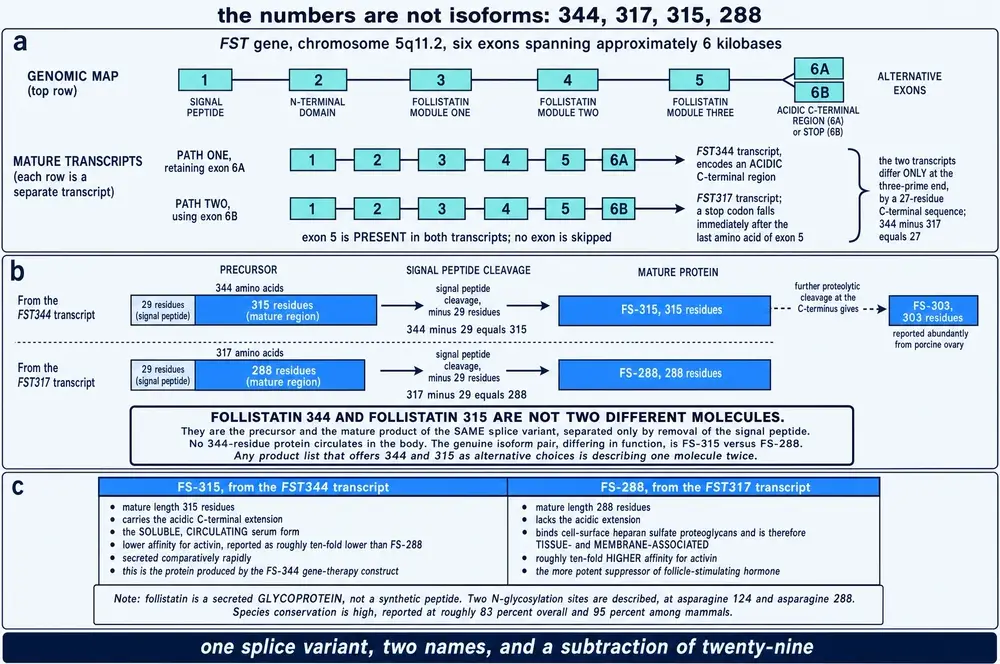
In 1988 the Salk group cloned the human gene. The result is stated in a single sentence of their abstract, and it is the origin of every number in this monograph’s title. They sequenced eight complementary DNA clones from a human testis library. Three of them predicted a precursor protein of 344 amino acids. The other five predicted a precursor of 317 amino acids, and the difference between the two came from alternative splicing of the same messenger RNA (Shimasaki et al., 1988).
A companion paper on the pig gene the same year filled in the mechanism. The gene has six exons. The first encodes a signal sequence; four more encode the protein’s four domains; and the last encodes an extra twenty-seven amino acids that are present on the long form and absent from the short one (Shimasaki et al., 1988). Two transcripts, differing only at the tail end, from one gene.
So there are two forms. There have only ever been two forms. The reason there appear to be four numbers is that each form can be counted in two different ways, and the literature — entirely reasonably, for its own purposes — uses both.
06The subtraction nobody does
A protein destined to leave the cell is made with a short leading sequence that acts as an address label. The ribosome makes the whole thing; the cell reads the label, threads the protein into the secretory machinery, and then cuts the label off. What gets secreted is shorter than what was built. Follistatin’s label is twenty-nine residues long.
Which gives, from the primary sequence record and by arithmetic rather than by quotation:
| Written as | What it is | Residues | Arithmetic |
|---|---|---|---|
| FST344 | long form, as built, label still on | 344 | — |
| FS-315 | the same molecule, secreted | 315 | 344 − 29 |
| FST317 | short form, as built, label still on | 317 | 344 − 27 |
| FS-288 | the same molecule, secreted | 288 | 317 − 29 |
| FS-303 | FS-315 after further trimming at the tail | 303 | — |
Read that table once and the market’s two products collapse into one. “Follistatin 344” and “follistatin 315” are not alternatives. They are the same splice variant before and after a step that happens automatically, inside the cell, on the way out. No 344-residue follistatin circulates in anybody. It cannot: the 344-residue form is what exists briefly inside the cell that made it, and by the time the molecule is in the bloodstream it is 315 residues long.
The genuine functional division in this family is the other one — 315 against 288 — and it is a real and consequential difference, which Section 07 is about.
07Twenty-seven residues that decide where the protein goes
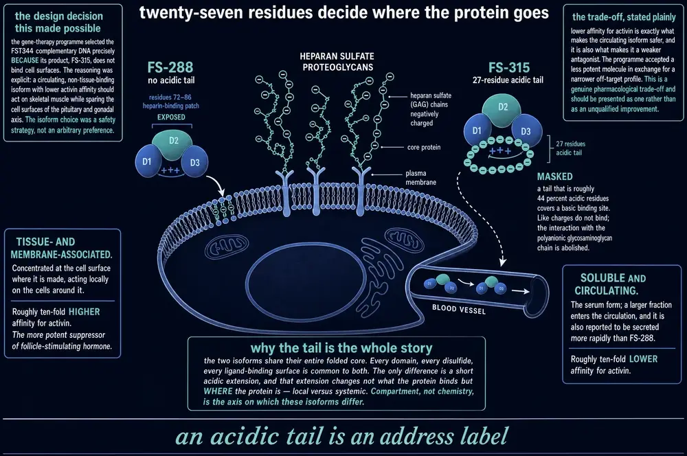
The long form carries a twenty-seven-residue extension on its tail that the short form lacks. Counted from the sequence record, twelve of those twenty-seven residues are aspartate or glutamate: the tail is 44 per cent acidic, which is to say strongly negatively charged.
Elsewhere on the molecule, inside its first follistatin domain, sits a short patch that is strongly positively charged — seven of fifteen residues are lysine or arginine. That patch is the heparin-binding site, and it is the reason the 1987 purification worked. Positively charged protein sticks to negatively charged sugar.
Cell surfaces are coated in exactly such sugars: heparan sulfate proteoglycans, densely negative, on nearly every cell in the body. So the short form, FS-288, with its basic patch exposed, binds the surface of the cell that made it and stays there. It is a local agent. The long form, FS-315, folds its acidic tail back over that basic patch and neutralises it; like charges do not attract, the interaction with the cell surface is abolished, and the molecule diffuses away into the blood. It is the circulating form.
Two proteins with an identical folded core, identical binding surfaces and identical chemistry, separated by a strip of negative charge that does nothing except decide where the molecule is allowed to be. Compartment rather than chemistry.
There is a price. Masking the basic patch also lowers the long form’s affinity for its targets — the short form is the tighter binder and the more potent suppressor of follicle-stimulating hormone. The circulating form is the weaker antagonist. That is not a defect to be engineered away; as Section 08 shows, it is precisely why the clinical programme chose it.
08Why the gene-therapy field really does say “FS344”
If 344 denotes something that never circulates, why does the clinical
literature use the number constantly? Because in that literature FS344 is
the name of a piece of DNA, not of a protein. The vector used in every
human follistatin trial is designated rAAV1.CMV.huFollistatin344:
an adeno-associated virus carrying the complementary DNA of the long splice
form, under a viral promoter.
The group that built it states the consequence explicitly. Their construct is “an alternatively spliced cDNA of follistatin (FS344)”, and its transgene product is a peptide of 315 amino acids that is secreted from the muscle and circulates in the serum, thus avoiding cell-surface binding sites (Rodino-Klapac et al., 2009). The 344 is the gene they put in. The 315 is the protein that comes out.
And the choice was deliberate. They wanted a form that would leave the muscle and act at a distance without plastering itself over the surfaces of every cell it met — in particular without accumulating on the pituitary and gonadal tissue where activin blockade has consequences nobody wanted. They took the weaker antagonist on purpose, in exchange for a narrower reach. On the evidence in Section 16, that trade appears to have worked.
So the trade name on a vial is not arbitrary nonsense. It is a designation lifted, accurately, from the gene-therapy literature, and then attached to a product that is not a gene. Section 24 returns to what that implies.
09A disulfide-rich glycoprotein, not a peptide
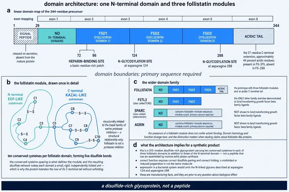
The mature protein is 315 residues arranged in four parts: an N-terminal domain, then three copies of a module so characteristic that it is named after this protein — the follistatin domain — then, on the long form, the acidic tail. Each module is a small, rigid, independently folded unit stitched together by disulfide bonds. Across the whole chain the sequence record carries thirty-eight cysteines and eighteen disulfide bonds, and two sites where sugar chains are attached.
Those are manufacturing facts, and they sit prior to any question of biological effect. A molecule of this kind is not made by peptide synthesis; it is expressed in living cells that fold it and glycosylate it. A preparation that has the right sequence but the wrong disulfide pairing is not this molecule in any functional sense, and a preparation made in bacteria has no sugars on it at all. Neither property is visible to a purchaser.
It is also worth being precise about what “follistatin domain” does and does not tell you, because the term is a live source of confusion in exactly the literature a reader is likely to encounter. The module occurs in several unrelated secreted proteins. Of the proteins that carry it, only follistatin-like 3 has been shown to bind transforming growth factor beta family ligands the way follistatin does. Carrying the module does not confer the activity, and a paper about a “follistatin-like” protein is generally not a paper about follistatin.
10Double-muscled
In 1997 a group at Johns Hopkins went looking for new members of the transforming growth factor beta family and found one expressed almost entirely in skeletal muscle. They called it growth and differentiation factor 8. Then they deleted it in mice.
The animals were not subtly different. Individual muscles weighed two to three times as much as in normal littermates, and the increase came from both more fibres and larger ones (McPherron et al., 1997). A single gene deletion had roughly doubled the muscle mass of a mammal. The gene got a second name almost immediately: myostatin.
What made the finding immediately credible rather than merely striking was that the phenotype was already familiar to anyone who had looked at cattle. Belgian Blue and Piedmontese cattle have been bred for a trait stockmen call double muscling, and the mutations responsible sit in the same gene (Bass et al., 1999; Bellinge et al., 2005). The animals also have a well-documented problem: calving is difficult, which is the sort of detail that gets left out when the phenotype is described as an unqualified improvement.
In 2004 a single human case was reported — a child with a myostatin mutation and gross muscle hypertrophy (Schuelke et al., 2004). It is cited more or less universally as proof that the pathway operates in people. It is a case report of one, and it should be read as the strong hint it is rather than the population evidence it is not.
11The idea of a chalone
Twenty-five years after describing the gene, Se-Jin Lee set out what it means. Myostatin is made by muscle fibres, circulates in the blood, and acts back on muscle fibres to limit their growth. It is a negative feedback loop whose purpose is to hold the size of a tissue where it belongs (Lee, 2023).
There is an old word for a hypothetical substance of exactly that kind — a circulating, tissue-specific growth inhibitor that tells an organ how big to be. The word is chalone, coined more than half a century ago for a class of molecules nobody could produce. Myostatin appears to be a real one.
This reframes what follistatin does. It is not adding a growth stimulus. It is removing a brake that evolution installed and maintains, and the interesting question about removing it is not whether muscle grows — it does — but what the brake was for.
12Why follistatin outperforms deleting myostatin
In 2001 Lee and McPherron compared several ways of interfering with the pathway in mice: the myostatin propeptide, a dominant-negative receptor, and follistatin, each expressed under a muscle-specific promoter. All three produced dramatic increases in muscle mass. The follistatin animals showed increases comparable to myostatin-knockout mice — and follistatin achieved that without touching the myostatin gene at all (Lee & McPherron, 2001).
Subsequent work found the effect can exceed what deleting myostatin alone achieves, and the explanation is the one Part One gave: follistatin does not only neutralise myostatin. It neutralises activin as well, and activin restrains muscle through the same receptors. Blocking two brakes beats blocking one.
Two qualifications belong with that, both from mouse work. The hypertrophy requires an intact insulin and insulin-like-growth-factor pathway: in animals made deficient in both, follistatin loses its anabolic effect, and restoring either restores it (Barbé et al., 2015). And the muscle that results is not simply more of the same — proteomic and transcriptomic analysis of follistatin-overexpressing muscle finds altered energy metabolism, fibre type, calcium handling and membrane repair alongside the extra mass (Barbé et al., 2017). Bigger is not a synonym for better, and the animal literature has been saying so for a decade.
A third finding sharpens the picture usefully. Follistatin’s N-terminal domain can be mutated or deleted without abolishing its effect on fat, while the effect on muscle disappears (Zheng et al., 2017). The muscle action and the metabolic action are separable at the level of protein architecture — which is an argument that a more selective molecule is possible, and simultaneously an admission that follistatin itself is not it.
13Encirclement, not competition
Most blocking drugs compete. An antibody sits on a binding surface; a receptor decoy soaks up ligand by offering an alternative. Follistatin does something structurally different, and crystallography settled it.
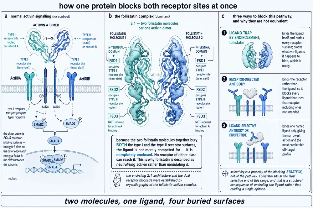
Activin is a dimer, and it presents four surfaces that receptors use: two outer sites for the type II receptors and two inner clefts for the type I receptors. Two follistatin molecules wrap around one activin dimer from opposite sides and bury all four. The N-terminal domain together with the first follistatin domain occupies the type I site; the second domain occupies the type II site (Harrington et al., 2006). The third domain is not required for activin binding at all — the minimal inhibitory fragment is the first two domains.
The consequence is that the ligand is not merely outcompeted; it is enclosed. No receptor of either class can reach it. This is why the literature describes follistatin as neutralising activin rather than modulating it, and why the complex is effectively irreversible once formed.
It also explains the selectivity problem in one sentence. A molecule that works by reading a single epitope can be made specific. A molecule that works by wrapping the whole ligand will wrap anything the right shape, and the transforming growth factor beta family contains a great many things of roughly the right shape. Follistatin’s breadth is a structural consequence of its mechanism, not an incidental impurity of its pharmacology. The same principle separates it from its own closest relative: the structure of the follistatin-like 3 complex with activin shows the two paralogues contacting the ligand differently through their N-terminal domains, which is much of why their ligand preferences diverge (Stamler et al., 2008; Cash et al., 2012).
14What it binds, and where it acts
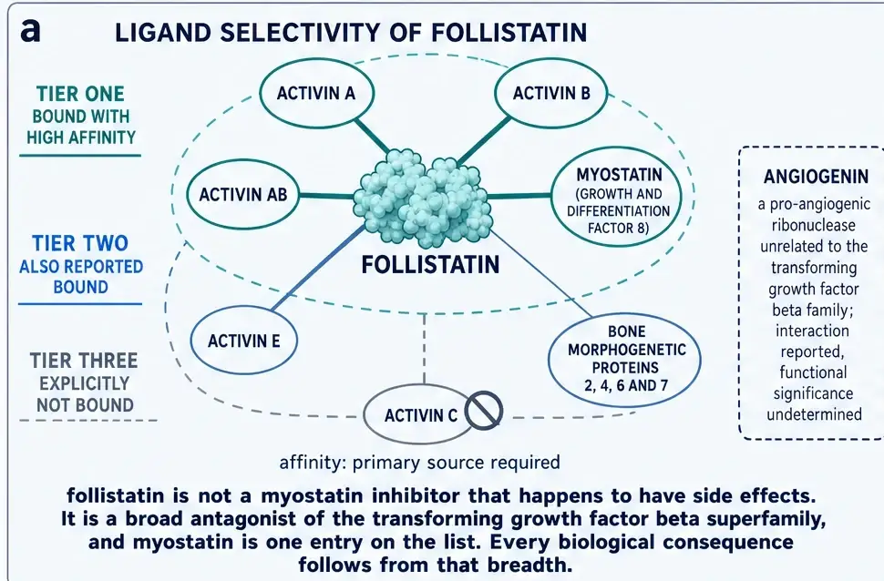
The list is long, and its length is the point. Follistatin binds activin A, activin B and activin AB with high affinity, and myostatin with high affinity. Activin E and several bone morphogenetic proteins are also reported bound. Activin C is specifically not bound. An interaction with angiogenin — a ribonuclease with no relationship to this family at all — has been reported without a settled functional significance.
Follistatin is therefore not a myostatin inhibitor that happens to have side effects. It is a broad antagonist of a signalling family, and myostatin is one entry on the list. Every biological consequence in Part Five follows from that breadth.
It matters also where in the pathway it acts. Myostatin is made as a precursor and has to be processed twice before it can signal: the signal peptide comes off, then a cleavage separates a propeptide from the business end, and the propeptide stays attached non-covalently, holding the mature dimer latent in the circulation until a tolloid-family protease frees it. Follistatin does nothing to any of that. It binds the finished, activated ligand. It does not stop myostatin being made, processed or released; it removes it after the fact.
15The road to a person
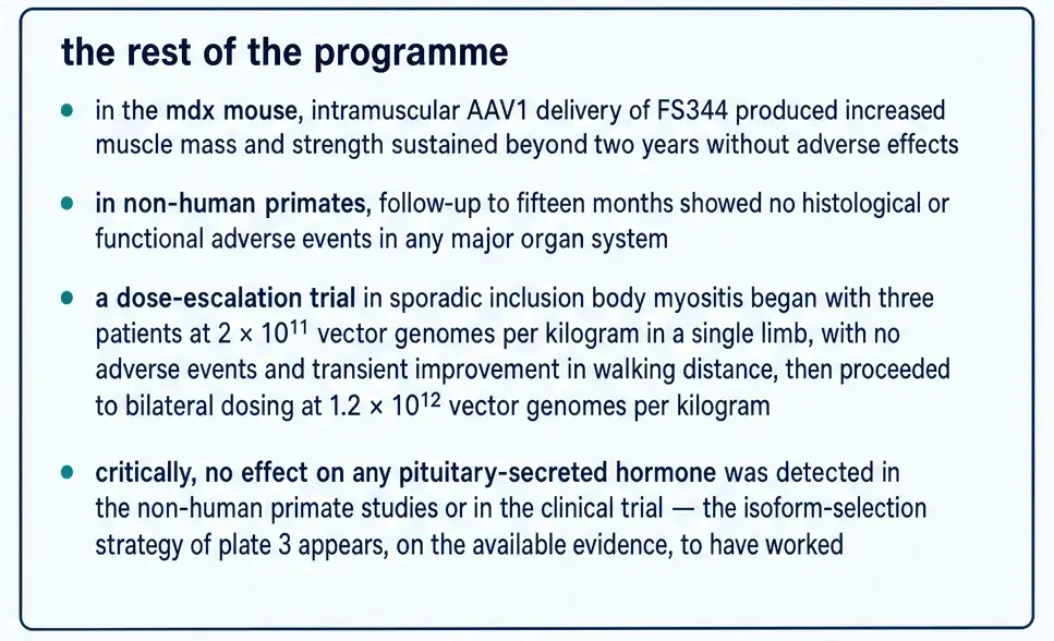
The animal case was built in stages, and the stages matter because each one narrowed what would be attempted in people. Mice overexpressing follistatin in muscle grew (Section 12). Delivering the gene by virus to the muscle of a dystrophic mouse produced increased mass and strength that persisted beyond two years. Then, in 2009, the same approach was taken to monkeys.
Kota and colleagues injected an adeno-associated virus carrying the FS344 complementary DNA into the quadriceps of cynomolgus macaques and reported pronounced and durable increases in muscle size and strength, with follow-up to fifteen months and no abnormal changes in the morphology or function of major organs (Kota et al., 2009). Crucially for what follows, they used the alternatively spliced form deliberately, describing it as one that affects skeletal muscle while having only minimal effects on non-muscle cells.
That is a well-constructed preclinical package: a mechanism, a rodent effect, a large-animal effect, and a safety window. It is more than most compounds in this series can show.
16Six patients
Two trials of follistatin gene transfer have been run in people, both at Nationwide Children’s Hospital in Columbus, and both are on the public registry. NCT01519349 was a phase 1 study in Becker muscular dystrophy and sporadic inclusion body myositis with a planned enrolment of fifteen, running from January 2012 to October 2017; its primary outcome measure is safety, and no results have been posted to the registry. NCT02354781 was a phase 1/2 study in Duchenne muscular dystrophy with an enrolment of three, completed in November 2017. Both records were read directly rather than through a summary of them.
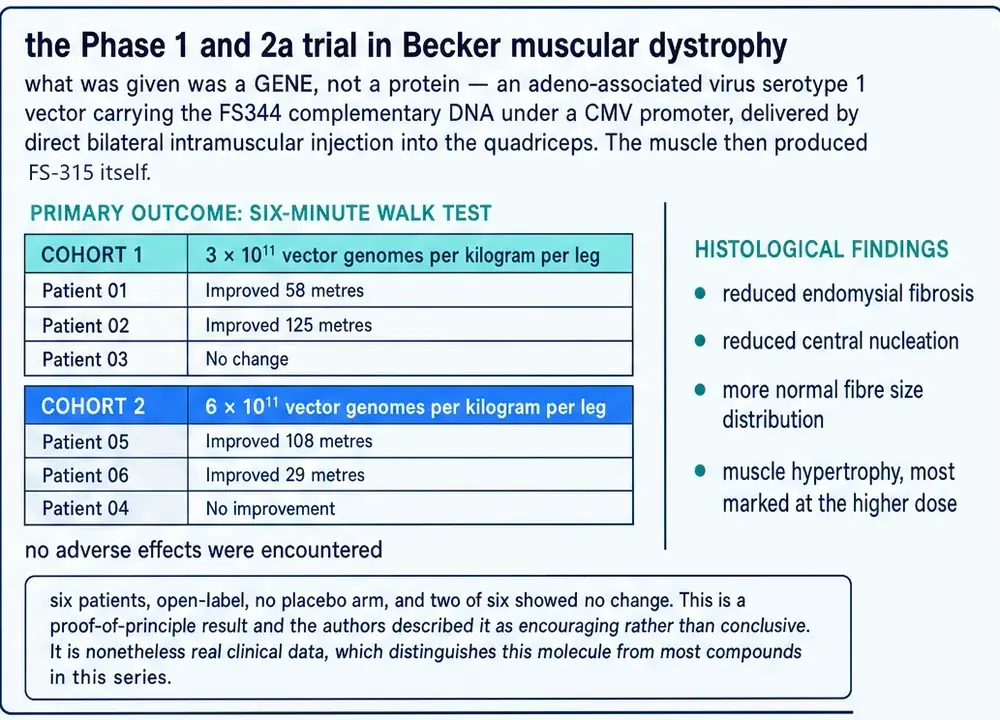
The Becker cohort was published in 2015 and is the substantive human result for this molecule. Six patients received direct bilateral intramuscular injection into the quadriceps. Three at the lower dose improved their six-minute walk distance by 58 metres, 125 metres, and not at all. Three at the higher dose improved by 108 metres, 29 metres, and not at all. Biopsies showed reduced endomysial fibrosis, reduced central nucleation, a more normal distribution of fibre sizes and muscle hypertrophy, most marked at the higher dose. No adverse effects were encountered (Mendell et al., 2015).
Four of six improved. Two did not. The authors described the result as encouraging rather than conclusive, which is the correct description.
17What six patients can and cannot establish
The honest reading of that trial requires holding two things at once, and most accounts of follistatin drop one of them.
The first is that this is real clinical data, reported per patient, including its own null results. A great many compounds sold in the research-chemical market have nothing of the kind — no trial, no registry record, no published human measurement at all. On the evidence base alone follistatin is among the best-supported compounds this series has examined.
The second is that six open-label patients with no placebo group cannot establish efficacy, and the specific reason is not a technicality. The outcome measure was a walking test, performed unblinded, in a slowly progressive disease, by patients who knew they had received an experimental gene therapy. Walking tests are effort-dependent and improve with familiarity. There is no control arm against which to ask how much of 58 metres is the treatment.
How much does that matter? The field supplies an unusually direct answer, because the same walking test was run against placebo in the same class of disease, at scale.
18The neighbours, and the largest trial in the field
Bimagrumab is a monoclonal antibody against the type IIB activin receptor. It blocks the same pathway from the receptor side rather than the ligand side, and it reliably increases muscle volume on imaging. In an early study of eleven patients with sporadic inclusion body myositis it produced increased muscle volume at eight weeks and a 14.6 per cent improvement in six-minute walk distance at sixteen weeks, reported as statistically significant. That result is the reason the programme went forward, and it is roughly the shape of the follistatin result.
It was then tested properly. RESILIENT randomised 251 patients across 38 sites in Australia, Europe, Japan and the United States to one of three bimagrumab doses or matching placebo, for at least 48 weeks, with six-minute walk distance at week 52 as the primary outcome. At week 52 the change in walking distance did not differ from placebo at any dose: +17.6 m at 10 mg/kg (99 per cent CI −19.6 to 54.8, p = 0.22), +18.6 m at 3 mg/kg (p = 0.19), and −1.3 m at 1 mg/kg (p = 0.93). Falls were the most frequent adverse event in every group including placebo. It remains the largest randomised controlled trial ever conducted in this disease (Hanna et al., 2019). A two-year extension did not change the conclusion (Amato et al., 2021).
Set the two results side by side and the comparison is uncomfortable but instructive. The uncontrolled follistatin gains are of the same order as the placebo-adjusted differences that a 251-patient trial found to be indistinguishable from nothing. That does not show follistatin does not work. It shows that the study design used to observe it cannot tell the difference between working and not working.
The wider class record points the same way. Myostatin-pathway agents have repeatedly and reproducibly increased muscle mass in humans and have repeatedly failed to improve what patients can do. The ligand traps from this family that did succeed — luspatercept and sotatercept — succeeded in anaemia and pulmonary arterial hypertension, indications that have nothing to do with muscle. The class lesson is that adding muscle mass is comparatively easy and is not the same thing as helping anybody, and it is the single most important piece of context for reading any follistatin claim.
19Two limits that are easy to miss
The follistatin trials excluded anyone whose neutralising antibody titre against the viral vector was too high. A substantial proportion of adults have been exposed to adeno-associated viruses naturally and carry such antibodies. Whatever this approach eventually achieves, it is not available to everyone, and the constraint is immunological rather than economic.
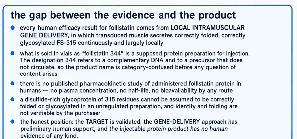
The second limit is more fundamental and is the hinge of Part Five. Every human efficacy result for follistatin comes from putting a gene into a specific muscle under imaging guidance, after which the transduced muscle manufactures correctly folded, correctly glycosylated protein continuously and largely where it was made. No human has been given follistatin protein systemically in a published, controlled study, and there is no published pharmacokinetic study of administered follistatin protein in people at all — no plasma concentration, no half-life, no bioavailability by any route.
That absence is not a gap in this monograph’s search. It is a statement about the literature, and it is what Section 24 is about.
20The job it was named for
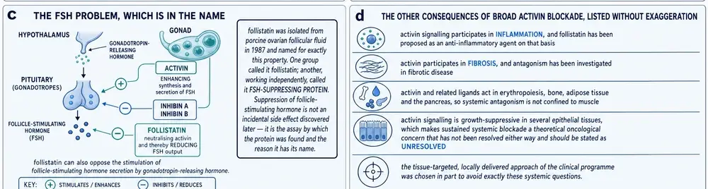
Follistatin’s original assay was suppression of follicle-stimulating hormone, and that action has never gone away. Activin, made in the gonad and in the pituitary itself, drives the synthesis and release of that hormone; the inhibins oppose it; follistatin neutralises activin and lowers the output. This is not a side effect discovered later. It is the reason the protein has its name.
How central it is to the animal is easiest to see from what happens when it is removed. Mice engineered to lack follistatin entirely are born with multiple developmental defects and die within hours of birth (Matzuk et al., 1995). Pushing the other way is not benign either: mice engineered to overexpress it develop reproductive defects (Guo et al., 1998). A molecule whose deletion is lethal and whose overexpression causes infertility is not one where more is straightforwardly better.
The clinical programme’s answer to this was the isoform choice of Section 08 combined with local delivery, and on the reported evidence it held: no effect on any pituitary-secreted hormone was detected in the primate studies or in the clinical trial. That is a genuine and underappreciated success of drug design. It is also an argument that applies only to a gene delivered into one muscle.
21Metabolism, fibrosis, and the rest of the list
Activin signalling is involved in inflammation, in fibrosis, in wound repair, in bone, in the pancreas, in erythropoiesis and in the growth control of several epithelial tissues. A broad activin antagonist reaches all of it, and the recent literature is accumulating findings on exactly that breadth — most of it in mice, and most of it by viral overexpression rather than by giving protein.
Follistatin promotes the browning of white adipose tissue and improves lipid handling. A 2026 study delivering AAV1-FST344 to a mouse model of atherosclerosis reported reduced aortic lesion area, improved plasma lipids, increased uncoupling protein 1 in white fat and raised arginase 1, with reduced inflammatory and adhesion markers in fat, liver and heart (Dirakvand et al., 2026). Keratinocyte-derived follistatin regulates epidermal homeostasis and wound repair in mice (Antsiferova et al., 2009). More broadly, follistatin behaves as a stress-response protein, induced across a range of insults, and has been reported to translocate into the nucleus in a manner independent of its ligand-trapping role (Zhang et al., 2018).
Cancer is where the two-sidedness is starkest. Follistatin has been reported as a metastasis suppressor in a mouse model of HER2-positive breast cancer. It is also elevated in people who are wasting: in a study of 55 patients, plasma follistatin and activin A were both higher in head and neck cancer than in controls while myostatin was lower, and the tumours themselves secreted all three into culture medium (Saroul et al., 2026). The authors read follistatin as protective against muscle loss in that setting. A molecule that suppresses metastasis in one model and marks cachexia in another does not have a simple story, and the honest position is that activin blockade’s long-term oncological implications are unresolved in either direction rather than reassuring.
22Exercise raises it, and that is the best human evidence there is
There is a body of human follistatin data that is properly controlled, and it is not about injecting anything. Follistatin rises with exercise, and the ratio of follistatin to myostatin is used as a readable index of whether the anabolic balance in muscle has shifted.
A 2026 randomised controlled trial put 96 healthy women aged 65 and over into four groups for twelve weeks: control, resistance exercise, essential amino acids, or both. The combined group gained muscle mass and improved across the senior fitness test battery. Myostatin fell and follistatin rose in both exercising groups, and the follistatin-to-myostatin ratio increased most in the combined group, significantly against both control and supplement-only (Jeong et al., 2026).
This is the best-controlled human follistatin finding in the corpus, and it concerns endogenous follistatin responding to training and protein intake. It is evidence that the axis is real and modifiable in people. It is not evidence for a vial, and it points in an awkward direction for anyone selling one: the intervention that reliably moved this axis in a randomised trial was lifting weights and eating adequate protein.
The axis can also be measured and found unmoved. In a randomised, placebo-controlled crossover study, eighteen days of liraglutide in adults with type 2 diabetes reduced total body mass and trunk and android fat with no measurable change in lean or bone compartments; among the myostatin-activin-follistatin-IGF-1 components measured, insulin-like growth factor 1 rose modestly and the others did not move (Gutierrez de Piñeres et al., 2026). Short studies of this axis frequently return nothing, which is worth knowing before reading a positive one.
23The specificity problem, stated plainly
Put Sections 13, 20 and 21 together and the structural argument closes. Follistatin works by wrapping its ligand rather than by reading a single epitope. That mechanism cannot be made selective, because selectivity would require discriminating between molecules that present the same surfaces. The breadth is not an impurity in the pharmacology; it is the pharmacology. A drug that neutralises activin systemically neutralises activin everywhere activin does a job, and activin does a great many jobs.
This is why the clinical programme is built the way it is. The isoform was chosen to avoid cell-surface accumulation; the delivery is local, into a named muscle, under imaging; the expression is continuous and largely confined to the tissue that was injected. Every one of those choices exists to keep a broad antagonist away from the places its breadth would cause trouble.
It follows that the safety record of the trials is a safety record for that arrangement. It says nothing about systemic exposure to follistatin protein, because no such study has been done in people.
The target is validated: myostatin restrains muscle in mice, in cattle and in at least one reported human. The mechanism is established at atomic resolution. The gene-delivery approach has preliminary human support in six patients, reported honestly, including two who did not respond.
What does not exist is any human evidence for administered follistatin protein — no controlled trial, no pharmacokinetic study, no measured plasma concentration, no half-life, and no bioavailability figure by any route. That is not an omission in this search; it is the state of the literature.
24What is actually in the vial
Both names have been on sale for more than a decade. This project’s vendor records carry “Follistatin 344 1mg” first captured in January 2014 and “Follistatin 315 1mg” first captured in August 2014, offered side by side by the same retailer as though they were alternatives, and current listings from other vendors carry the 344 in one-milligram vials. The catalogue this project benchmarks lists them as two separate compounds with different priorities.
Three observations follow from the preceding twenty-three sections, and none of them depends on knowing what any particular vial contains.
The name is a category error. The designation 344 belongs to a complementary DNA and to a precursor that exists only briefly inside a cell. Nothing that circulates in a body is 344 residues long. A product offering 344 and 315 as choices is offering the same molecule twice, and the fact that this has gone uncorrected in the market for twelve years is the first indication that the product and the science are not closely connected.
It is not a peptide, and the manufacturing question comes before the biology question. This is a 315-residue glycoprotein with eighteen disulfide bonds and two glycosylation sites. Its activity depends on the disulfides being paired correctly and the chain being folded correctly. Neither is verifiable by a purchaser, and neither can be assumed of an unregulated preparation. A molecule with the right sequence and the wrong folding is not this molecule.
The human evidence is for a different intervention. Every efficacy result in Part Four came from injecting a gene into a muscle, not a protein into a person. The trials do not transfer.
Two further episodes belong on the record. Offshore plasmid-based follistatin gene therapy has been offered commercially and registered: one study of 43 healthy subjects completed in August 2023 with no results posted and no peer-reviewed report located, and a combination study began recruiting in December 2025. A separate AAV-follistatin study in age-related muscle decline opened in June 2026. None has reported.
And in May 2022 a paper in a leading journal reported that delivering follistatin by cytomegalovirus vector extended median mouse lifespan by 32.5 per cent (Jaijyan et al., 2022). It was retracted in August 2025, at the request of the originating university’s research regulatory office, following an internal review of data discrepancies in two figures; two of the senior academic authors agreed with the retraction and several of the company-affiliated authors did not. The claim is recorded here as a claim that was made and withdrawn. It supports nothing, and it is mentioned only because it circulated widely and continues to.
The picture that leaves is unusual for this series, and it is worth stating without softening in either direction. Follistatin has the strongest evidence base of any compound covered so far — a validated target, atomic-level mechanism, large-animal work, and a real if tiny human trial that reported its own failures. It also has the widest gap between that evidence and the thing being sold. Both statements are true, and the second is not a reason to dismiss the first.
One last measurement makes the point about the literature itself. Of the 9,386 full texts retrieved for this document, more than two thousand named a follistatin-like protein and not follistatin, and the identity gate that separated them was written before any of them were read. A compound whose name is a prefix of an entire protein family is one where a reader — or a vendor, or a search engine — can accumulate a great deal of confident material about the wrong molecule without ever making an obvious mistake.
This document reports published research about follistatin. It does not recommend human use of follistatin or of any other compound named in it, and it specifies no dose, route or schedule for any person. Reported study parameters appear only as descriptions of what was done in the study being described, always with the species, the route and the duration attached.
25References
Generated from verified NCBI records rather than from recall. Author lists, journal names, volumes, pages and identifiers are taken from the PubMed record for each citation, and the build refuses to run if any identifier fails to resolve. Reading the resolved author, journal and year line back against the prose caught five year discrepancies in the drafting of this document, all of them electronic-publication dates differing from the issue year.
- Amato AA, Hanna MG, Machado PM, Badrising UA, Chinoy H, Benveniste O, et al.. Efficacy and Safety of Bimagrumab in Sporadic Inclusion Body Myositis: Long-term Extension of RESILIENT. Neurology. 2021;96(12):e1595-e1607.
PMID 33597289 · doi:10.1212/WNL.0000000000011626 · PMC8032371 - Antsiferova M, Klatte JE, Bodó E, Paus R, Jorcano JL, Matzuk MM, et al.. Keratinocyte-derived follistatin regulates epidermal homeostasis and wound repair. Lab Invest. 2009;89(2):131-41.
PMID 19079322 · doi:10.1038/labinvest.2008.120 · PMC4087116 - Barbé C, Kalista S, Loumaye A, Ritvos O, Lause P, Ferracin B, et al.. Role of IGF-I in follistatin-induced skeletal muscle hypertrophy. Am J Physiol Endocrinol Metab. 2015;309(6):E557-67.
PMID 26219865 · doi:10.1152/ajpendo.00098.2015 · PMC4572457 - Barbé C, Bray F, Gueugneau M, Devassine S, Lause P, Tokarski C, et al.. Comparative Proteomic and Transcriptomic Analysis of Follistatin-Induced Skeletal Muscle Hypertrophy. J Proteome Res. 2017;16(10):3477-3490.
PMID 28810121 · doi:10.1021/acs.jproteome.7b00069 - Bass J, Oldham J, Sharma M, Kambadur R. Growth factors controlling muscle development. Domest Anim Endocrinol. 1999;17(2-3):191-7.
PMID 10527122 · doi:10.1016/s0739-7240(99)00036-3 - Bellinge RH, Liberles DA, Iaschi SP, O'brien PA, Tay GK. Myostatin and its implications on animal breeding: a review. Anim Genet. 2005;36(1):1-6.
PMID 15670124 · doi:10.1111/j.1365-2052.2004.01229.x - Cash JN, Angerman EB, Keutmann HT, Thompson TB. Characterization of follistatin-type domains and their contribution to myostatin and activin A antagonism. Mol Endocrinol. 2012;26(7):1167-78.
PMID 22593183 · doi:10.1210/me.2012-1061 · PMC3385792 - Dirakvand G, Pervin S, Villa B, Le C, Yohanna K, Grijalva V, et al.. Follistatin Mitigates Atherosclerosis Through Activation of Arginine Metabolism and Adipose Browning. Cells. 2026;15(13).
PMID 42439678 · doi:10.3390/cells15131205 · PMC13360072 - Guo Q, Kumar TR, Woodruff T, Hadsell LA, DeMayo FJ, Matzuk MM. Overexpression of mouse follistatin causes reproductive defects in transgenic mice. Mol Endocrinol. 1998;12(1):96-106.
PMID 9440814 · doi:10.1210/mend.12.1.0053 - Gutierrez de Piñeres V, Tamayo-Torres CS, Ramirez-Cisneros A, Kavelidou M, Stefanakis K, Mantzoros CS. Effects of liraglutide treatment for 18 days on metabolic parameters, regional body composition and the myostatin-activin-follistatin-IGF-1 axis: Results from an exploratory, randomized, placebo-controlled, crossover study. Diabetes Obes Metab. 2026;28(2):1258-1265.
PMID 41287185 · doi:10.1111/dom.70313 - Hanna MG, Badrising UA, Benveniste O, Lloyd TE, Needham M, Chinoy H, et al.. Safety and efficacy of intravenous bimagrumab in inclusion body myositis (RESILIENT): a randomised, double-blind, placebo-controlled phase 2b trial. Lancet Neurol. 2019;18(9):834-844.
PMID 31397289 · doi:10.1016/S1474-4422(19)30200-5 - Harrington AE, Morris-Triggs SA, Ruotolo BT, Robinson CV, Ohnuma S, Hyvönen M. Structural basis for the inhibition of activin signalling by follistatin. EMBO J. 2006;25(5):1035-45.
PMID 16482217 · doi:10.1038/sj.emboj.7601000 · PMC1409725 - Jaijyan DK, Selariu A, Cruz-Cosme R, Tong M, Yang S, Stefa A, et al.. New intranasal and injectable gene therapy for healthy life extension. Proc Natl Acad Sci U S A. 2022;119(20):e2121499119.
PMID 35537048 · doi:10.1073/pnas.2121499119 · PMC9171804 - Jeong D, Valentine RJ, Jeong H, Sung JY, Lim H, Kang S. Combined resistance exercise and essential amino acid intake enhance follistatin/myostatin ratio and muscle fitness in older women: a randomized controlled trial. J Int Soc Sports Nutr. 2026;23(1):2646626.
PMID 41863133 · doi:10.1080/15502783.2026.2646626 · PMC13007441 - Kota J, Handy CR, Haidet AM, Montgomery CL, Eagle A, Rodino-Klapac LR, et al.. Follistatin gene delivery enhances muscle growth and strength in nonhuman primates. Sci Transl Med. 2009;1(6):6ra15.
PMID 20368179 · doi:10.1126/scitranslmed.3000112 · PMC2852878 - Lahouti AH, Amato AA, Christopher-Stine L. Inclusion body myositis: update. Curr Opin Rheumatol. 2014;26(6):690-6.
PMID 25215417 · doi:10.1097/BOR.0000000000000116 - Lee SJ. Myostatin: A Skeletal Muscle Chalone. Annu Rev Physiol. 2023;85:269-291.
PMID 36266260 · doi:10.1146/annurev-physiol-012422-112116 · PMC10163667 - Lee SJ, McPherron AC. Regulation of myostatin activity and muscle growth. Proc Natl Acad Sci U S A. 2001;98(16):9306-11.
PMID 11459935 · doi:10.1073/pnas.151270098 · PMC55416 - Matzuk MM, Lu N, Vogel H, Sellheyer K, Roop DR, Bradley A. Multiple defects and perinatal death in mice deficient in follistatin. Nature. 1995;374(6520):360-3.
PMID 7885475 · doi:10.1038/374360a0 - McPherron AC, Lawler AM, Lee SJ. Regulation of skeletal muscle mass in mice by a new TGF-beta superfamily member. Nature. 1997;387(6628):83-90.
PMID 9139826 · doi:10.1038/387083a0 - Mendell JR, Sahenk Z, Malik V, Gomez AM, Flanigan KM, Lowes LP, et al.. A phase 1/2a follistatin gene therapy trial for becker muscular dystrophy. Mol Ther. 2015;23(1):192-201.
PMID 25322757 · doi:10.1038/mt.2014.200 · PMC4426808 - Rodino-Klapac LR, Haidet AM, Kota J, Handy C, Kaspar BK, Mendell JR. Inhibition of myostatin with emphasis on follistatin as a therapy for muscle disease. Muscle Nerve. 2009;39(3):283-96.
PMID 19208403 · doi:10.1002/mus.21244 · PMC2717722 - Saroul N, Dissard A, Pereira B, Mom T, Boirie Y, Gilain L, et al.. Myostatin, activin-A and follistatin are produced by the tumor in head and neck cancer and likely contribute to sarcopenia: A case-control, cross-sectional exploratory study. Clin Nutr ESPEN. 2026;71:102839.
PMID 41338451 · doi:10.1016/j.clnesp.2025.11.159 - Schuelke M, Wagner KR, Stolz LE, Hübner C, Riebel T, Kömen W, et al.. Myostatin mutation associated with gross muscle hypertrophy in a child. N Engl J Med. 2004;350(26):2682-8.
PMID 15215484 · doi:10.1056/NEJMoa040933 - Shimasaki S, Koga M, Esch F, Cooksey K, Mercado M, Koba A, et al.. Primary structure of the human follistatin precursor and its genomic organization. Proc Natl Acad Sci U S A. 1988;85(12):4218-22.
PMID 3380788 · doi:10.1073/pnas.85.12.4218 · PMC280398 - Shimasaki S, Koga M, Esch F, Mercado M, Cooksey K, Koba A, et al.. Porcine follistatin gene structure supports two forms of mature follistatin produced by alternative splicing. Biochem Biophys Res Commun. 1988;152(2):717-23.
PMID 3365249 · doi:10.1016/s0006-291x(88)80097-4 - Stamler R, Keutmann HT, Sidis Y, Kattamuri C, Schneyer A, Thompson TB. The structure of FSTL3.activin A complex. Differential binding of N-terminal domains influences follistatin-type antagonist specificity. J Biol Chem. 2008;283(47):32831-8.
PMID 18768470 · doi:10.1074/jbc.M801266200 · PMC2583307 - Ueno N, Ling N, Ying SY, Esch F, Shimasaki S, Guillemin R. Isolation and partial characterization of follistatin: a single-chain Mr 35,000 monomeric protein that inhibits the release of follicle-stimulating hormone. Proc Natl Acad Sci U S A. 1987;84(23):8282-6.
PMID 3120188 · doi:10.1073/pnas.84.23.8282 · PMC299526 - Walker RG, Kattamuri C, Goebel EJ, Zhang F, Hammel M, Tainer JA, et al.. Heparin-mediated dimerization of follistatin. Exp Biol Med (Maywood). 2021;246(4):467-482.
PMID 33197333 · doi:10.1177/1535370220966296 · PMC7885052 - Walsh FS, Celeste AJ. Myostatin: a modulator of skeletal-muscle stem cells. Biochem Soc Trans. 2005;33(Pt 6):1513-7.
PMID 16246158 · doi:10.1042/BST0331513 - Ying SY. Inhibins, activins, and follistatins: gonadal proteins modulating the secretion of follicle-stimulating hormone. Endocr Rev. 1988;9(2):267-93.
PMID 3136011 · doi:10.1210/edrv-9-2-267 - Zhang L, Liu K, Han B, Xu Z, Gao X. The emerging role of follistatin under stresses and its implications in diseases. Gene. 2018;639:111-116.
PMID 29020616 · doi:10.1016/j.gene.2017.10.017 - Zheng H, Qiao C, Tang R, Li J, Bulaklak K, Huang Z, et al.. Follistatin N terminus differentially regulates muscle size and fat in vivo. Exp Mol Med. 2017;49(9):e377.
PMID 28912572 · doi:10.1038/emm.2017.135 · PMC5628274 - [No authors listed]. Retraction for Jaijyan et al., New intranasal and injectable gene therapy for healthy life extension. Proc Natl Acad Sci U S A. 2025;122(33):e2519570122.
PMID 40789040 · doi:10.1073/pnas.2519570122 · PMC12377719
Sources without a PubMed record
Trial registry records and the primary sequence record have no PubMed entry and are listed separately, so the generated list above remains wholly machine-verified. Each registry record below was read directly at clinicaltrials.gov rather than through a summary of it.
- NCT01519349. Follistatin gene transfer to patients with Becker muscular dystrophy and sporadic inclusion body myositis. Phase 1, rAAV1.CMV.huFollistatin344, Nationwide Children's Hospital with Parent Project Muscular Dystrophy; enrolment 15; January 2012 to October 2017; status Completed; primary outcome Safety; no results posted.
https://clinicaltrials.gov/study/NCT01519349 - NCT02354781. Phase I/II intramuscular gene transfer of rAAV1.CMV.huFollistatin344 to patients with Duchenne muscular dystrophy. Sponsor Jerry R. Mendell; enrolment 3; January 2015 to November 2017; status Completed.
https://clinicaltrials.gov/study/NCT02354781 - NCT01925209. RESILIENT: efficacy and safety of bimagrumab at 52 weeks on physical function, muscle strength and mobility in patients with sporadic inclusion body myositis. The registry record for the trial reported in reference [RESILIENT].
https://clinicaltrials.gov/study/NCT01925209 - NCT06411366. A phase I, open label, single dose study to evaluate the safety and efficacy of an injectable follistatin plasmid gene therapy in healthy subjects. Sponsor Minicircle; conditions Frailty, Aging; enrolment 43; August 2022 to August 2023; status Completed; no results posted and no peer-reviewed report located.
https://clinicaltrials.gov/study/NCT06411366 - NCT07285629. Evaluating the safety and efficacy of injectable combination klotho and follistatin plasmid gene therapy in humans. Sponsor Minicircle; Early Phase 1; enrolment 30; start 16 December 2025; status Recruiting.
https://clinicaltrials.gov/study/NCT07285629 - NCT07443826. CALM-AF-AI: counteracting age-related loss of muscle with AAV-follistatin combined with angiogenesis-inducing VEGF plasmid gene therapy. Sponsor Unlimited Biotechnology LLC; Phase 1/2; enrolment 12; start 1 June 2026; status Recruiting.
https://clinicaltrials.gov/study/NCT07443826 - UniProt P19883. FST_HUMAN, follistatin. Sequence length 344; signal peptide 1-29; mature chain 30-344; isoform 2 (FS288) missing residues 318-344. Read directly and recomputed by arithmetic in pipeline/01b_verify_identity.py.
https://www.uniprot.org/uniprotkb/P19883/entry
26How this document was assembled
The corpus was built against project 05, the Therapeutic Peptide Research Library, together with a four-arm external harvest. The interesting part of the arithmetic is how much had to be thrown away, and why.
The identity problem, in numbers. This compound's name is a literal prefix of the names of a whole family of proteins that are not it — follistatin-like 1, follistatin-like 3, and the “follistatin domain” that occurs in unrelated secreted proteins. The colliding string is not a homograph; it is the subject's own name, used correctly, about a different molecule. Separately, the gene symbol FST is the standard abbreviation for the forced swim test, whose literature at 8,755 records is nearly three times the size of this compound's own 2,947.
The matcher therefore blanks every follistatin-like occurrence before any other test runs, refuses a bare symbol whose text carries a known non-subject expansion, and admits an uncorroborated bare symbol only with compound-level corroboration in a window around the hit. It was break-tested against twenty constructed traps before the first sweep — nine that had to be admitted, eleven that had to be refused — and all twenty passed.
Over the 9,386 full texts retrieved, the gate refused 2,053 documents that named only a follistatin-like protein and 1,529 that carried no designation at all. Those two numbers mean opposite things and are never pooled: the first says the identity gate is earning its keep, the second says the query reached wider than the subject.
The substantive-use problem, in numbers. Follistatin is a standard laboratory reagent for neutralising activin in cell culture, so it is named in the methods of a large literature that is not about it. Classifying the retrieved documents by structure rather than by title — does the article have a real methods section, and does it name the protein inside it — retained 769 primary papers naming it in methods, 304 further experimental papers and 277 substantive reviews, and discarded 3,515 passing mentions and 208 documents with no retrievable body text.
| Stage | Records |
|---|---|
| PubMed surface, follistatin[Title/Abstract] | 2,947 |
| harvested across four named arms | 3,671 |
| passing the relevance gate | 3,143 |
| … of those, about follistatin itself | 2,371 |
| PubMed Central body-text sweep surface | 9,159 |
| unique full texts fetched | 9,386 |
| retained after the substantive-use screen | 1,350 |
| local project-05 documents admitted | 49 |
| present in both stores, counted once | 24 |
| reading corpus | 1,375 |
| printed-page equivalents | ~17,908 |
The two stores are keyed by accession and the union counted once; a naive sum would have reported 1,399 documents. The surface and the corpus are reported as different numbers deliberately — a corpus figure of 9,159 would be a claim about coverage this document does not have.
An unusual local finding. Every one of the 49 documents admitted from the project's own library is a peer-reviewed full text. There is no vendor-snapshot tier at all. Previous monographs in this series measured local commercial-to-scientific ratios of twenty, fifty and sixty-three to one; here it is zero. The market for this molecule exists — the vendor tables carry one-milligram vials of both names from 2014 onward — but it has generated almost no captured literature, which is itself worth recording.
27Evidence handling
Study type is named in the sentence that reports the finding. The great majority of what is known about follistatin's effect on muscle comes from mice, and much of the rest from a single macaque study; those are described as such. The human efficacy evidence is one open-label trial in six people, and every reference to it in this document carries the number six and the absence of a placebo arm.
Recency is weighted, but not above design. The most recent findings here — adipose browning and atherosclerosis in mice, the tumour-secretion study in head and neck cancer, the exercise trial in older women — are given full weight where they are well designed, and the 2026 randomised exercise trial is treated as the strongest human measurement in the document precisely because of its design. A 2019 result is not superseded by a 2026 one merely for being older: RESILIENT remains the largest randomised controlled trial in its disease and its null result stands.
Conflicts are presented as conflicts. Three are live here. Follistatin is reported as a metastasis suppressor in one mouse model and as elevated in human cancer cachexia in another setting, and this document does not resolve that. The oncological implication of sustained systemic activin blockade is described as unresolved in either direction rather than as reassuring. And the relationship between muscle mass and muscle function in this drug class is treated as the open question the trial record says it is.
Values that could not be traced are named as untraceable. Two figures printed on the commissioned artwork — a cytogenetic location and a pair of cross-species conservation percentages — are carried as supplied because no primary source for them was located, and they are captioned to say so. A third, the approximately ten-fold affinity difference between the two isoforms, is well supported in direction and is captioned as approximate because the evidence base does not carry it as a single measured constant.
Commissioned artwork was audited, and two panels were withheld. Six plates were supplied with a caption file; the caption file was identity-checked against the plates before use. Twenty printed values were checked against primary records by arithmetic. Five verified, four were qualified in the caption, two were untraceable, and three were wrong — all three in one panel, which was withheld. A second panel was withheld because it reported a favourable early result for a competing agent and named the confirmatory trial that followed without reporting that the trial failed; shipping it would have placed a contradiction of this document's own Part Four a few pages away. Both withheld originals travel with the delivery bundle so the decisions remain checkable against the artwork.
Publication status was verified against PubMed itself. Seven records in the harvest carry a retraction or expression-of-concern notice. One is directly relevant and is discussed in Section 24; it is described only as a claim that was made and withdrawn, and no factual assertion in this document rests on it.

Continue exploring