
GDF-11 The near-twin of myostatin that was briefly sold as young blood
For most of its published life, growth differentiation factor 11 was a developmental biologist’s molecule — a TGF-β family ligand that helps an embryo put its vertebrae in the right order. Then, for a few years in the mid-2010s, it was asked to explain why sewing a young mouse to an old one seemed to reverse pieces of aging. The claim made headlines, spawned a controversy about immunoassays that could not tell GDF-11 from myostatin, and left the field with a cleaner methods problem than it started with. This monograph follows that arc: discovery, mechanism, the young-blood episode, the assay collapse, and what human biomarker studies can and cannot say. Nothing here is a recommendation for human use.
Findings are labelled by the kind of evidence that produced them. A result in aged C57BL/6 mice is not a human pharmacology claim. An association between circulating GDF-11 immunoreactivity and a clinical outcome is not proof that the protein caused the outcome — especially when the assay may have counted myostatin as well.
GDF-11 and myostatin (GDF-8) share roughly ninety percent of their mature amino-acid sequence and the same receptor family. Where a paper cannot tell them apart, this monograph says so. Doses appear only as reported study parameters. Nothing here recommends use by any person.
01What GDF-11 is
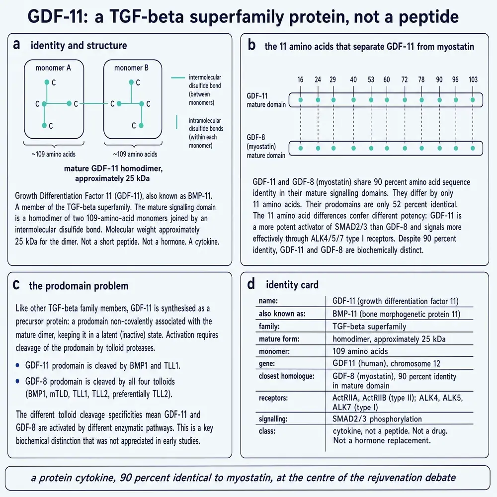
GDF-11 is a secreted member of the transforming growth factor-β superfamily, also catalogued as bone morphogenetic protein 11 (BMP-11). In humans the gene sits on chromosome 12q13.2 and encodes a 407-residue precursor (UniProt O95390). Like its relatives, the protein is cut twice: once by a proprotein convertase that separates the N-terminal prodomain from the C-terminal growth-factor domain, and again in steps that release an active disulfide-linked homodimer. The mature monomer is on the order of a hundred residues; the working species in tissue is the dimer (Jamaiyar2017; Walker2016; UniProtO95390).
The fact that matters for almost every later argument is structural kinship. Mature GDF-11 is about ninety percent identical to mature myostatin (GDF-8 / MSTN) — the better-known muscle brake discovered by the same laboratory two years earlier. Both ligands bind activin type II receptors (ActRIIA/B) and type I receptors in the ALK4/5/7 group, and both drive SMAD2/3 signaling. Extracellular proteins such as follistatin and the GDF-associated serum proteins (GASP-1/2) can hold either ligand off its receptors. On paper, they look like siblings hired for similar jobs. In the body, the jobs diverge — and the assays that tried to count them in blood did not always notice (McPherron1999; Walker2016).
Project 05’s local open-access store holds forty JATS files that name GDF-11 and two hundred that name myostatin. The catalogue database has a myostatin compound row whose aliases mention “growth differentiation factor,” and fifty-six chunks that mention GDF-11 — mostly in myostatin papers. The local library is not empty, but it is myostatin-shaped. This build therefore expands from NCBI rather than pretending the shelf was already full.
02Discovery, 1999
GDF-11 entered the literature the way many TGF-β family members did: by homology hunting. Alexandra McPherron, Ann Lawler and Se-Jin Lee, having cloned myostatin in 1997, used it as a probe and pulled out a closely related sequence they named growth/differentiation factor 11. Their mouse genetics tied the gene to anterior–posterior patterning of the axial skeleton: animals lacking GDF-11 showed homeotic transformations along the vertebral column and an elongated trunk — a developmental address, not a gerontology story (McPherron1999).
In the same year, Masaki Nakashima and colleagues cloned the factor from rat incisor pulp and mapped its embryonic expression, with early signal strongest in the tail bud and later spread through the embryo (Nakashima1999). L. W. Gamer and colleagues likewise reported human and mouse BMP-11/GDF-11 sequences and argued for roles in mesodermal and neural patterning (Gamer1999). Three independent cloning routes, one molecule, one clear developmental phenotype. Human and mouse proteins are essentially interchangeable by sequence (Jamaiyar2017).
Context matters. The late 1990s were the decade in which myostatin made “double-muscled” cattle and dogs into molecular celebrities. GDF-11 arrived as the quieter sibling — same fold, different tissue script. For roughly fifteen years that is how the field treated it: a patterning ligand with interesting knockout phenotypes in skeleton and kidney, not a systemic youth factor (Esquela2003; Oh2002). The identity and biochemistry of that molecule are summarised in Figure 1.
03The job it was hired for
The developmental job is still the firmest chapter in the dossier. Without GDF-11, mice transform cervical vertebrae toward thoracic identity, delay trunk-to-tail transition, and can fail to form kidneys properly when the ureteric bud never launches — a defect linked to GDNF expression in metanephric mesenchyme (McPherron1999; Esquela2003; Jamaiyar2017). GDF-11 signaling through ALK5/SMAD routes helps set Hox gene programmes that tell segments of the embryo who they are (Oh2002; Jamaiyar2017).
Those phenotypes do not require a story about aging blood. They require a ligand that is present at the right time in the posterior growth zone and in organ primordia. When later papers recruited GDF-11 as a circulating rejuvenation factor, they were asking a developmental specialist to moonlight in systemic physiology. Moonlighting is allowed in biology. It is not free of burden of proof. The three cloning routes that put the ligand on the map are laid out in Figure 2.
04How it signals
On a cell that expresses the right receptors, GDF-11 behaves like a canonical activin/TGF-β ligand: type II receptors phosphorylate type I receptors; SMAD2/3 partner with SMAD4 and move to the nucleus; transcriptional programmes for growth arrest, differentiation, or patterning shift. Non-SMAD branches (MAPK, PI3K) are reported as well. Brakes exist at every layer — soluble traps (follistatin, FSTL3, GASP proteins), decoy co-receptors such as BAMBI, and inhibitory SMADs inside the cell (Walker2016; Jamaiyar2017). Receptor logic is summarised in Figure 3.
Because myostatin uses the same hardware, any drug or decoy that binds ActRIIB will usually touch GDF-11 too. That fact will return when this monograph reaches ligand-trap biologics: sequestering “myostatin” in a human trial is rarely a GDF-11-specific experiment, even when GDF-11 appears on the ligand list.
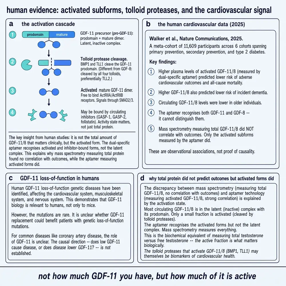
One further biochemical detail foreshadows the human epidemiology later in this dossier. Circulating GDF-11, like myostatin, spends much of its life in latent complexes; the mature domain that engages receptors is released by tolloid-family proteases. Papers that report “total” immunoreactivity and papers that report activated subforms are not measuring the same thing, even when both print the letters GDF-11 (Walker2016; Walker2025).
With identity, developmental job and receptor logic in place, the next question is historical rather than structural: how a patterning ligand became the named molecule of the young-blood years — and what had to be true in serum for that story to hold.
05Parabiosis before the molecule
Long before anyone named GDF-11 a youth factor, physiologists had a stranger experiment: join the circulations of a young mouse and an old mouse so that they share blood. Heterochronic parabiosis repeatedly suggested that something in young blood could improve aspects of aged tissue — muscle repair, neurogenesis, cardiac structure — and that something in old blood could do the reverse. The technique is brutal, the shared variables are many, and the temptation to name a single protein as the cause is almost irresistible (Loffredo2013; Jamaiyar2017).
By the early 2010s the field was hunting circulating candidates. GDF-11 was an attractive suspect on paper: present in blood, structurally a growth-factor ligand, already known to act on mesenchymal lineages. What nobody had firmly established was that its blood levels fell with age in a way that explained the parabiosis phenotypes — or that restoring those levels would be safe and sufficient.
062013 — the heart
Francesco Loffredo, Amy Wagers, Richard Lee and colleagues reported in Cell that circulating GDF-11 declined with age in mice, and that restoring it — including by recombinant protein — reversed age-related cardiac hypertrophy toward a more youthful morphology (Loffredo2013). The paper did what high-impact aging papers do: it offered a named molecule for a named hope. Cardiac hypertrophy with age is a real clinical problem; a circulating factor that thinned an old heart in mice was always going to travel.
Read carefully, the claim was still a mouse claim. The assays used to track “GDF-11” in serum would later become the battleground. But in 2013 the narrative was simple enough for everyone outside the methods section: young blood carries more GDF-11; old hearts listen; the protein is the message (Loffredo2013; Walker2016).
072014 — muscle and brain
A year later the same intellectual circle published two Science papers that widened the franchise. Sinha and colleagues reported that GDF-11 improved satellite-cell function and muscle repair in aged mice (Sinha2014). Katsimpardi and colleagues reported that GDF-11 enhanced neurogenesis and cerebral vasculature in aged mice (Katsimpardi2014). Commentaries reached for fountain language. For a brief window, GDF-11 was not merely a developmental ligand. It was the leading molecular explanation for why young blood seemed to remake old tissues (Jamaiyar2017; Suh2019).
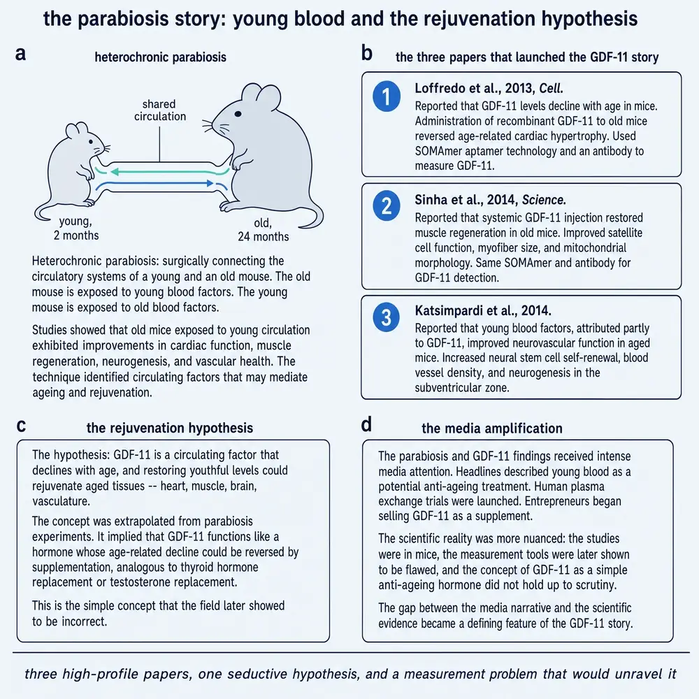
The muscle claim was the sharpest paradox. Myostatin, GDF-11’s near-twin, is the textbook negative regulator of skeletal muscle mass. Losing myostatin makes animals heavily muscled; adding myostatin signaling tends to shrink the fibre programme. Asking GDF-11 to improve aged muscle regeneration meant asking two proteins that bind the same receptors to pull in opposite directions depending on age and context. That is not impossible — dose, latency state, and co-receptors can split twins — but it demanded better tools than the field initially brought (Walker2016; Egerman2015). The claim wave is laid out in Figure 4.
08Why the claim was surprising
Surprise is data. When a result contradicts the closest related molecule’s job description, either biology is more interesting than the homology suggested, or the measurement is wrong. Both can be true at once. The Wagers/Lee programme argued for interesting biology: GDF-11 as an age-dependent restorative signal distinct from myostatin’s brake. The counter-programme, led most visibly by work from Novartis and collaborators around Egerman and Glass, argued that the measurements — and therefore the direction of the age trend — could not be trusted (Egerman2015; Walker2016).
For a common reader, the stakes are easy to state. If GDF-11 truly falls with age and restoring it rejuvenates heart, muscle and brain, then a therapeutic path exists in principle. If the immunoassays were largely counting myostatin, or if raising GDF-11 harms muscle stem cells, then the path is a mirage built from cross-reactive ink. The next part of this monograph is about which way the ink ran.
Before that quarrel is summarised, hold the chronological order in mind. The claim wave of 2013–2014 named a protein; the objections of 2015–2016 named the assays; the later human papers inherited both the hope and the measurement problem. Skipping any of those layers turns GDF-11 into a slogan. Keeping all three is what a research monograph is for (Loffredo2013; Egerman2015; Walker2016; Walker2025).
092015 — Egerman, Glass and the assay
In 2015, Egerman, Glass and colleagues published a direct challenge in Cell Metabolism. Using reagents and platforms that could better separate the twins — or at least expose where they could not be separated — they argued that GDF-11 does not decline with age in the way the rejuvenation story required, and that elevated GDF-11 could impair rather than improve muscle regeneration (Egerman2015). Nature’s news coverage put the dispute in plain language: the young-blood mechanism was back in play, and GDF-11 was no longer an uncontested answer.
The disagreement was not a polite difference of emphasis. One side’s therapeutic rationale was the other side’s methodological artifact. That is why the controversy lasted: it was about whether the field had been measuring the protein it named (Walker2016; Schafer2016).
10What could not be told apart
Mature GDF-11 and mature myostatin are so similar that many antibodies and aptamer reagents bind both. A Western blot band or a SOMAmer signal labelled “GDF-11” can be a mixture, a myostatin-dominated signal, or something else that shares an epitope. Schafer and colleagues, among others, pushed mass-spectrometry approaches that distinguish the two by unique peptides — the kind of method that should have been load-bearing before anyone sold a fountain story (Schafer2016; Walker2016).
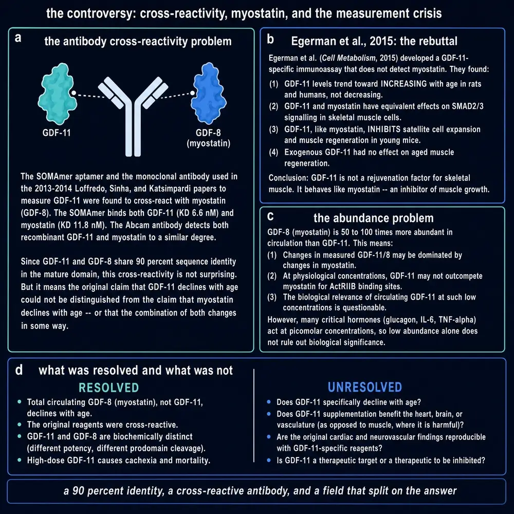
Walker, Thompson, and colleagues’ 2016 Circulation Research review remains one of the most useful documents in the file: it treats GDF-11 and myostatin as a joint biochemistry problem, catalogues where tools fail, and refuses to let either the rejuvenation narrative or the total dismissal stand without mechanism (Walker2016). The adult supervision arrived after the headlines. That is common. It is still costly.
11Dose, source protein, and replication
Even when the protein is real, dose and reagent quality decide the phenotype. Follow-up cardiac work suggested that some anti-hypertrophic effects attributed to GDF-11 were sensitive to how much protein was given and whether the preparation was what the label claimed (Smith2015; Poggioli2016; Harper2016). Too little does nothing detectable; too much engages the same pathways that make myostatin a muscle suppressor. A U-shaped or narrow window is not a conspiracy. It is ordinary ligand biology — and it is exactly the pattern that makes sloppy assays fatal.
Replication attempts split by tissue and by lab. Some groups preserved pieces of the cardiac story under constrained conditions; muscle regeneration became the loudest negative; neural and vascular claims were harder to settle in public view because fewer groups ran the same exact protocol (Walker2016; Jamaiyar2017; Zhang2017). The fair summary is not “GDF-11 does nothing.” It is that systemic rejuvenation by restoring youthful GDF-11 is not an established fact, and several high-quality challenges made the original measurement story unsafe to lean on.
12Weighing the animal data
Put the animal literature on a scale that prefers recent, method-aware work without erasing the developmental bedrock:
- Development (strong). Axial patterning and organogenesis phenotypes from loss of GDF-11 are reproducible and mechanistically coherent (McPherron1999; Esquela2003; Oh2002).
- Cardiac aging (contested, dose-bound). Early reversal claims in mice met assay and dosing objections; some later work supports context-specific effects rather than a simple youth hormone (Loffredo2013; Smith2015; Poggioli2016; Harper2016).
- Skeletal muscle (contested, often adverse at high ligand). The 2014 regeneration claim collided with 2015 evidence that GDF-11 can suppress the muscle stem-cell programme — the myostatin-like behaviour the homology predicted (Sinha2014; Egerman2015; Walker2016).
- Brain / neurovascular (promising, thinner). Fewer independent replications than the cardiac/muscle fight; still active as a research question rather than a settled therapy rationale (Katsimpardi2014; Jamaiyar2017).
- Erythropoiesis and other organs. Additional phenotypes appear in reviews; they widen the map without rescuing the fountain narrative (Jamaiyar2017; Zhang2017).
None of those rows, alone, restores a youth-factor marketing claim. Taken together they define how much weight each tissue chapter can still carry.
Preponderance, as of this compilation: GDF-11 is a real ligand with a real developmental job and context-dependent adult effects. The mid-2010s claim that it is the circulating youth factor that declines with age and broadly rejuvenates mammalian tissues does not survive contact with assay-aware evidence. Recency helps where newer specific assays and careful dosing refine earlier reports; it does not license ignoring the cross-reactivity problem that broke the first wave.
13Circulating GDF-11 in people
Once the mouse fight was public, human papers arrived measuring “GDF-11” in blood and asking whether levels tracked cardiovascular events, frailty, cancer, or survival (Jamaiyar2017; Suh2019; Ma2021). Some associations are intriguing. None of them, alone, establish that giving people GDF-11 would help, and many inherit the same immunoassay ambiguity that destabilised the mouse literature. When a clinical paper reports GDF-11 without a method that separates it from myostatin, the honest caption is “GDF-11/myostatin immunoreactivity,” not a named youth factor (Schafer2016; Walker2016).
The most substantial recent human dataset in this corpus is Walker et al. 2025 in Nature Communications: across 11,609 participants in six cohorts, higher plasma levels of activated GDF-11/8 measured by a dual-specific aptamer associated with lower risk of adverse cardiovascular outcomes, all-cause mortality and incident dementia, while mass-spectrometry measures of total GDF-11/8 did not show the same outcome relationships (Walker2025). That is still an observational association. The aptamer recognises activated forms of both ligands; it does not grant a GDF-11-only human pharmacology claim.
Activation state, not total protein, is the hinge of that epidemiology — latent complexes dominate the circulation, and only the cleaved forms the aptamer sees track outcomes. Biomarker language remains the right language. Drug language is not.
14What ClinicalTrials.gov actually holds
A registry search for GDF-11 / GDF11 / BMP-11 returns studies that collect the protein as a biomarker during aging, exercise, frailty, chemotherapy, or unrelated drug programmes. It does not surface a clean first-in-human trial of recombinant GDF-11 protein as the investigational product under that name. That absence is itself a finding. A molecule can be famous in Science and still never become a named clinical candidate (registry scan, August 2026).
This monograph therefore has no human dose table to cite as practice, and will not invent one. Where animal studies report milligrams per kilogram, those numbers stay inside the animal sentence that produced them. Figure 6 summarises the activation cascade and the 2025 meta-cohort framing against that empty registry shelf.
15Ligand traps and the sibling problem
Humans have been exposed to drugs that bind GDF-11 — indirectly. ActRIIB ligand traps such as ACE-031 (ramatercept) and related decoy-receptor biologics sequester myostatin, activins, GDF-11 and other TGF-β family members. Their clinical stories are about muscle, anemia, or safety signals from broad ligand blockade, not about selective GDF-11 pharmacology. Reading those trials as “human GDF-11 experiments” would repeat the twin error in a new key: many ligands moved at once (Walker2016; Zhang2017).
Luspatercept and related agents in hematology likewise act in a neighborhood where GDF-11 biology is discussed; they are not GDF-11 replacement therapy. The distinction matters for anyone trying to import mouse rejuvenation rhetoric into a clinic schedule. This document will not make that import.
So the human chapter closes without a dose table and without a named first-in-human GDF-11 product. What remains to ask is narrower: after the assay war and the empty registry shelf, which parts of the GDF-11 file still stand on their own evidence.
16Developmental biology still stands
Strip away the fountain language and GDF-11 remains what McPherron, Nakashima and Gamer described in 1999: a patterning ligand that helps an embryo build a body axis and, in related programmes, a kidney. Those conclusions do not depend on serum Western blots from aged mice. They depend on genetics, expression maps, and phenotypes that multiple laboratories can still see (McPherron1999; Nakashima1999; Gamer1999; Esquela2003).
If the rejuvenation chapter had never been written, GDF-11 would still deserve a monograph as a TGF-β family case study — especially as the twin that shows how homology misleads when tissues diverge.
17The address book after the war
The useful afterlife of the controversy is methodological. Specific MS assays, better antibodies, attention to latent versus mature forms, and honest reporting of cross-reactivity are now table stakes (Schafer2016; Walker2016). Genetic tools — conditional alleles, tissue-specific deletions — can ask whether GDF-11 acts mainly as a circulating hormone or as a local signal, a distinction the first rejuvenation papers blurred (Jamaiyar2017; Ma2021).
Recent reviews (2021–2025) tend to treat GDF-11 as multifunctional and context-dependent: sometimes protective, sometimes harmful, rarely simple. That tone matches the evidence better than either 2014’s optimism or a total write-off (Ma2021; Jing2022). Recency earns weight here because newer papers are more likely to cite the assay problem; it does not automatically validate every new positive claim.
18What is sold, and what is known
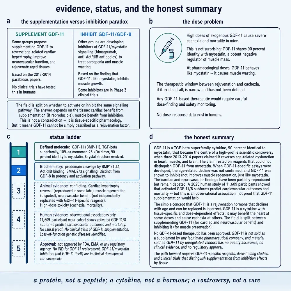
Research vendors list GDF-11 among aging and “rejuvenation” proteins. Set beside that shelf label is the ledger this monograph has been building: a solid developmental literature; a contested systemic-aging literature; human data that are mostly biomarker associations — including the 2025 activated-subform epidemiology (Walker2025) — no approved GDF-11 drug; and a famous measurement failure that still echoes whenever an immunoassay claims to count only one of two near-identical ligands. The field’s split between pathway inhibition for muscle wasting and speculative supplementation for other tissues is itself evidence that GDF-11 is not a simple youth hormone.
That ledger is the whole argument in miniature: development first, contested adult physiology second, biomarker associations third, and no approved therapeutic last. Figure 7 draws the same ladder without adding a use recommendation.
This document is a research monograph. It does not recommend that any person use GDF-11, young plasma, or any product marketed with those words. It does not provide a dose, route, or schedule for human administration. Animal doses and clinical biomarker protocols appear only as reported in their sources. Research use only.
19References
Generated from verified NCBI records rather than from recall. Author lists, titles, journals, years and identifiers below were fetched from PubMed before the build was permitted to finish.
- Collet G, Szade K, Nowak W, Klimkiewicz K, El Hafny-Rahbi B, Szczepanek K, et al.. Endothelial precursor cell-based therapy to target the pathologic angiogenesis and compensate tumor hypoxia. Cancer letters. 2016;370(2):345-57.
PMID 26577811 · doi:10.1016/j.canlet.2015.11.008 - Ferrero S, Grados-Torrez RE, Leivar P, Antolín-Llovera M, López-Iglesias C, Cortadellas N, et al.. Proliferation and Morphogenesis of the Endoplasmic Reticulum Driven by the Membrane Domain of 3-Hydroxy-3-Methylglutaryl Coenzyme A Reductase in Plant Cells. Plant physiology. 2015;168(3):899-914.
PMID 26015445 · doi:10.1104/pp.15.00597 · PMC2115167 - Jamaiyar A, Wan W, Janota DM, Enrick MK, Chilian WM, Yin L. The versatility and paradox of GDF 11. Pharmacology & therapeutics. 2017.
PMID 28223232 · PMC419886 - Katsimpardi L, Litterman NK, Schein PA, Miller CM, Loffredo FS, Wojtkiewicz GR, et al.. Vascular and neurogenic rejuvenation of the aging mouse brain by young systemic factors. Science (New York, N.Y.). 2014.
PMID 24797482 · PMC4104429 - Kolodin D, van Panhuys N, Li C, Magnuson AM, Cipolletta D, Miller CM, et al.. Antigen- and cytokine-driven accumulation of regulatory T cells in visceral adipose tissue of lean mice. Cell metabolism. 2015;21(4):543-57.
PMID 25863247 · doi:10.1016/j.cmet.2015.03.005 · PMC2884187 - Li W, Nagineni CN, Efiok B, Chepelinsky AB, Egwuagu CE. Interferon regulatory transcription factors are constitutively expressed and spatially regulated in the mouse lens. Developmental biology. 1999;210(1):44-55.
PMID 10364426 · doi:10.1006/dbio.1999.9267 - Liuzzo G, Rolandi S, Serraino A, Piva S, Giacometti F. Pre-packaged food products Business to Consumer (B2C) distance selling and information obligations in Italian mass market retailers. Italian journal of food safety. 2019;8(3):7713.
PMID 31632926 · doi:10.4081/ijfs.2019.7713 · PMC6784586 - Loffredo FS, Steinhauser ML, Jay SM, Gannon J, Pancoast JR, Yalamanchi P, et al.. Growth differentiation factor 11 is a circulating factor that reverses age-related cardiac hypertrophy. Cell. 2013.
PMID 23663781 · PMC3170097 - Long F, Zhang XM, Karp S, Yang Y, McMahon AP. Genetic manipulation of hedgehog signaling in the endochondral skeleton reveals a direct role in the regulation of chondrocyte proliferation. Development (Cambridge, England). 2001;128(24):5099-108.
PMID 11748145 · doi:10.1242/dev.128.24.5099 - Luu M, Jenike E, Vachharajani N, Visekruna A. Transcription factor c-Rel is indispensable for generation of thymic but not of peripheral Foxp3. Oncotarget. 2017;8(32):52678-52689.
PMID 28881761 · doi:10.18632/oncotarget.17079 · PMC3479579 - McPherron AC, Lawler AM, Lee SJ. Regulation of anterior/posterior patterning of the axial skeleton by growth/differentiation factor 11. Nature genetics. 1999.
PMID 10391213 - Mohan A, Guerron AD, Karam PA, Worley S, Seifarth FG. Laparoscopic Extracorporeal Appendectomy in Overweight and Obese Children. JSLS : Journal of the Society of Laparoendoscopic Surgeons. 2016;20(2).
PMID 27186069 · doi:10.4293/JSLS.2016.00020 · PMC3625558 - Müller U. Ten years of gene targeting: targeted mouse mutants, from vector design to phenotype analysis. Mechanisms of development. 1999;82(1-2):3-21.
PMID 10354467 · doi:10.1016/s0925-4773(99)00021-0 - Raymond MA, Mollica L, Vigneault N, Désormeaux A, Chan JS, Filep JG, et al.. Blockade of the apoptotic machinery by cyclosporin A redirects cell death toward necrosis in arterial endothelial cells: regulation by reactive oxygen species and cathepsin D. FASEB journal : official publication of the Federation of American Societies for Experimental Biology. 2003;17(3):515-7.
PMID 12514115 · doi:10.1096/fj.02-0500fje - Shirakabe A, Ikeda Y, Sciarretta S, Zablocki DK, Sadoshima J. Aging and Autophagy in the Heart. Circulation research. 2016;118(10):1563-76.
PMID 27174950 · doi:10.1161/CIRCRESAHA.116.307474 · PMC4487780 - Shu Y, Yang N, Cheng N, Zou Z, Zhang W, Bei Y, et al.. Intervening pyruvate carboxylase stunts tumor growth by strengthening anti-tumor actions of tumor-associated macrophages. Signal transduction and targeted therapy. 2022;7(1):34.
PMID 35105865 · doi:10.1038/s41577-019-0127-6 · PMC7339861 - Sinha M, Jang YC, Oh J, Khong D, Wu EY, Manohar R, et al.. Restoring systemic GDF11 levels reverses age-related dysfunction in mouse skeletal muscle. Science (New York, N.Y.). 2014.
PMID 24797481 · PMC3714794 - Tawde M, McLaughlin S. Implementing a Virtual Midterm to Identify Unknown Bacteria in a Microbiology Lab Course. Journal of microbiology & biology education. 2021;22(1).
PMID 34012500 · doi:10.1111/j.1541-4329.2010.00108.x · PMC1637024 - Turk AS, Johnston SC, Hetts S, Mocco J, English J, Murayama Y, et al.. Geographic Differences in Endovascular Treatment and Retreatment of Cerebral Aneurysms. AJNR. American journal of neuroradiology. 2016;37(11):2055-2059.
PMID 27390314 · doi:10.3174/ajnr.A3755 · PMC7965822 - Walker RG, Kato T, Ben Driss L, Williams SA, Hinterberg MA, Janjic N, et al.. Activated GDF11/8 subforms predict cardiovascular events and mortality in humans. Nature communications. 2025.
PMID 40664633 · PMC5976624
Registry and database sources
- UniProt Consortium. Growth/differentiation factor 11 (GDF11_HUMAN), UniProtKB O95390. Protein database record. Accessed 5 August 2026.
https://www.uniprot.org/uniprotkb/O95390
Human precursor 407 aa; gene GDF11 on 12q13.2; also named BMP-11.
20How this document was assembled
Project 05’s open-access JATS store was swept first. Of 10,110 local JATS files, 40 named GDF-11 and the identity gate admitted 28 documents (~1,179.4 printed pages). Myostatin appears far more often locally (202 files); the gate treats myostatin-only specificity footnotes as disqualified unless GDF-11 is a dense subject.
PubMed harvest used 14 queries and returned a de-duplicated union of 733 records. PMC body-text search for GDF11/GDF-11 returned 4,060 documents (designation union 4,663). Full texts were fetched for local hits, PubMed-OA PMCIDs, and a capped PMC-union top-up; 667 bodies were readable (16,191 printed pages at 1,800 characters per page). The identity gate admitted 179 documents (4,529 pp), of which 145 were subject-tier.
Series number 77 was assigned as the lowest unused integer in the filed Desktop register at scaffold time, then re-checked immediately before LIBRARY copy (A38). Re-read the filed-PDF folder before any export.
21Evidence handling
Findings are labelled in-sentence by study kind and species or population. Immunoassay results that cannot separate GDF-11 from myostatin are not treated as GDF-11-specific human pharmacology. Recency is favoured for assay-aware and dose-aware work when it does not contradict the preponderance of developmental and mechanistic evidence. Conflicts and absences are printed rather than smoothed. No dose, route or schedule is recommended for any person.
Commissioned plates in this monograph are treated as schematic evidence summaries, not as primary data. Every numeric annotation on those plates was checked against the cited literature or captioned as a literature-reported value; none is a re-assay performed for this build. Authored SVG figures carry the same rule: they encode relationships the prose already argues, and they do not invent endpoints.
Where the corpus is silent — no clean recombinant-GDF-11 first-in-human trial under that name; no GDF-11-only human pharmacology claim from the 2025 aptamer epidemiology — the silence is reported as a finding. Marketing language that outruns those limits is refused rather than paraphrased into a softer claim.
Continue exploring