
MGF and PEG-MGF A splice variant, a twenty-four-residue peptide, and the distance between them
In 1996 a rabbit muscle was held under stretch for a few days and found to be reading its own growth-factor gene differently. Resting muscle made one version; loaded muscle made a second one nobody had described. It was a genuine discovery, and a heterodox one: it said that a muscle could sense mechanical work and write itself a local growth instruction without waiting for the liver or the pituitary. Thirty years later the same three letters are printed on vials. The distance between those two facts is what this document is about, because three different objects wear this name — a splice variant, a fragment of the protein it encodes, and a chemically modified product sold as a research chemical — and the evidence behind each is thinner than the evidence behind the one before it. The most consequential thing known about the fragment is a negative: it does not activate the receptor its parent hormone uses, and after three decades nobody has found the receptor it does use.
Findings are labelled by the kind of study that produced them, in the sentence that reports them. A result in a rat is called a result in a rat. That labelling carries more weight here than in most monographs in this series, because of a specific hazard: the word MGF is used in the literature for at least three different things, and the strongest evidence attaches to the one that is not sold. Where a finding belongs to the transcript rather than to the peptide, it says so. Where it belongs to a chemically stabilised analogue rather than to the native sequence, it says so.
A second rule applies throughout. Rodent IGF-1Eb and human IGF-1Ec are the same molecule under two names, while human IGF-1Eb is a different molecule that shares one of those names. Most of the experimental work is done in rodents. Every finding in this document is attributed to the species it was obtained in, and the naming is set out in Section 06 before any cross-species claim is made.
No dose, route or schedule for human use is recommended anywhere here. Where a study parameter is reported it is reported as what that study did, with its species, its population and its duration attached.
Section 01What MGF is, and what it is not
Mechano growth factor is not a separate molecule from insulin-like growth factor 1. It is one of the forms that the IGF-1 gene can be read into, and the thing sold under its name is a fragment of that form's tail. Getting this straight at the outset removes most of the confusion that surrounds the compound, because almost every claim made for it depends on which of the three objects is meant.
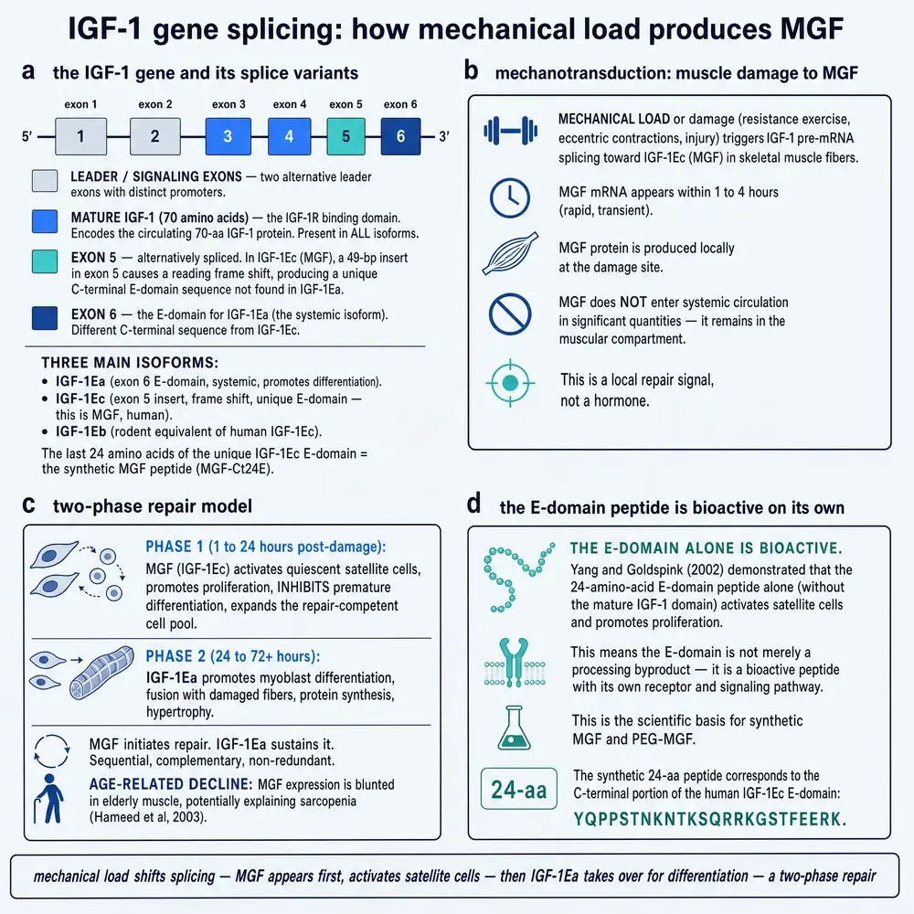
The IGF-1 gene is spliced — that is, the cell can assemble its messenger RNA from different combinations of the gene's coding blocks. Every combination produces the same mature IGF-1 hormone, the well-studied seventy-residue protein that circulates in blood under growth-hormone control. What differs between them is a tail, called the E domain, which is cut off during processing. Include a particular piece of the fifth coding block and the reading frame shifts, so that the tail comes out as an entirely different sequence. That variant is IGF-1Ec in humans, and its tail — twenty-four amino acids long — is what people mean when they say MGF the peptide.
The confusion this produces is not accidental, and it is worth naming before it does any damage. A vendor page describing MGF can quite truthfully cite decades of peer-reviewed work on mechanical loading and muscle regeneration, because that work exists and is about the splice variant. The reader is then invited to conclude that the vial has been studied for decades. It has not. The transcript has. Nothing in this document should be read as disputing the science of IGF-1 splicing, which is solid; the question throughout is which of the three objects a given piece of evidence is about.
So there are three things, and they are not interchangeable. The splice variant is a transcript and a pro-protein; it is what almost all the physiology measures, and the evidence for its existence and its regulation is strong. The E-domain peptide is a synthetic twenty-four-residue fragment; it is what almost all the pharmacology administers, and the evidence for it is substantial but is nearly all in cells and animals. PEG-MGF is that fragment with a polyethylene-glycol group attached to slow its clearance; it is what is sold, and the indexed literature contains one record naming it.
A claim that begins “MGF increases after resistance training” is a claim about the first object and is well supported. A claim that begins “MGF builds muscle” is a claim about the second object and is supported by cell and animal work whose translation is untested. A claim about what PEG-MGF does in a person is supported by nothing at all. This document keeps the three apart, and the reader who keeps them apart will find that most of the disagreement about this compound dissolves.
Section 02A rabbit's back, held under tension
The discovery belongs to a laboratory that had spent a decade doing something unfashionable: making muscles grow by force, surgically, and then asking what had changed inside them. Geoffrey Goldspink's group, working at the Royal Free in London, used a preparation in which a rabbit's tibialis anterior or latissimus dorsi was held under stretch — by casting a limb in a lengthened position, or by an implanted device — sometimes with electrical stimulation added. Held that way, a muscle gains mass within days. The question the laboratory kept returning to was what carried the instruction.
The default answer in the early 1990s was that it did not need to be carried locally at all. Insulin-like growth factor 1 was understood as an endocrine hormone: the liver makes it under growth-hormone control, it circulates, and tissues respond. A review of the field written from that laboratory in the year before the discovery sets out the problem plainly — muscle demonstrably grows in response to mechanical stimuli, and the local signal that converts force into growth had not been identified (Goldspink DF, 1995).
In 1996 Shi Yang, working with Marwan Alnaqeeb, Helen Simpson and Goldspink, extracted RNA from stretched and control rabbit muscle and probed it with a fragment spanning the third and fourth coding blocks of the IGF-1 gene (Yang et al., 1996). Resting muscle yielded a single IGF-1 sequence, the familiar one. Stretched muscle yielded that one and a second. The second carried a fifty-two-base-pair insertion that changed the reading frame of the tail, producing a precursor protein the field had not seen. They called it IGF-1Eb, after the exon composition, and proposed — carefully — that it was probably the isoform responsible for local growth regulation in response to mechanical stimulation.
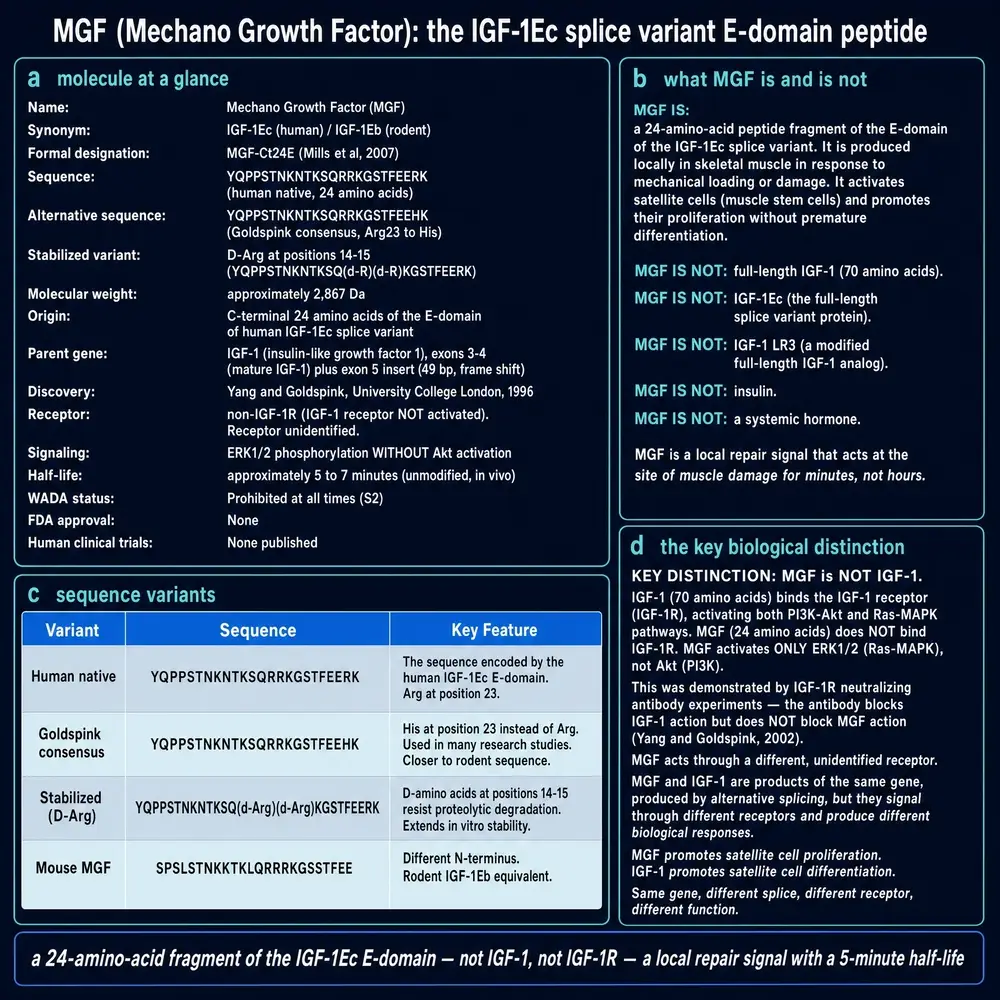
YQPPSTNKNTKSQRRKGSTFEEHK (His23); the plate also prints Arg23 (…FEERK) — both are named, not collapsed. Half-life is stated in the prose as minutes; the plate’s “5–7 minute” card is artwork framing, not a PK study reproduced here. WADA / non-approval language is status framing.Two features of that paper are worth holding on to. The first is that the molecule was discovered as a transcript, not as a protein and certainly not as a peptide: what was cloned was a messenger RNA. Everything downstream — the peptide, the vial — is an inference from that sequence, made later. The second is that the name everyone now uses does not appear in it.
Section 03How it got its name
“Mechano growth factor” arrives three years later. In 1999 Geraldine McKoy and colleagues built an assay that could distinguish the two transcripts directly and applied it to rabbit muscle under three conditions: stretch alone, electrical stimulation alone, and both together (McKoy et al., 1999). Stretch raised both IGF-1 forms; stretch with stimulation raised them further; and stimulation on its own, without stretch, did not raise them above sham-operated controls. It is that dissociation that earns the name. The transcript is not responding to muscle activity. It is responding to mechanical strain.
The paper describes the second isoform as apparently designed for an autocrine or paracrine action — acting on the cell that made it, or its neighbours — and gives it the name mechano-growth factor, MGF. Goldspink set the programme out for a wider readership the following year (Goldspink, 2000).
It is worth being explicit that the name is a claim, made of two parts. The first part, that mechanical strain is what elicits it, is the part the 1999 experiment actually supports and it has held up well. The second part, that the thing is a growth factor — a molecule that binds a receptor and instructs a cell to grow — was an inference from the family it belongs to, not a measurement. Section 09 is about what happened when that second half was finally measured, twenty years later. A great deal of what is claimed for this compound rests on a reader taking the name as a description rather than as a hypothesis.
Section 04One gene, three messages
The splicing is worth understanding properly, because it explains both why the peptide is unusual and why the naming is such a mess.
The human IGF1 gene's third and fourth coding blocks encode the mature IGF-1 hormone. Those are present in every variant, which is why every variant delivers the same growth factor. What follows them is the E domain, and its composition is what the splicing decides: exons four and six give the E domain called Ea, exons four and five give Eb, and exons four, five and six together give Ec. The first sixteen residues of all three are identical, because they come from exon four. After that they diverge completely.
The divergence is not gradual. Splicing in a forty-nine-base-pair piece of exon five — forty-nine is not divisible by three — shifts the reading frame, so every codon downstream is read out of step. The result is not a variant tail but an unrelated one. This is the mechanism that produces IGF-1Ec, and the twenty-four residues it produces are the subject of most of this document.
Two consequences follow immediately. The first is that the same gene, under mechanical load, produces a hormone plus a peptide that has no sequence relationship to anything else in the IGF system. The second is that because the insert differs in size between species — forty-nine base pairs in humans, fifty-two in rodents — the resulting peptides differ in length, twenty-four residues against twenty-five, and the species use different names for the same thing. That is the trap set out in Section 06, and it is not a pedantic one: it decides which findings belong to the molecule under discussion.
Section 05The last twenty-four residues
The peptide at the centre of this compound's commercial life is short enough to print in a line:
Twenty-four residues, six of them lysine or arginine, which makes the peptide strongly positively charged at physiological pH. It corresponds to the carboxy-terminal portion of the human IGF-1Ec E domain, and it is synthesised to order rather than purified from tissue. The sequence given above was verified character by character against four independent full texts in the corpus assembled for this document, which is worth stating because vendor listings for this compound are not consistent with one another.
Two things about it are unusual. The first is that it is a fragment of a pro-protein — a piece that is cut off during processing and discarded — rather than a signalling molecule in its own right, at least as classically conceived. Whether the E domain has an independent life after cleavage, or is simply a spacer that gets degraded, is the question the whole field turns on, and it is not settled. The second is that this fragment is extremely short-lived. Native, unmodified, it is destroyed by plasma proteases within minutes, which is why almost nothing in the animal literature uses it in that form.
It is also worth noting what the peptide is not. It is not a growth hormone, it is not IGF-1, and it contains none of the IGF-1 sequence: the mature hormone comes from exons three and four and is removed before this fragment begins. A vial of MGF, if it contains what its label says, contains none of the hormone whose gene it is named after.
Section 06The species problem
This section exists because a naming collision in this literature is capable of reversing the meaning of a citation, and it is not obvious from the outside.
Human IGF1 produces three E-domain variants: Ea, Eb and Ec. Rat and mouse Igf1 produce only two: Ea and Eb. The rodent Eb is the mechanically responsive one, and it is the orthologue of the human Ec. So rodent IGF-1Eb and human IGF-1Ec are the same molecule, both called MGF — while human IGF-1Eb is a different molecule that happens to share the rodent's name.
The practical consequence is large, because most of the experimental work on this compound is done in rodents, and a substantial part of it is published under the Eb designation. A reader who maps names to names rather than orthologue to orthologue will make one of two errors: attributing a rodent finding about MGF to the wrong human variant, or discarding a rodent MGF finding on the grounds that it concerns Eb. The founding 1996 paper is itself an example — it was done in rabbit, it names the isoform IGF-1Eb, and it is the discovery paper for what humans call Ec (Yang et al., 1996). The reviews in the corpus that set the pairing out explicitly are the ones worth following on this point; several sources that do not set it out get it wrong.
Nothing in this document attributes a finding across species without naming both the species and the designation the original used.
Section 07What is actually in the vial
The native twenty-four-residue peptide is not, in general, what has been studied, and it is not what is sold. This is the single most under-reported fact about the compound, and it is recoverable only from methods sections.
The cardiac literature — which is the most methodologically careful body of in-vivo work on the E peptide — does not use the native sequence. Mavrommatis and colleagues describe their construction precisely: the peptide is amidated at the carboxy terminus, and the two adjacent arginines at positions fourteen and fifteen are replaced with their D-stereoisomers, both changes made to resist proteolysis (Mavrommatis et al., 2013). The same construction carries through the subsequent infarct work (Shioura et al., 2014; Peña et al., 2015). That is a chemically distinct molecule from the sequence printed in Section 05, and every result obtained with it belongs to it.
The same papers also define two controls that matter for reading the literature. A scrambled peptide, containing the same amino acids in random order, is the in-vivo negative control; and an S18A peptide, with the serine at position eighteen replaced by alanine, is the inactive control for the cell work. Both contain the compound's designation in their names, and both generate null results. Any automated summary of this literature that counts mentions rather than reading them will attribute those nulls to the active peptide. The corpus assembly for this document strips both before counting, for exactly that reason.
A third construction appears in the anti-doping literature rather than the academic one. MGF R23H, with the arginine at position twenty-three replaced by histidine, was identified in confiscated vials by a laboratory working on detection methods; it is stabilised in plasma sufficiently that its metabolites should be findable in urine (Cox et al., 2017). And PEG-MGF is a fourth, in which a polyethylene-glycol group is attached to slow renal clearance further.
The reason to lay all four out side by side is that they are routinely discussed as though they were one substance. They are not. A result obtained with a D-amino-acid-containing amidated analogue delivered by osmotic pump into a mouse is not a result about a PEGylated peptide injected by a person, and neither is a result about the native sequence. Where this document reports a finding, it names the construction the source used.
The modifications exist because the native peptide does not last long enough to do anything. That is not a technicality: it is a finding about the native peptide, and it constrains what can be expected of any preparation that has not been characterised. A product sold as “MGF” whose chemistry is unstated could be the native sequence, in which case its circulating life is measured in minutes, or a modified form, in which case the animal literature on the native sequence does not describe it.
Section 08Proliferate, but do not become muscle yet
The most consistently replicated biological finding about the E peptide is also the one least often stated correctly in material written to sell it.
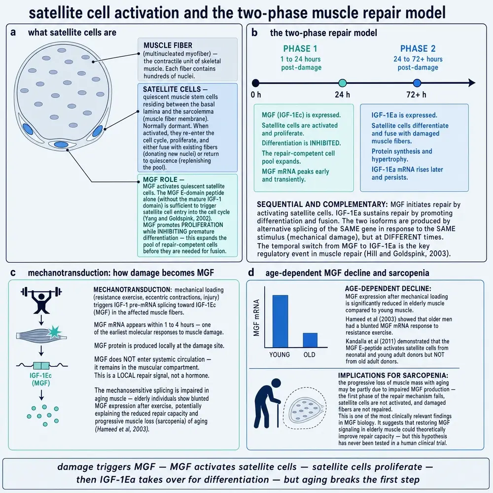
Skeletal muscle repairs itself using satellite cells: a reserve population sitting quiescent against the muscle fibre, which on injury or loading divide, and whose daughters either fuse into the damaged fibre or return to the reserve. IGF-1 acts on that system in a well-characterised way — it drives the cells towards differentiation and fusion. The E peptide does something different, and in one respect opposite.
Ates, Yang and Orrell cultured primary human muscle cells from healthy donors and from patients with muscular dystrophy and amyotrophic lateral sclerosis, and compared the two signals directly (Ates et al., 2007). The E peptide raised the number of mononucleated progenitor cells in all three groups, including the diseased muscle where baseline progenitor numbers were lower. IGF-I enhanced myogenic differentiation; the E peptide blocked that pathway. Their conclusion is the useful formulation: the result is an increased progenitor pool rather than immediate fibre repair.
Kandalla, Goldspink and Butler-Browne extended this across donor age (Kandalla et al., 2011). In cultures from neonatal and young adult muscle the twenty-four-residue peptide increased proliferative lifespan and delayed senescence; in cultures from old adults it did not. The fusion capacity of the cells rose in all the cultures.
What follows from this is worth stating carefully, because it is where most of the overstatement enters. Expanding a pool of progenitor cells while holding them back from differentiating is not the same as building muscle. It is, if anything, an argument for the peptide as one half of a two-stage process in which something else — IGF-1, or the withdrawal of the E peptide — supplies the second half. The primary papers describe it that way. A claim that the peptide causes hypertrophy is not what these experiments show, and no experiment in the corpus assembled for this document shows it in a person.
Section 09The receptor that is not there
This is the mechanistic heart of the compound, and it is a negative result.
The name “growth factor” carries an implicit model: the molecule binds a receptor on the cell surface, the receptor's kinase domain activates, and a signalling cascade follows. For the IGF system that receptor is IGF-1R. Because MGF is an IGF-1 variant, the assumption ran for years that its activity was IGF-1R activity. In 2016 that assumption was tested directly.
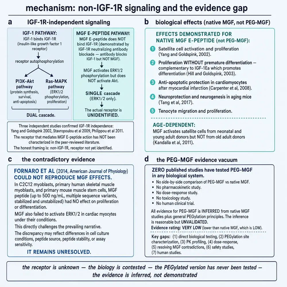
Janssen, Hofland and Strasburger used kinase receptor activation assays — which measure autophosphorylation of the receptor itself on ligand binding — for the human IGF-1 receptor and both insulin receptor isoforms, and ran four ligands across them (Janssen et al., 2016). Full-length MGF, meaning the whole pro-protein including the mature IGF-1 sequence, did activate IGF-1R: its maximal effect at high equimolar concentration was comparable to IGF-I, eighty-nine-fold against seventy-seven-fold, but its half-maximal concentration was 7.83 nmol/L against 0.86 for IGF-I. That is roughly a ninefold loss of potency, which is what one would expect of a molecule carrying the same hormone with a different tail attached. It is a weaker agonist of the same receptor, not a different kind of signal.
The E peptide is the finding. Neither human MGF — meaning the E-domain peptide — nor the stabilised analogue produced any detectable IGF-1R activation in that assay.
Two other laboratories arrived at compatible conclusions from different directions. Mavrommatis and colleagues, tracking a fluorescently tagged peptide into cardiac cells, reported rapid cellular uptake that did not involve IGF-1 receptor activation, and localisation to the nucleus (Mavrommatis et al., 2013). Armakolas and colleagues silenced the IGF-1 receptor and the insulin receptor with interfering RNA in prostate cancer cells and found that the E peptide's mitogenic activity and its intracellular signalling persisted (Armakolas et al., 2010).
Three routes, one conclusion: whatever the peptide does, it does without the receptor its parent hormone uses. That is genuinely interesting. It is also, thirty years after the transcript was cloned, an admission that the mechanism is unknown. Candidate explanations exist in the literature — direct entry to the cell and action in the nucleus, an unidentified surface receptor, effects mediated through cytoskeletal and 14-3-3 protein interactions (Solís et al., 2022) — and they are candidates. No binding constant for the E peptide at any named target is carried by the evidence base assembled for this document.
A molecule with a known receptor can be reasoned about before it is tested in people: the receptor's distribution predicts which tissues will respond, its other ligands predict what it will interact with, and a dose-response curve predicts what a given exposure will do. None of that reasoning is available here. The peptide is taken up by many cell types in culture and does something in most of them, which is the profile of a molecule whose selectivity nobody can yet state.
Section 10Everything else it has been tried on
The E peptide has been applied to a striking range of tissues, and the breadth is easy to mistake for depth.
In the heart, the stabilised analogue delivered at the time of coronary artery ligation in mice reduced apoptosis and preserved contractile function at two weeks, and delivery from polymeric microstructures placed on the infarct improved function similarly (Mavrommatis et al., 2013; Shioura et al., 2014; Peña et al., 2015). Different regions of the prohormone act differently on human mesenchymal stem cells, which is an argument that the E domain is not merely a spacer (Collins et al., 2010).
In nerve, plasmid-delivered MGF into the hindlimb muscles of SOD1(G93A) mice at symptom onset improved hindlimb strength and motor unit survival, with significantly more motoneurons surviving than with IGF-I delivered the same way (Riddoch-Contreras et al., 2009). The peptide promotes neurogenesis in the ageing mouse brain (Tang et al., 2017).
In connective tissue, it accelerates tenocyte migration by reducing cell stiffness (Zhang et al., 2014), improves healing of injured rat tendon (Zhang et al., 2016), improves mobility of mechanically injured human anterior cruciate ligament fibroblasts through Rac1 and RhoA pathways (Sha et al., 2022), and reduces cartilage degeneration in a knee osteoarthritis model (Song et al., 2018). In bone it promotes osteoblast proliferation and defect healing in rabbits (Deng et al., 2011), and it has proangiogenic activity on vascular endothelial cells (Deng et al., 2012). In periodontal ligament stem cells it acts through Fyn, RhoA and YAP (Feng et al., 2024).
The most recent in-vivo work in this corpus is an arthritis model: in HLA-B27 transgenic rats given MGF for seven weeks after inoculation, the onset of arthritis symptoms was delayed in a dose-dependent manner, from fourteen days in controls to twenty-three days in the high-dose group, with anti-inflammatory cytokines rising and tumour necrosis factor alpha falling (Moro et al., 2025).
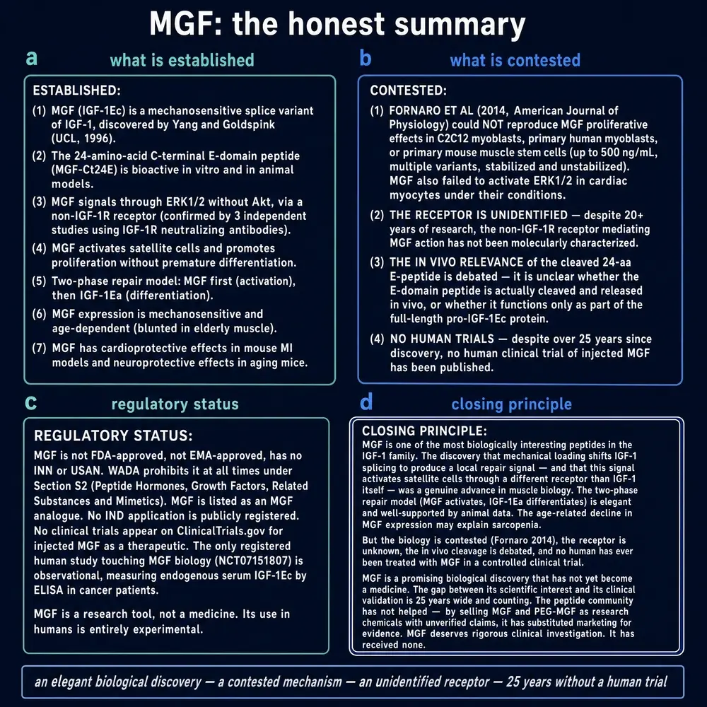
Read as a list, that is impressive. Read as evidence, three qualifications apply and they are not minor. Almost every entry is a single laboratory's work without independent replication. The great majority is in vitro or in rodents. And a molecule that produces a beneficial-looking result in every tissue it is applied to is either extraordinarily useful or is producing a generic response — increased proliferation, reduced apoptosis — that looks beneficial in any short-term injury model and whose long-term consequences a short-term injury model cannot see. Section 15 is about what that second possibility looks like when someone goes looking for it.
Section 11The response that fades
One further finding from the founding laboratory deserves its own section, because it is the one that gave this molecule a therapeutic story.
The problem it speaks to is real and large. Skeletal muscle mass declines from roughly the fifth decade onwards, and the decline is not merely cosmetic: it predicts falls, frailty, loss of independence and mortality. What makes sarcopenia hard is that ageing muscle does not simply have less of everything. It responds differently. Load an old muscle and it hypertrophies less than a young one given the same stimulus, and the question of why has occupied the field for thirty years. Any molecule that could be shown to be the missing step in that response would be genuinely important, which is why this particular result has been cited far outside its original context.
The experiment behind it is a demanding one. In synergist ablation the muscles that share a tendon's work are removed, so that the muscle under study must carry the whole load; it is a much larger and more sustained mechanical stimulus than exercise, and it produces rapid hypertrophy in a young animal. That is the stimulus the old animals failed to respond to.
Owino, Yang and Goldspink overloaded rat plantaris and soleus muscles by ablating their synergists, and compared young animals with old (Owino et al., 2001). Young muscle hypertrophied markedly and activated its satellite cells. Old muscle did neither, and the molecular correlates were specific: considerably lower expression of MGF, together with a failure to upregulate the IGF-1 receptor and MyoD.
The reading that made the molecule famous is that ageing muscle loses the ability to make its own local growth signal, and that supplying the signal might restore the response. It is a reasonable hypothesis and it is why most readers have heard of this compound. Three things should be said alongside it. The result is a transcript measurement in rats. It reports a whole programme failing to respond — receptor and transcription factor as well as ligand — rather than one missing molecule, so restoring the one need not restore the programme. And in human cells the peptide's proliferative effect was present in young donors and absent in old ones (Kandalla et al., 2011), which is the opposite of what a straightforward replacement model predicts.
Section 12Measured, not administered
There is a fact about the human evidence for this compound that ought to be stated once, plainly, without hedging, before anything else in this Part:
No published study located for this document has administered MGF or PEG-MGF to a human being, by any route, at any dose. Every human study in this corpus measured the endogenous transcript in muscle biopsies.
This is not a gap of the kind that a monograph normally reports — an absent phase III, a missing long-term safety study. It is the absence of the first step. The peptide has never been given to a person in a study that reported what happened.
The distinction is easy to lose because the human literature is real, competent and reasonably large. People have been biopsied before and after resistance exercise, after eccentric loading, after weeks of training, after therapeutic ultrasound, and while receiving recombinant growth hormone, and their IGF-1Ec transcript has been quantified each time. Those are proper experiments and Section 13 reports what they found. But the thing measured is the body's own response, and the intervention is exercise or a different drug. A study showing that muscle makes more of this transcript after training is evidence that the transcript responds to training. It is not evidence that injecting a fragment of the resulting protein does anything, and no amount of restatement converts one into the other.
Section 13What the human loading studies show
Taken on their own terms, the human studies establish that the response is real, fast and variable.
Greig and colleagues biopsied healthy women after isometric exercise and found the isoforms differentially expressed (Greig et al., 2006). Hameed and colleagues compared young and elderly people after eccentric cycling, the contraction type that produces the most mechanical strain per unit of work (Hameed et al., 2008). Popov and colleagues varied resistance exercise intensity and metabolic stress in the same participants and tracked anabolic signalling alongside myogenic gene expression (Popov et al., 2015). Ahtiainen and colleagues followed resistance training in younger and older men (Ahtiainen et al., 2016). Delgado-Diaz and colleagues showed that therapeutic ultrasound — mechanical energy delivered without voluntary contraction — affects splice variant expression in human muscle (Delgado-Diaz et al., 2011), which is a neat confirmation of the mechanical rather than metabolic trigger.
Two studies come at it from the endocrine side. Aperghis and colleagues gave healthy young men recombinant growth hormone and measured both serum IGF-I and the splicing in muscle (Aperghis et al., 2009); Velloso and colleagues ran two weeks of the same and tracked the response (Velloso et al., 2013). And Gharahdaghi and colleagues did something more pointed: they suppressed testosterone pharmacologically in young non-hypogonadal men and put them through six weeks of supervised resistance training, showing that the molecular transducers of exercise-induced growth are impaired without it (Gharahdaghi et al., 2022). That is a demonstration that this transcript sits inside a hormonal context and does not operate as an independent switch.
The honest summary of this literature is that IGF-1Ec expression in human muscle rises with mechanical loading, that the size and timing of the rise depend on age, training status, contraction type and hormonal state, and that it is one member of a coordinated programme rather than the programme's controller. None of these studies administered the peptide, and none of them was designed to test whether administering it would do anything.
Section 14The registry, read directly
House style for this series requires every registry claim to be verified against the registry itself rather than against a summary of it, so this section reports a query rather than a belief.
ClinicalTrials.gov was queried on 3 August 2026 through its API, on the intervention field, for “mechano growth factor”, “MGF”, “PEG-MGF” and “IGF-1Ec”. The result is easily stated: no interventional registration of MGF or PEG-MGF as an intervention was returned by any query. The records that free-text searching surfaces are studies of recombinant IGF-1, of growth hormone and its pegylated forms, and of unrelated agents whose descriptions happen to contain the search terms.
One registration names the molecule. NCT07151807, sponsored by Çanakkale Onsekiz Mart University, began on 1 April 2025 and was active and not recruiting when checked. It is observational, with a hundred participants, and it measures serum IGF-1 and IGF-1Ec by ELISA in patients with solid tumours, correlating them with stage, size, metastasis and lymph node involvement, to see whether the IGF-1Ec level and the IGF-1Ec/IGF-1 ratio track tumour progression.
That is the entire registered human presence of this molecule: one observational study, investigating it as a marker of how far a cancer has advanced. The direction is worth sitting with. The compound is sold on the proposition that more of it is good for you; the only place it currently appears in the clinical research record is as a candidate index of tumour burden.
Section 15The signal that runs the other way
The finding in Section 09 — that the E peptide acts without the IGF-1 receptor — is usually presented as the interesting thing about the molecule. It is also the reason the following body of work is difficult to dismiss.
In 2010 Armakolas and colleagues, working in Athens, examined IGF-1Ec in human prostate tissue and cell lines (Armakolas et al., 2010). They reported that it was markedly overexpressed in prostate cancer and in prostatic intraepithelial neoplasia relative to normal prostate, and that normal prostate epithelial cells did not express it at all. They then applied the synthetic E peptide to prostate cancer cells and found that it stimulated their growth and activated ERK1/2 — and that silencing the IGF-1 receptor and the insulin receptor did not abolish either effect. Their conclusion names the peptide as possessing mitogenic activity through mechanisms independent of IGF-1R, the insulin receptor and their hybrids.
The same laboratory has continued the work, and its most recent output is the fact that most clearly indicates how the molecule is now regarded in oncology. In 2024 they published the development and characterisation of an anti-Ec-peptide monoclonal antibody as a candidate prostate cancer therapy (Armakolas et al., 2024). In cell lines the antibody reduced proliferation, migration and invasion; in tumours in immunodeficient mice it reduced tumour size and metastasis rate, reversed a mesenchymal phenotype, and inhibited the mobilisation of host stem cells towards the tumour. A parallel review from the same group sets out the roles of the IGF-1 isoforms in breast cancer and treats their defining E peptides as therapeutic targets (Kotsifaki et al., 2024).
It does not establish that the peptide causes cancer, and this document does not say so. The human data are associative — a molecule overexpressed in a tumour may be a consequence of the tumour's biology rather than a driver — and the interventional work is in cell lines and in mice.
What it does establish is that a serious research programme, sustained over fifteen years and now at the stage of developing a blocking antibody, proceeds on the premise that less of this peptide is better in at least one human disease. The most advanced translational effort built on this molecule is an effort to neutralise it. A reader deciding what weight to give the regenerative literature should know that the same property — proliferation driven independently of the usual receptors — is what both literatures are describing.
No study in the corpus assembled for this document has looked for neoplastic outcomes after administering the peptide to an animal for a long period, and none has looked in a person at all. The absence of such a study is not evidence of safety; it is the absence of the experiment that would address the question.
Section 16Why a two-minute peptide invites a coat
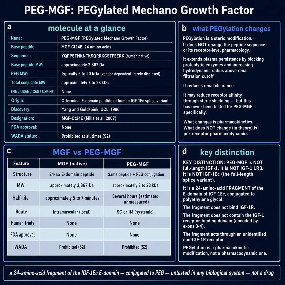
The marketed form of this molecule is not the native twenty-four-residue peptide. It is almost always sold as PEG-MGF — the same fragment with a polyethylene-glycol chain attached. That is not a branding flourish. It is an attempt to solve a pharmacokinetic problem that the research literature itself keeps rediscovering: the unmodified E peptide does not last long enough to be a useful circulating agent. The cardiac series that provides the most careful in-vivo work on the fragment did not even try. It redesigned the molecule first — amidating the carboxy terminus and replacing the adjacent arginines at positions fourteen and fifteen with their D-stereoisomers — precisely because proteolysis otherwise wins in minutes (Mavrommatis et al., 2013). PEGylation is the other answer to the same problem, chosen by the market rather than by the laboratories that published the biology.
So PEGylation is not a side note to the PEG-MGF story. It is the PEG-MGF story. Without the coat, what is sold would be a peptide the research literature already treats as too short-lived to study without chemical stabilisation. With the coat, what is sold is a conjugate that has never been characterised in public. The rest of this Part holds those two facts apart: the platform is real and mature; the product that borrows its name is not.
Section 17What PEGylation actually is
Polyethylene glycol is a repeating ether polymer, water-soluble and largely inert in biological systems. In 1977 Abraham Abuchowski, Frank Davis and colleagues at Rutgers showed that covalently attaching methoxypolyethylene glycol chains to a protein could blunt its immunogenicity, protect it from proteases, and extend its circulating life. In acatalasemic mice, PEG-modified bovine catalase lasted longer in blood across repeated intravenous doses and resisted trypsin, chymotrypsin and a bacterial protease while retaining most of its enzymatic activity (Abuchowski et al., 1977). That pair of results — keep the activity, lose the clearance and the immune recognition — is the founding claim of the field.
Three decades of refinement turned the claim into a delivery platform. Attaching one or more PEG chains to a peptide or protein prolongs residence in the body, reduces enzymatic degradation, and can reduce or eliminate immunogenicity; the polymer itself is non-toxic, non-immunogenic and water-soluble, and conjugates built on it have reached regulatory approval across several therapeutic classes (Veronese & Pasut, 2005). The chemistry is not a single reaction. Site of attachment, PEG molecular weight, linear versus branched architecture, and whether the linker is stable or releasable all change the conjugate’s size, activity and clearance, and the choices are the subject of a literature of their own (Roberts et al., 2002).
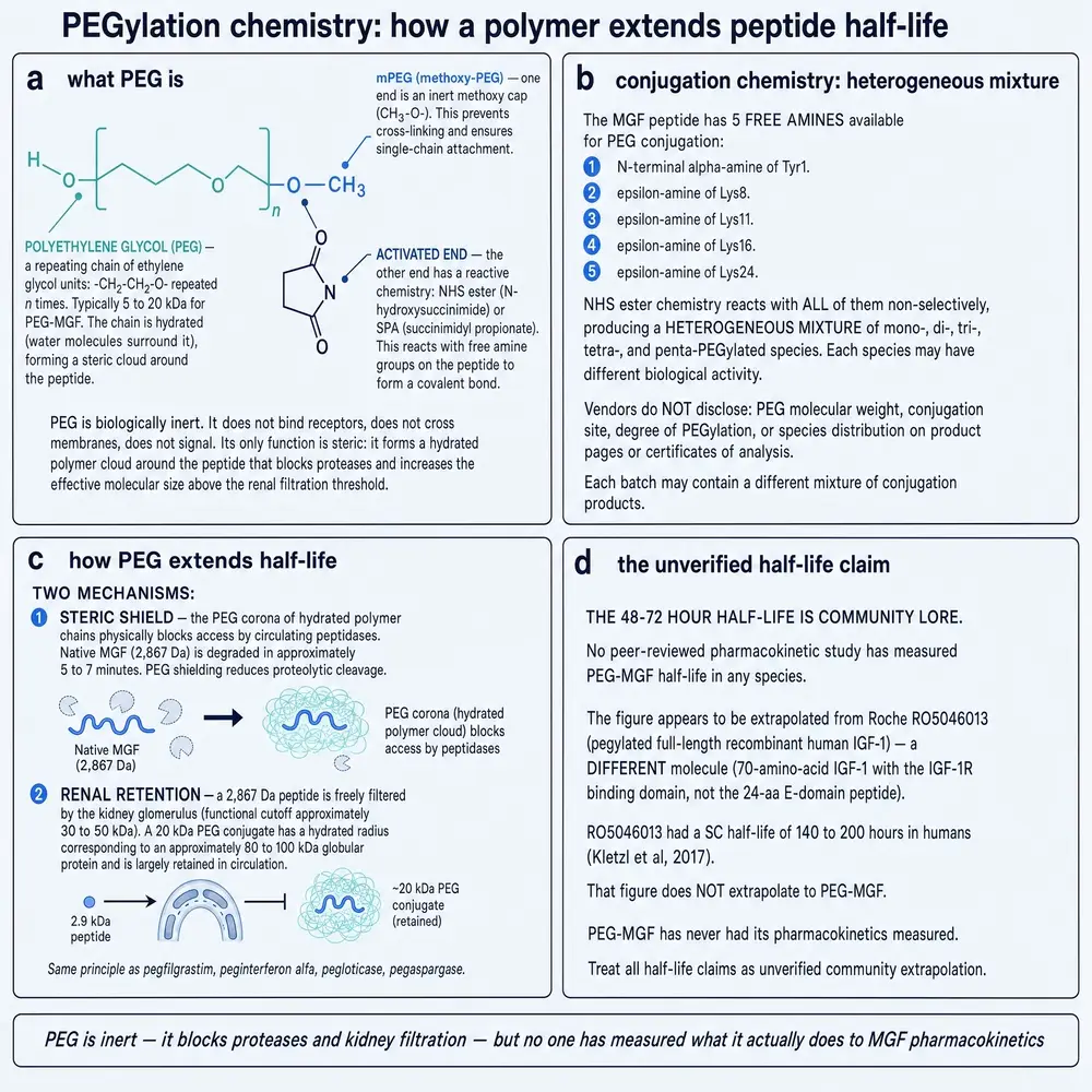
For a short peptide the intended gains are easy to state in ordinary language. A twenty-four-residue fragment is small enough to be filtered by the kidney almost as soon as it reaches the glomerulus. A PEG chain of several kilodaltons increases the hydrodynamic radius of the conjugate, so the kidney sees something larger and clears it more slowly. The same chain sterically hinders proteases, which is a second, independent reason the unmodified fragment disappears in minutes. Neither effect creates a new biology. Both buy time — time for whatever the peptide does to happen before the molecule is gone.
That is why PEGylation is the obvious industrial answer to the proteolysis problem in Section 07. The academic literature answered it with D-amino acids and amidation, which change the peptide itself. The market answered it with a polymer coat, which leaves the peptide sequence notionally intact and changes its size. Both answers are chemically coherent. Only one of them has a published body of work attached to this sequence, and it is not the coat.
Section 18What a PEGylated MGF would have to specify
“PEG-MGF” is not a complete chemical name. It is a class label. A characterised PEGylated peptide states at least the peptide sequence, the molecular weight of the PEG, the attachment site, the architecture, and the linker chemistry. Without those fields, two vials with the same trade abbreviation can contain different molecules, and a pharmacokinetic claim made for one does not transfer to the other (Roberts et al., 2002; Veronese & Pasut, 2005).
For the twenty-four-residue Ec E peptide the attachment-site question is
especially sharp. The sequence
YQPPSTNKNTKSQRRKGSTFEEHK carries six lysine or arginine
residues. Random lysine PEGylation would produce a mixture of mono- and
multi-PEGylated species with different activities; site-specific N-terminal
PEGylation would produce one species. Branched twenty-kilodalton PEG and
linear two-kilodalton PEG are not interchangeable coats. None of this is
exotic chemistry. It is the ordinary specification of every PEGylated
medicine that has been approved. None of it was located for material sold as
PEG-MGF.
There is a further requirement that PEGylation reviews state plainly and that vendors of research chemicals often omit: the conjugate must still do what the parent did. A PEG chain large enough to slow clearance can also sterically block the face of the peptide that contacts its target, and retained activity is an empirical result, not a promise that travels with the polymer (Veronese & Pasut, 2005). For this molecule the requirement is stricter than usual. The parent fragment’s target is itself unidentified (Section 09). A demonstration that PEG-MGF retains “the activity of MGF” would first have to say which activity, in which assay, against which construction of the parent. No such demonstration was located.
Section 19What the literature holds for PEG-MGF
The PEGylation platform is therefore well documented. The PEGylated form of this peptide is not.
Searched on 5 August 2026, PubMed returns one record for “PEG-MGF”, and the union of every PEGylated-MGF phrasing returns seven, most of which name it only in passing. The single most relevant is a 2026 review of performance-enhancing peptides that modulate the growth hormone and IGF-1 axis, which lists PEG-MGF among the agents encountered in clinical practice and in online self-administration protocols, alongside the growth hormone secretagogues and IGF-1 analogues, and records the classes of adverse effect reported across that whole group — endocrine and metabolic disturbance, fluid retention, musculoskeletal symptoms, injection-site reactions (Dominikowski et al., 2026). It is a description of a market, not a study of a conjugate.
Adjacent work on PEGylated IGF-1 family agents exists and is worth naming so that it is not silently borrowed. Prabhath and colleagues delivered a pegylated IGF-1 mimic from a polycaprolactone-based matrix into a rat rotator-cuff repair model and reported improved enthesis structure and tendon tensile properties at eight weeks relative to unpegylated and repair controls (Prabhath et al., 2022). That is evidence that PEGylation can matter for an IGF-1-related biotherapeutic in an animal injury model. It is not evidence about the twenty-four-residue Ec E peptide, not evidence about material sold as PEG-MGF, and not a characterisation of any commercial PEG-MGF vial. The molecules share a gene family and a delivery idea. They are not the same conjugate.
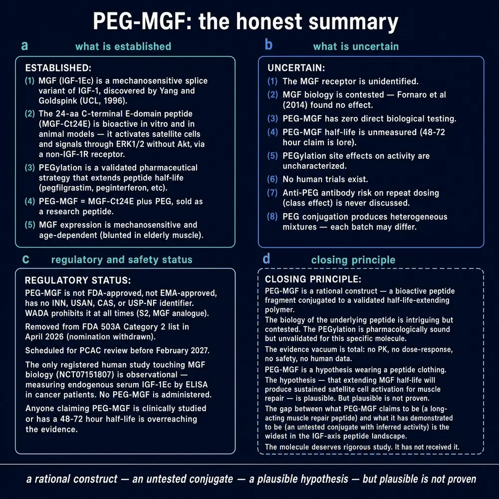
So the position on PEG-MGF is this. No published characterisation of marketed material was located: no mass, no purity, no confirmation of the PEG attachment site or molecular weight, no demonstration that the peptide inside is the sequence in Section 05. No pharmacokinetic study in any species. No efficacy study of the PEGylated form in any model. No human administration. No registered trial. The claim that pegylation extends this peptide’s half-life is a chemically well-founded expectation drawn from the PEGylation platform (Abuchowski et al., 1977; Veronese & Pasut, 2005) that, for this molecule, has not been shown.
Stating what would have to exist for the question to become answerable is more useful than restating the absence: an analytical characterisation of marketed material that reports sequence, PEG mass, attachment site and architecture; a pharmacokinetic study in one species; and a demonstration that the PEGylated form retains a defined activity of the native fragment. None of the three is chemically difficult. None has been published for PEG-MGF.
The absence that matters is not an absence of PEGylation science. That science is mature, cited above, and underwrites medicines people already take. The absence that matters is an absence of any public bridge from that platform to this twenty-four-residue sequence. Until that bridge is built, every claim about what PEG-MGF does is a claim about a molecule whose chemistry has not been shown.
Section 20Confiscated vials
The most informative literature on what is actually being sold under this name was not written by the people studying the biology. It was written by anti-doping laboratories, whose job is to find out what is in a vial and whether its use can be detected.
Thevis and colleagues characterised a biotechnologically produced full-length mechano growth factor by mass spectrometry, specifically for doping-control purposes (Thevis et al., 2014) — that is, material presented as MGF was obtained and its identity established analytically rather than assumed from the label. Cox, Miller and Eichner went further: they identified the peptides in confiscated vials, finding MGF R23H alongside BPC 157, worked out the analogue's metabolism in plasma, and established that it is stable enough that it should be detectable in urine (Cox et al., 2017). The same laboratory has since extended detection methods to large peptides above two kilodaltons (Cox et al., 2022).
Two things are worth drawing out. The first is the R23H substitution itself. The material in those vials was not the native sequence; it was an engineered analogue chosen for stability, which means somebody in the supply chain understood the proteolysis problem in Section 07 and engineered around it. The second is what this implies for reading vendor claims: the compound circulating commercially is heterogeneous, and its chemistry is not generally stated.
This document takes no position on doping regulation and offers no guidance on use. The anti-doping literature is cited here for one reason: it is the only body of work that has analysed the material people actually obtain, and it finds that the material is not necessarily what the research literature studied.
Section 21Weighing it
Pulling the evidence together, claim by claim, with the level of support each has in the corpus assembled for this document.
The criteria are stated so that a reader can disagree with a particular row rather than with the exercise. A claim scores on four things: the kind of study behind it, whether it has been replicated, whether the replication came from an independent group, and whether the species and preparation used bear on the claim as worded. A result obtained once, in one laboratory, in cultured cells, supporting a claim phrased about human physiology scores low not because the experiment was poor but because three separate inferential steps sit between it and the claim.
Two things this table deliberately does not do. It does not weight by journal, and it does not treat a large number of small studies as equivalent to a replication, because in this literature the same laboratory frequently supplies several of them. Where a row rests on a single group's work, that is recorded in the row rather than averaged away.
Where recency and precedence conflict, this document weights the newer result — provided it is not contradicted by a preponderance of older evidence — and two conflicts in this literature are decided that way.
The first is mechanism. Between 1996 and roughly 2010 the working model was that MGF was an IGF-1 variant acting through IGF-1R. The 2016 receptor measurement contradicts that for the E peptide directly, using an assay built for the purpose, and it is corroborated by two independent lines of evidence (Janssen et al., 2016; Mavrommatis et al., 2013; Armakolas et al., 2010). The newer result supersedes, and this document treats the E peptide's mechanism as unidentified.
The second is the therapeutic framing. The ageing literature from 1999–2007 points towards replacement therapy for sarcopenia; the oncology literature from 2010–2024 points towards blockade. These are not strictly contradictory — a molecule can be beneficial in one tissue and harmful in another — but they cannot both be assumed, and the blockade programme is the one that has progressed to a therapeutic candidate. The document reports both and treats neither as settled.
What emerges is a molecule whose biology is well established and whose pharmacology is almost entirely unexamined. The gene is real, the splicing is real, the mechanical trigger is real and demonstrated in humans, the peptide has reproducible effects on cultured cells, and animal injury models respond to it. Past that point the evidence stops. There is no human administration, no registered trial, no receptor, no dose-response in any human system, no long-term animal safety study, no characterisation of the marketed product, and one active human research question, which is whether the molecule marks cancer progression.
One further measurement belongs here, because it bears on how much of that literature a reader can expect to find unaided. This compound's three-letter abbreviation is among the most heavily over-subscribed in the series: it returns roughly ten records for every one about the molecule, and the largest single competitor is not an abbreviation at all but the chemical formula of magnesium fluoride.
The practical consequence is that a straightforward literature search on this compound returns mostly other subjects, and a reader who does not notice will conclude either that the evidence base is enormous or that it is incoherent. It is neither. It is small, specific, and mostly preclinical, and finding it requires knowing that the name is shared.
Section 22One retraction
One paper in this compound's literature has been withdrawn, and this series identifies what was retracted rather than quietly omitting it.
A 2019 paper in Bioscience Reports reported that mechano growth factor attenuated apoptosis in intervertebral disc nucleus pulposus cells subjected to mechanical overload, through inhibition of the p38 MAPK pathway (Xu et al., 2019). It was retracted in 2024; the journal notice is PMID 39171815. This document does not rely on it for any factual claim about the compound, and it is described here only as a claim that was made and withdrawn.
Setting it aside changes little, which is worth saying explicitly. It was a single in-vitro study in a tissue not otherwise central to this document, and no other conclusion here depends on it. The publication status of every record in the corpus was checked against PubMed's own publication-type and comments-corrections fields rather than against any secondary report; this was the one hit.
Section 23What would settle it
It is more useful to end by naming the experiments that would move this compound out of its current position than by restating the position.
An identified target. The single largest gap is mechanistic. A binding partner for the twenty-four-residue peptide, with an affinity constant, would convert a list of tissue effects into a pharmacology and would make selectivity and dose predictable rather than empirical.
An analytical characterisation of marketed material. Mass, purity, sequence confirmation and, for PEG-MGF, the attachment chemistry. The anti-doping laboratories have shown this is straightforward (Thevis et al., 2014; Cox et al., 2017), and until it is done routinely, no result in the research literature can be assumed to describe what is sold.
A long-term animal study with neoplastic endpoints. The prostate work raises a question that short-term injury models cannot answer, and it has not been asked in the form that would answer it.
A first-in-human study. Not an efficacy trial — a characterisation. What happens to the peptide, how long it lasts, what tolerability looks like. Every other compound discussed in this monograph series that reached clinical use began there. This one has not begun.
This document describes published research. It does not recommend human use of MGF, PEG-MGF, or any related peptide, and it specifies no dose, route or schedule for any person. Study parameters reported in these pages are reported as what a particular study did, in a particular species, for a particular duration, and are not transferable to human use. Neither MGF nor PEG-MGF is an approved medicine in any jurisdiction identified in the preparation of this document, and no registered interventional trial of either was located.
Section 21References
Generated from verified NCBI records rather than from recall. Author lists, journal names, volumes, pages and identifiers are taken from the PubMed record for each citation, and the build refuses to run if any identifier fails to resolve. This is not a formality. During the preparation of this monograph three identifiers recalled from memory were checked against PubMed and two resolved to real but entirely unrelated papers — one on Notch signalling in breast cancer, one on coronary stents. That is the eighth consecutive monograph in this series in which recalled identifiers were wrong, and it is why nothing in the list below was typed.
A second identifier defect was reproduced live during this build and is worth recording because it is invisible to every count. An ad-hoc script written to check these records collected each paper's PubMed Central accession with a walk over every ArticleId element in the record. That walk also traverses the reference list, so what came back was the accession of the last work each paper cited. Every value looked well formed. It was caught only because a 2025 paper cannot carry a 2008-era accession. The reference generator reads the record's own identifier list and does not have the defect.
- Abuchowski A, McCoy JR, Palczuk NC, van Es T, Davis FF. Effect of covalent attachment of polyethylene glycol on immunogenicity and circulating life of bovine liver catalase. J Biol Chem. 1977;252(11):3582-6.
PMID 16907 - Ahtiainen JP, Hulmi JJ, Lehti M, Kraemer WJ, Nyman K, Selänne H, et al.. Effects of resistance training on expression of IGF-I splice variants in younger and older men. Eur J Sport Sci. 2016;16(8):1055-63.
PMID 27231807 · doi:10.1080/17461391.2016.1185164 - Aperghis M, Velloso CP, Hameed M, Brothwood T, Bradley L, Bouloux PM, et al.. Serum IGF-I levels and IGF-I gene splicing in muscle of healthy young males receiving rhGH. Growth Horm IGF Res. 2009;19(1):61-7.
PMID 18799338 · doi:10.1016/j.ghir.2008.07.002 - Armakolas A, Philippou A, Panteleakou Z, Nezos A, Sourla A, Petraki C, et al.. Preferential expression of IGF-1Ec (MGF) transcript in cancerous tissues of human prostate: evidence for a novel and autonomous growth factor activity of MGF E peptide in human prostate cancer cells. Prostate. 2010;70(11):1233-42.
PMID 20564425 · doi:10.1002/pros.21158 - Armakolas A, Alevizopoulos N, Stathaki M, Petraki C, Agrogiannis G, Samiotaki M, et al.. Anti-PEc: Development of a novel monoclonal antibody against prostate cancer. Br J Cancer. 2024;131(3):551-564.
PMID 38902531 · doi:10.1038/s41416-024-02713-8 · PMC11300853 - Ates K, Yang SY, Orrell RW, Sinanan AC, Simons P, Solomon A, et al.. The IGF-I splice variant MGF increases progenitor cells in ALS, dystrophic, and normal muscle. FEBS Lett. 2007;581(14):2727-32.
PMID 17531227 · doi:10.1016/j.febslet.2007.05.030 - Collins JM, Goldspink PH, Russell B. Migration and proliferation of human mesenchymal stem cells is stimulated by different regions of the mechano-growth factor prohormone. J Mol Cell Cardiol. 2010;49(6):1042-5.
PMID 20875825 · doi:10.1016/j.yjmcc.2010.09.017 · PMC2989540 - Cox HD, Miller GD, Eichner D. Detection and in vitro metabolism of the confiscated peptides BPC 157 and MGF R23H. Drug Test Anal. 2017;9(10):1490-1498.
PMID 28035768 · doi:10.1002/dta.2152 - Cox HD, Knussmann GN, Moore C, Eichner D. Detection of insulin analogues and large peptides >2 kDa in urine. Drug Test Anal. 2022;14(7):1264-1272.
PMID 35261185 · doi:10.1002/dta.3249 - Delgado-Diaz DC, Gordon BS, Dompier T, Burgess S, Dumke C, Mazoué C, et al.. Therapeutic ultrasound affects IGF-1 splice variant expression in human skeletal muscle. Am J Sports Med. 2011;39(10):2233-41.
PMID 21785002 · doi:10.1177/0363546511414857 - Deng M, Zhang B, Wang K, Liu F, Xiao H, Zhao J, et al.. Mechano growth factor E peptide promotes osteoblasts proliferation and bone-defect healing in rabbits. Int Orthop. 2011;35(7):1099-106.
PMID 21057789 · doi:10.1007/s00264-010-1141-2 · PMC3167400 - Deng M, Wang Y, Zhang B, Liu P, Xiao H, Zhao J. New proangiogenic activity on vascular endothelial cells for C-terminal mechano growth factor. Acta Biochim Biophys Sin (Shanghai). 2012;44(4):316-22.
PMID 22382131 · doi:10.1093/abbs/gms012 - Dominikowski A, Rękoś Z, Olejarz M, Szczepanek-Parulska E, Domin R, RuchaŁa M. The emerging landscape of performance-enhancing peptides modulating GH-IGF1 axis: bridging the gap between clinical evidence and patient self-administration. Front Endocrinol (Lausanne). 2026;17:1822475.
PMID 42395176 · doi:10.3389/fendo.2026.1822475 · PMC13322892 - Feng F, Tu T, Wang H, Song R, Li J, Zhu Y, et al.. Mechano-growth factor regulates periodontal ligament stem cell proliferation and differentiation through Fyn-RhoA-YAP signaling. Biochem Biophys Res Commun. 2024;733:150450.
PMID 39067248 · doi:10.1016/j.bbrc.2024.150450 - Fornaro M, Hinken AC, Needle S, Hu E, Trendelenburg AU, Mayer A, et al.. Mechano-growth factor peptide, the COOH terminus of unprocessed insulin-like growth factor 1, has no apparent effect on myoblasts or primary muscle stem cells. Am J Physiol Endocrinol Metab. 2014;306(2):E150-6.
PMID 24253050 · doi:10.1152/ajpendo.00408.2013 - Gharahdaghi N, Rudrappa S, Brook MS, Farrash W, Idris I, Aziz MHA, et al.. Pharmacological hypogonadism impairs molecular transducers of exercise-induced muscle growth in humans. J Cachexia Sarcopenia Muscle. 2022;13(2):1134-1150.
PMID 35233984 · doi:10.1002/jcsm.12843 · PMC8977972 - Goldspink DF, Cox VM, Smith SK, Eaves LA, Osbaldeston NJ, Lee DM, et al.. Muscle growth in response to mechanical stimuli. Am J Physiol. 1995;268(2 Pt 1):E288-97.
PMID 7532362 · doi:10.1152/ajpendo.1995.268.2.E288 - Goldspink G. Cloning of local growth factors involved in the determination of muscle mass. Br J Sports Med. 2000;34(3):159-60.
PMID 10854011 · doi:10.1136/bjsm.34.3.159 · PMC1763256 - Greig CA, Hameed M, Young A, Goldspink G, Noble B. Skeletal muscle IGF-I isoform expression in healthy women after isometric exercise. Growth Horm IGF Res. 2006;16(5-6):373-6.
PMID 17107821 · doi:10.1016/j.ghir.2006.09.005 - Hameed M, Orrell RW, Cobbold M, Goldspink G, Harridge SD. Expression of IGF-I splice variants in young and old human skeletal muscle after high resistance exercise. J Physiol. 2003;547(Pt 1):247-54.
PMID 12562960 · doi:10.1113/jphysiol.2002.032136 · PMC2342624 - Hameed M, Toft AD, Pedersen BK, Harridge SD, Goldspink G. Effects of eccentric cycling exercise on IGF-I splice variant expression in the muscles of young and elderly people. Scand J Med Sci Sports. 2008;18(4):447-52.
PMID 18067523 · doi:10.1111/j.1600-0838.2007.00714.x - Hill M, Goldspink G. Expression and splicing of the insulin-like growth factor gene in rodent muscle is associated with muscle satellite (stem) cell activation following local tissue damage. J Physiol. 2003;549(Pt 2):409-18.
PMID 12692175 · doi:10.1113/jphysiol.2002.035832 · PMC2342958 - Janssen JA, Hofland LJ, Strasburger CJ, van den Dungen ES, Thevis M. Potency of Full-Length MGF to Induce Maximal Activation of the IGF-I R Is Similar to Recombinant Human IGF-I at High Equimolar Concentrations. PLoS One. 2016;11(3):e0150453.
PMID 26991004 · doi:10.1371/journal.pone.0150453 · PMC4798685 - Kandalla PK, Goldspink G, Butler-Browne G, Mouly V. Mechano Growth Factor E peptide (MGF-E), derived from an isoform of IGF-1, activates human muscle progenitor cells and induces an increase in their fusion potential at different ages. Mech Ageing Dev. 2011;132(4):154-62.
PMID 21354439 · doi:10.1016/j.mad.2011.02.007 - Kotsifaki A, Maroulaki S, Karalexis E, Stathaki M, Armakolas A. Decoding the Role of Insulin-like Growth Factor 1 and Its Isoforms in Breast Cancer. Int J Mol Sci. 2024;25(17).
PMID 39273251 · doi:10.3390/ijms25179302 · PMC11394947 - Mavrommatis E, Shioura KM, Los T, Goldspink PH. The E-domain region of mechano-growth factor inhibits cellular apoptosis and preserves cardiac function during myocardial infarction. Mol Cell Biochem. 2013;381(1-2):69-83.
PMID 23712705 · doi:10.1007/s11010-013-1689-4 · PMC3720995 - McKoy G, Ashley W, Mander J, Yang SY, Williams N, Russell B, et al.. Expression of insulin growth factor-1 splice variants and structural genes in rabbit skeletal muscle induced by stretch and stimulation. J Physiol. 1999;516 ( Pt 2)(Pt 2):583-92.
PMID 10087355 · doi:10.1111/j.1469-7793.1999.0583v.x · PMC2269271 - Moro A, Qin H, Yuan S, Li H, Liao S, Yang J. Mechanical growth factor inhibited syndesmophyte formation and the progression of osteoarthritis and ankylosing spondylitis-like symptoms in HLA-B27/Hu-β2m transgenic rats. Arthritis Res Ther. 2025;27(1):218.
PMID 41272763 · doi:10.1186/s13075-025-03677-7 · PMC12639975 - Owino V, Yang SY, Goldspink G. Age-related loss of skeletal muscle function and the inability to express the autocrine form of insulin-like growth factor-1 (MGF) in response to mechanical overload. FEBS Lett. 2001;505(2):259-63.
PMID 11566187 · doi:10.1016/s0014-5793(01)02825-3 - Peña JR, Pinney JR, Ayala P, Desai TA, Goldspink PH. Localized delivery of mechano-growth factor E-domain peptide via polymeric microstructures improves cardiac function following myocardial infarction. Biomaterials. 2015;46:26-34.
PMID 25678113 · doi:10.1016/j.biomaterials.2014.12.050 · PMC4328136 - Popov DV, Lysenko EA, Bachinin AV, Miller TF, Kurochkina NS, Kravchenko IV, et al.. Influence of resistance exercise intensity and metabolic stress on anabolic signaling and expression of myogenic genes in skeletal muscle. Muscle Nerve. 2015;51(3):434-42.
PMID 24916884 · doi:10.1002/mus.24314 - Prabhath A, Vernekar VN, Esdaille CJ, Eisenberg E, Lebaschi A, Badon M, et al.. Pegylated insulin-like growth factor-1 biotherapeutic delivery promotes rotator cuff regeneration in a rat model. J Biomed Mater Res A. 2022;110(7):1356-1371.
PMID 35253991 · doi:10.1002/jbm.a.37378 · PMC10324960 - Riddoch-Contreras J, Yang SY, Dick JR, Goldspink G, Orrell RW, Greensmith L. Mechano-growth factor, an IGF-I splice variant, rescues motoneurons and improves muscle function in SOD1(G93A) mice. Exp Neurol. 2009;215(2):281-9.
PMID 19038252 · doi:10.1016/j.expneurol.2008.10.014 - Roberts MJ, Bentley MD, Harris JM. Chemistry for peptide and protein PEGylation. Adv Drug Deliv Rev. 2002;54(4):459-76.
PMID 12052709 · doi:10.1016/s0169-409x(02)00022-4 - Sha Y, Zhang B, Chen L, Hong H, Chi Q. Mechano Growth Factor Accelerates ACL Repair and Improves Cell Mobility of Mechanically Injured Human ACL Fibroblasts by Targeting Rac1-PAK1/2 and RhoA-ROCK1 Pathways. Int J Mol Sci. 2022;23(8).
PMID 35457148 · doi:10.3390/ijms23084331 · PMC9026312 - Shioura K, Pena J, Goldspink P. Administration of a Synthetic Peptide Derived from the E-domain Region of Mechano-Growth Factor Delays Decompensation Following Myocardial Infarction. Int J Cardiovasc Res. 2014;3(3):1000169.
PMID 25606570 · doi:10.4172/2324-8602.1000169 · PMC4297642 - Solís C, Thompson WC, Peña JR, McDermott-Roe C, Langa P, Warren CM, et al.. Mechano-growth factor E-domain modulates cardiac contractile function through 14-3-3 protein interactomes. Front Physiol. 2022;13:1028345.
PMID 36467694 · doi:10.3389/fphys.2022.1028345 · PMC9709209 - Song Y, Xu K, Yu C, Dong L, Chen P, Lv Y, et al.. The use of mechano growth factor to prevent cartilage degeneration in knee osteoarthritis. J Tissue Eng Regen Med. 2018;12(3):738-749.
PMID 28599103 · doi:10.1002/term.2493 - Tang JJ, Podratz JL, Lange M, Scrable HJ, Jang MH, Windebank AJ. Mechano growth factor, a splice variant of IGF-1, promotes neurogenesis in the aging mouse brain. Mol Brain. 2017;10(1):23.
PMID 28683812 · doi:10.1186/s13041-017-0304-0 · PMC5501366 - Thevis M, Thomas A, Geyer H, Schänzer W. Mass spectrometric characterization of a biotechnologically produced full-length mechano growth factor (MGF) relevant for doping controls. Growth Horm IGF Res. 2014;24(6):276-80.
PMID 25466910 · doi:10.1016/j.ghir.2014.10.004 - Velloso CP, Aperghis M, Godfrey R, Blazevich AJ, Bartlett C, Cowan D, et al.. The effects of two weeks of recombinant growth hormone administration on the response of IGF-I and N-terminal pro-peptide of collagen type III (P-III-NP) during a single bout of high resistance exercise in resistance trained young men. Growth Horm IGF Res. 2013;23(3):76-80.
PMID 23433656 · doi:10.1016/j.ghir.2013.01.002 - Veronese FM, Pasut G. PEGylation, successful approach to drug delivery. Drug Discov Today. 2005;10(21):1451-8.
PMID 16243265 · doi:10.1016/S1359-6446(05)03575-0 - Xu Q, Fang H, Zhao L, Zhang C, Zhang L, Tian B. Mechano growth factor attenuates mechanical overload-induced nucleus pulposus cell apoptosis through inhibiting the p38 MAPK pathway. Biosci Rep. 2019;39(3).
PMID 30858307 · doi:10.1042/BSR20182462 · PMC6438874 - Yang S, Alnaqeeb M, Simpson H, Goldspink G. Cloning and characterization of an IGF-1 isoform expressed in skeletal muscle subjected to stretch. J Muscle Res Cell Motil. 1996;17(4):487-95.
PMID 8884603 · doi:10.1007/BF00123364 - Yang SY, Goldspink G. Different roles of the IGF-I Ec peptide (MGF) and mature IGF-I in myoblast proliferation and differentiation. FEBS Lett. 2002;522(1-3):156-60.
PMID 12095637 · doi:10.1016/s0014-5793(02)02918-6 - Yi Q, Feng J, He L, Wan R, Zeng H, Yang L, et al.. The structure-function relationships of insulin-like growth factor 1 Ec in C2C12 cells. Cell Adh Migr. 2018;12(1):47-55.
PMID 28471324 · doi:10.1080/19336918.2017.1318240 · PMC5810762 - Zhang B, Luo Q, Mao X, Xu B, Yang L, Ju Y, et al.. A synthetic mechano-growth factor E peptide promotes rat tenocyte migration by lessening cell stiffness and increasing F-actin formation via the FAK-ERK1/2 signaling pathway. Exp Cell Res. 2014;322(1):208-16.
PMID 24434354 · doi:10.1016/j.yexcr.2014.01.005 - Zhang B, Luo Q, Kuang D, Ju Y, Song G. Mechano-growth factor E peptide promotes healing of rat injured tendon. Biotechnol Lett. 2016;38(10):1817-25.
PMID 27334712 · doi:10.1007/s10529-016-2162-8 - [No authors listed]. Retraction: Mechano growth factor attenuates mechanical overload-induced nucleus pulposus cell apoptosis through inhibiting the p38 MAPK pathway. Biosci Rep. 2024;44(8).
PMID 39171815 · doi:10.1042/BSR-2018-2462_RET · PMC11345658
Sources without a PubMed record
Registry records and sequence-database entries have no PubMed record and are listed separately, so that the generated list above remains wholly machine-verified. Each was read directly rather than through a summary of it.
- United States National Library of Medicine. ClinicalTrials.gov — intervention-field queries for “mechano growth factor”, “MGF”, “PEG-MGF” and “IGF-1Ec”. Queried 3 August 2026 through the API v2 studies endpoint. No interventional registration of MGF or PEG-MGF as an intervention was returned by any query. This is the source for the statement in Section 14 that the compound has no registered interventional trial anywhere.
https://clinicaltrials.gov/ - Canakkale Onsekiz Mart University. NCT07151807 — Serum IGF-1 and IGF-1Ec in Malignancy Assessment. Observational, 100 participants, start 1 April 2025, active and not recruiting as at 3 August 2026. The only registration naming IGF-1Ec, and it measures the peptide as a serum biomarker of tumour burden rather than administering it.
https://clinicaltrials.gov/study/NCT07151807 - UniProt Consortium. P05019 — IGF1_HUMAN, insulin-like growth factor I. Used for the exon composition and isoform structure of the human IGF1 gene product.
https://www.uniprot.org/uniprotkb/P05019/entry
Section 22How this document was assembled
The corpus was built against project 05, the Therapeutic Peptide Research Library, and against PubMed and PubMed Central. As with every compound in this series the interesting arithmetic is not what was collected but what had to be refused, and for this compound the refusals are unusually large in proportion to the subject.
The identity problem, which is this compound's largest single obstacle
The three-letter abbreviation MGF returned 2,001 PubMed records when this document was compiled, against 205 for “mechano growth factor” by name — roughly ten to one against the subject. Ten distinct non-subject meanings were identified by reading a title sample of the rejected set rather than by assuming them, and two of them are larger than anything a peptide chemist would predict.
The largest is not an abbreviation at all. MgF2 is magnesium fluoride, and it accounts for 824 records: optical anti-reflection coatings, solid-electrolyte interphases in lithium and sodium batteries, surface-plasmon-resonance biosensors, radioluminescent dosimetry. Letter case is a genuine discriminator here for once — Mg is not MG — but it cannot be relied on alone, because indexers render the subscript inconsistently and any case-insensitive matching collapses the distinction entirely.
The second is more dangerous, because subject-matter corroboration is inert against it. In African swine fever virus genomics MGF expands to multigene family, and the gene clusters are written exactly as this compound is written: MGF 110, MGF 360, MGF 505, MGF_110-3L. It is a large and very active literature, and it is about interferon signalling, innate immunity, inflammation and host response — which is the same vocabulary that would corroborate a growth factor. Nothing in the expansion contains the word “growth”, so a reader of a hit never sees the problem.
The remainder: mangiferin, a plant xanthone abbreviated MGF throughout natural-product pharmacology (1,347 records); mast cell growth factor, which is stem cell factor and kit ligand, and whose mouse gene locus is itself named Mgf; macrophage, myeloid and milk growth factor; Mycoplasma gallisepticum F-strain in poultry vaccination; Mascot Generic Format, the mass spectrometry file; the moment generating function in statistics; and recent machine-learning coinages built on “multi-granularity fusion”.
Because the collisions share the subject's vocabulary, the gate could not work by looking for supporting terms. It is inverted: a bare code is refused outright when the text carries a known non-subject expansion, and admitted only on an unambiguous designation — the coined name, either species' splice designation, the PEGylated form, or the twenty-four-residue sequence — or on compound-level corroboration in the absence of any disqualifying expansion.
The matcher was break-tested before the first sweep, against twenty-seven constructed traps drawn from real records. The first run passed twenty-six of twenty-seven and was wrong: the reason column showed that every refusal — swine fever, mangiferin, the mass-spectrometry format, the moment generating function — was being attributed to magnesium fluoride, because the fluoride pattern had been written case-insensitively and was matching the subject's own designation. The pass count was clean and the gate was broken. Only naming each rejection exposed it.
| Stage | What it did | Result |
|---|---|---|
| local sweep | every full-text store in project 05 | 45,975 files opened |
| identity gate | applied to each proposed match | 422 proposed, 97 refused, 325 admitted |
| source classification | by document kind, before any corpus figure | 21 peer-reviewed full texts of 325 |
| PubMed harvest | four-arm scoped query | 1,599 records, 1,161 retained |
| PMC body-text sweep | full text, not title and abstract | 494 matches, 81 indexed as subject |
| full-text fetch | the whole body-text surface, not the screened subset | 773 documents |
| substantive-use screen | applied on the far side of the fetch | 166 substantive, 90 tool-use, 154 passing mentions discarded |
| keyed merge | local and external union, keyed by identifier | 98 duplicate documents removed |
Reading corpus: 264 unique scientific full texts, ~5,668 printed-page equivalents. The body-text sweep was fetched in full rather than screened first: at 494 documents the surface was small enough to read, and screening it down to the 81 whose abstracts name the compound would have discarded exactly the papers this document most needed — the physiology that measures the splice variant among a panel of transcripts and never names it outside a methods section. 98 documents present in both the local and external routes were counted once.
What the local stores actually contain
Classifying the local sweep by document kind before quoting any corpus figure is a standing requirement in this series, and for this compound the ratio is itself a finding. Of 325 local assets admitted as being about MGF, 21 were peer-reviewed full texts. The remaining 303 were 290 vendor page snapshots and product listings, 8 pieces of trade and affiliate copy, and this project's own earlier write-ups — a ratio of about 14.4 to one in favour of commerce. For a compound with a substantial grey market and a small, mostly preclinical literature, that split describes where the compound now lives, and reporting the raw match count as a corpus would have described the market while appearing to describe the science.
Publication integrity
Every record in the harvest was checked against PubMed's own publication type and comments-corrections fields rather than against any secondary report. One retracted paper was found and is identified in Section 19. No claim in this document rests on it.
Figures
All 23 figures in this document are original works, authored as inline SVG and generated from the values in the evidence dossier. No commissioned artwork exists for this compound and no third-party figure has been reproduced. Each generator derives any total it prints from the data it is drawing and refuses to emit otherwise; the first draft of the sequence figure claimed nine basic residues and was refused at six, which also confirmed the positions this document cites for the stabilising modifications.
Section 23Evidence handling
Study type is named in the sentence that reports the finding. A result in a rat is called a result in a rat, and a result in cultured cells is called that. Nothing in this document phrases an animal or in-vitro result so as to imply a human outcome.
The three objects are held apart. This is the rule that does the most work here. “MGF” denotes a splice variant, a synthetic twenty-four-residue peptide, and a PEGylated product, and the evidence is strongest for the first and absent for the third. Where a finding belongs to the transcript it is attributed to the transcript. Where it belongs to a chemically modified analogue — the amidated, D-arginine-substituted peptide of the cardiac studies, or MGF R23H — it is attributed to that analogue, because those are different molecules and the modification is usually stated only in a methods section.
Negative controls are stripped before counting. The cardiac series uses a scrambled peptide as its in-vivo control and an S18A substitution as its inactive control. Both carry the compound's designation in their names and both generate null results, so any procedure that counts mentions rather than reading them will attribute those nulls to the active peptide. Both were removed before any mention count was taken.
Species and designation travel together. Rodent IGF-1Eb and human IGF-1Ec are the same molecule under two names; human IGF-1Eb is a different molecule sharing one of them. Every cross-species statement in this document names both the species and the designation its source used.
Recency is weighted, and where it conflicts with precedence the conflict is reported as a conflict. The 2016 receptor measurement supersedes the earlier assumption that the E peptide acts through IGF-1R, because it tested the question directly and is corroborated by two independent lines of evidence. The regenerative literature of 1999–2011 and the oncology literature of 2010–2024 point in opposite directions about whether more of this peptide is desirable; both are reported, neither is treated as settled, and the reader is told which has progressed to a therapeutic candidate.
Absence is reported as a finding rather than passed over. No human administration study, no registered interventional trial, no identified receptor, no characterisation of marketed material, and no long-term animal study with neoplastic endpoints were located. Each of those absences is stated where it bears on a claim, and the registry position was established by querying the registry directly on the date given rather than by citing a review.
No human use, dose, route or schedule is recommended anywhere in this document. Study parameters are reported as what a particular study did, in a particular species, for a particular duration.

Continue exploring