
The Semax Family A Soviet neuropeptide, four products, and the distance between them
Semax is a real pharmacological achievement. In the 1980s a Moscow laboratory took a fragment of a stress hormone, bolted a three-residue tail onto it, and produced a molecule that kept the hormone's effects on behaviour while shedding its effects on the adrenal gland. It has been a registered medicine in Russia for decades. But the evidence supporting it is narrower than its reputation, and of the four compounds now sold under its name, three have almost no scientific literature at all — while the one direct test of the most popular modification found that the modification made the molecule worse.
01What the four names mean
Four products circulate in the research-peptide market under closely related names: Semax, N-acetyl Semax, N-acetyl Semax amidate, and Adamax. They are priced differently and described differently, and a buyer could reasonably infer that each represents a refinement of the last. The chemistry behind the names is straightforward. The evidence behind them is not, and the gap between the four is the subject of this monograph.
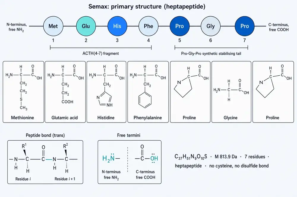
Semax itself is a heptapeptide — seven amino acids — with the sequence methionine, glutamate, histidine, phenylalanine, proline, glycine, proline. Written in single letters it is MEHFPGP. The first four residues are lifted directly from adrenocorticotropic hormone, the pituitary signal that tells the adrenal cortex to make cortisol; specifically they are residues four through seven of that hormone. The last three, proline–glycine–proline, are not from ACTH at all. They were added.
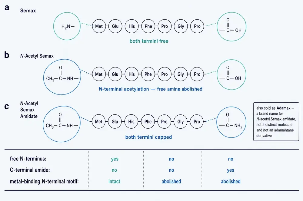
The three modified products differ from Semax only at its ends. N-acetyl Semax carries an acetyl group on the nitrogen at the front of the molecule. N-acetyl Semax amidate carries that acetyl group and, at the other end, replaces the terminal acid group with an amide. Both changes are standard medicinal chemistry, and both are made for the same stated reason: the enzymes that chew up peptides in the body work inward from the ends, so capping the ends should slow them down.
Adamax is the difficult one. It is catalogued as a "Semax-related peptide" and sold as such, but no molecular formula, no molecular weight, no registry number and no amino-acid sequence is attached to it in any source examined for this monograph, and it appears nowhere in the scientific literature. Section 23 deals with it at length, because what happened when an automated system tried to assemble evidence about it is instructive about this entire market.
02Moscow, 1977: memory, pain, and a mealworm beetle
The idea that became Semax is older than the molecule. In 1977 Igor Ashmarin, working in Leningrad and Moscow, published a paper in the Journal of Evolutionary Biochemistry and Physiology arguing that short chains of amino acids — oligopeptides — act as modulators of memory and pain, and speculating about where in evolution such a system might have come from (Ashmarin, 1977). This was a period when the peptide-regulation idea was in the air internationally, but the Soviet programme pursued it with unusual institutional weight: laboratories devoted to the chemistry of natural compounds had been standing up across the USSR since the late 1960s, among them the Institute of Molecular Genetics of the Academy of Sciences, which is where Semax was eventually made (Deigin et al., 2022).
The first experimental step in the published record is charmingly unlike what came after. In 1978 Ashmarin's group tested adrenocorticotropic hormone and two tripeptides against imprinting behaviour — in Tenebrio molitor, the mealworm beetle (Sheĭman et al., 1978). One of the co-authors on that paper is V. N. Nezavibat'ko, the peptide chemist whose name appears on every foundational Semax paper for the next thirteen years.
The choice of ACTH as a starting point was not arbitrary. By the 1970s it was established that fragments of ACTH influenced learning and attention in animals without needing the hormone's action on the adrenal gland, which is carried by a different part of the molecule. The behavioural activity sat in a short stretch near the N-terminus. If you could isolate that stretch, you would have a compound that acted on the brain without switching on the stress axis. The problem was that a four-residue peptide is destroyed almost immediately in the body.
03The tail
The solution was to attach something the body's peptide-cleaving enzymes handle badly. Proline is awkward for many proteases: its side chain loops back and bonds to its own backbone nitrogen, which distorts the local geometry and frustrates enzymes that expect a normal extended chain. A proline–glycine–proline tripeptide on the C-terminal end of ACTH(4–7) gives the molecule a rear guard.
That, at least, is the design rationale as it is stated across the literature — that Pro-Gly-Pro was appended "to ensure the resistance of Semax to peptidases" and "to increase the duration of action of ACTH(4–7)". It is worth being precise about the status of that claim. It is stated as background in essentially every modern paper on the compound, but none of the papers examined here presents the stability measurement that would demonstrate it for Semax specifically. The nearest direct evidence arrived much later, from a different angle, and it turned out to complicate the picture considerably: Pro-Gly-Pro is not an inert handle. It is itself biologically active, it is the dominant species recovered from tissue after Semax is administered, and it does some of what Semax does and conspicuously not the rest. Section 11 takes that up.
04What was engineered out
The result that justifies the whole design appeared in 1986, in the Bulletin of Experimental Biology and Medicine, under the title "Hormonal activity of the ACTH(4–10) analog — a prolonged-action stimulant of learning" (Ponomareva-Stepnaia et al., 1986). The authors set out to test whether their long-acting analogue had retained any of the parent hormone's endocrine activity. It had not. Unlike ACTH(5–10), the peptide showed no steroidogenic activity and no melanocyte-stimulating activity.
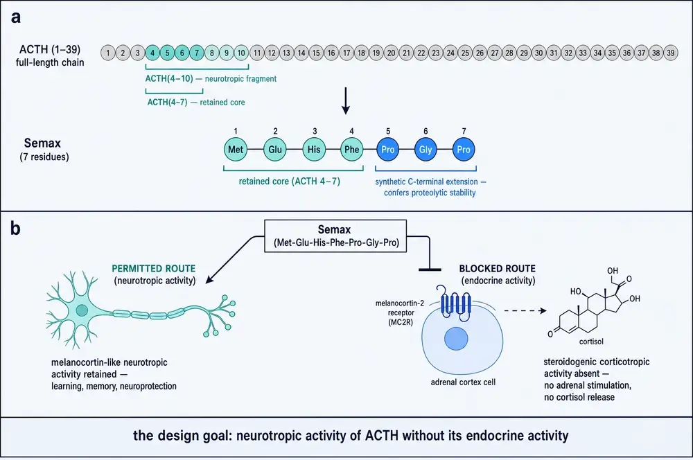
That is a negative result, and it is the most important single finding in the compound's history. A molecule that stimulated learning but also drove cortisol release would have been a stress hormone with a side effect, not a nootropic. Removing the endocrine arm is what made the thing a drug candidate rather than a curiosity. Every modern description of Semax as "devoid of hormonal activity" traces back to this measurement.
A second 1986 paper from the same circle — Antonova and colleagues, with Nezavibat'ko again on the author line — reported that the heptapeptide altered open-field behaviour in rats at 0.015 mg/kg and abolished amphetamine-induced stimulation, without changing forebrain concentrations of noradrenaline, dopamine or serotonin (Antonova et al., 1986). Neither 1986 paper uses the name "Semax". The name first appears in the indexed record in 1991, on a study of how the peptide is degraded by rat blood enzymes (Potaman et al., 1991b), and in a 1992 paper on resistance to hypoxia with Ashmarin as senior author (Kaplan et al., 1992).
A 1997 review by the founding group is titled, in translation, "A nootropic adrenocorticotropin analog 4-10-semax (15 years experience in its design and study)" (Ashmarin et al., 1997). Counting back fifteen years from 1997 places the start of the programme around 1982 — before any of the published papers, which is what one would expect of work conducted inside a Soviet institute and written up afterwards.
05Getting to the brain: two numbers, thirty years apart
A peptide taken into the body faces two problems: enzymes, and the blood–brain barrier. The Semax literature contains two quantitative measurements of the second problem, and they frame everything else.
The first came in 1991, in Neuroscience Letters, from a group including Myasoedov and Nezavibat'ko (Potaman et al., 1991a). They made a tritium-labelled version of the peptide, injected it intravenously into rats, and used high-performance liquid chromatography to look for it in blood and brain extracts. They found it. The brain-to-blood ratios were significantly higher than for labelled bovine serum albumin measured the same way, which matters because albumin does not cross the barrier — so the signal was genuine tissue entry rather than blood trapped in brain capillaries. The number they reported is the one worth remembering: the quantity of peptide in the brain did not exceed 0.01% of the injected dose.
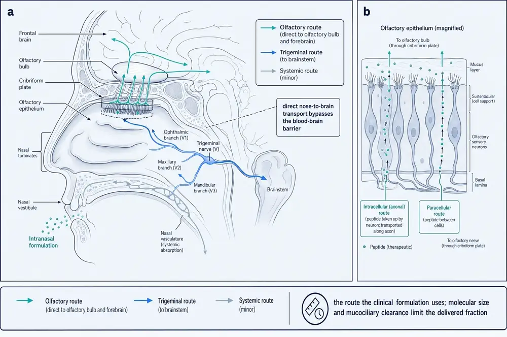
The second came fifteen years later, from Shevchenko and colleagues, and it tested the route the compound is actually given by (Shevchenko et al., 2006). Tritium-labelled at the C-terminal proline, molar radioactivity 56 Ci/mmol, administered intranasally at 50 µg/kg in 20 µL to outbred white rats of 200–250 g. Two minutes after administration, 0.093% of the total administered radioactivity per gram was recovered from brain, and 80% of that radioactivity was intact Semax. The rest was metabolites.
Those two figures are not directly comparable — one is total brain content after intravenous dosing, the other is per-gram at a single early timepoint after intranasal dosing — and the monograph will not pretend they are. What they establish jointly is the shape of the problem: a very small fraction of any administered amount reaches brain tissue, and the intranasal route was chosen because it does better than the intravenous one. The nose-to-brain argument for this compound rests on measurement, not on marketing.
The same 2006 study reported something else that turned out to matter more. The peptide is degraded rapidly, and the species that predominates in biological samples — relative to the total content of Semax plus all its metabolites — is the tripeptide Pro-Gly-Pro. The tail that was added to protect the molecule survives the molecule. Whatever Semax does after administration, a substantial part of what is actually present in the tissue is PGP.
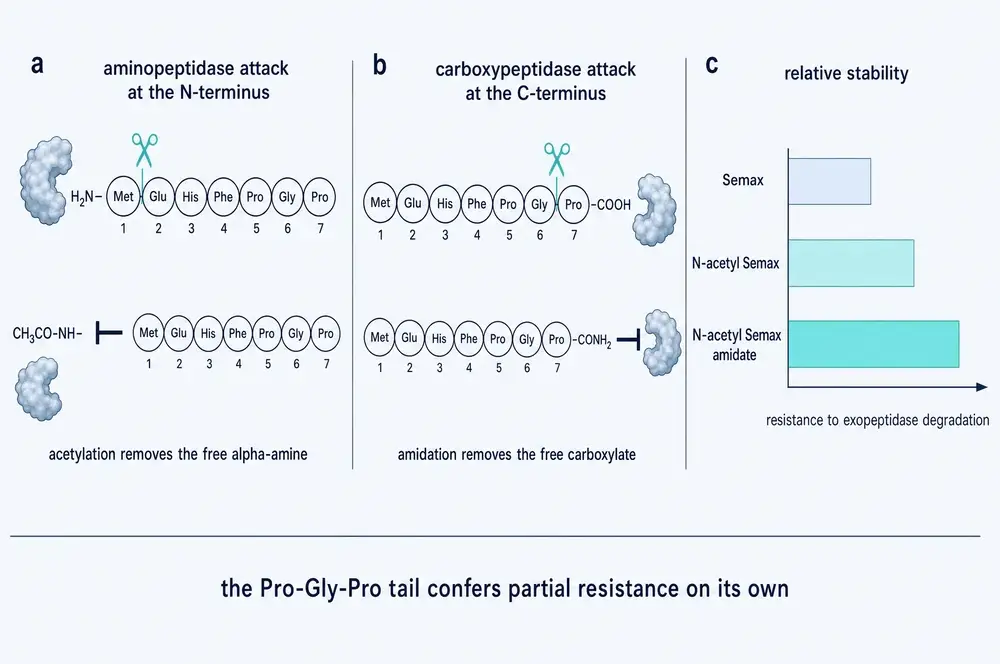
One further point about the ends of the molecule, because it becomes the hinge of Part Five. The same laboratory that measured the degradation went on to ask what happens if you change the residue at the front. Substituting alanine, glycine or threonine for the N-terminal methionine produced analogues more stable to aminopeptidase attack than Semax itself, and their first degradation product was His-Phe-Pro-Gly-Pro (Shevchenko et al., 2011). Capping the amine with an acetyl group is the same idea applied to the chemical group rather than the residue, and the group published a study of exactly that in 2013 (Shevchenko et al., 2013a). The commercial rationale for N-acetyl Semax is therefore real chemistry with a real laboratory pedigree. What nobody appears to have asked at the time is what capping that particular amine costs. Section 21 gives the answer, and it is not what the market assumes.
06Neurotrophins: the founding mechanism
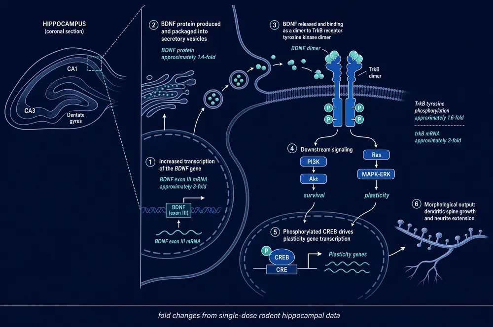
If Semax has a signature mechanism in the public imagination, it is BDNF. The claim that this peptide raises brain-derived neurotrophic factor is repeated in every consumer description of it, and the claim has a real origin.
In 2001, Shadrina and colleagues published a short paper in Neuroscience Letters reporting what happened when they applied Semax to glial cells taken from the basal forebrain of newborn rats (Shadrina et al., 2001). They measured messenger RNA for nerve growth factor and for BDNF. Both rose, and the effect was fast. At thirty minutes — the peak — BDNF messenger RNA was eight times control and NGF messenger RNA five times control. That is a large effect, and it arrived quickly enough to suggest a direct transcriptional action rather than a slow downstream consequence.
Three qualifications belong in the same breath as that number, and they are routinely dropped when it is quoted. The measurement is messenger RNA, not protein. It is in cultured glial cells from newborn rats, not in a brain. And it is a single timepoint in a single preparation. It is a good result. It is not a demonstration that taking Semax raises BDNF in a person.
A companion finding came in 2008, when Grivennikov and colleagues, working with an American group, looked at whether Semax kept cholinergic neurons alive in primary cultures from rat basal forebrain (Grivennikov et al., 2008). Survival of cholinergic neurons rose approximately 1.5 to 1.7-fold, and 100 nM Semax stimulated choline acetyltransferase activity. The nulls in that paper are as informative as the positives: the number of GABAergic neurons did not change, the total number of neuron-specific-enolase-positive neurons did not change, and across a thousand-fold concentration range from 1 nM to 10 µM the peptide did not affect glial proliferation at all. The authors closed by saying that implications for Alzheimer's disease "remain to be clarified" — a level of restraint that has not survived into the secondary literature.
The most-cited neurotrophin study in the modern papers examined whether the effect requires injury, and its answer is the single most useful thing in this section. Working in rats with permanent middle-cerebral-artery occlusion, and comparing three groups — animals with no surgery, sham-operated animals, and occluded animals — Dmitrieva and colleagues found that Semax selectively affected neurotrophin transcription in the ischemic cortex, while the effect of Pro-Gly-Pro alone was mainly non-specific (Dmitrieva et al., 2010). Semax raised BDNF, TrkC and TrkA transcription at three hours and NT-3 and NGF at twenty-four hours; PGP raised a broader receptor set at twenty-four hours but without regard to whether the animal was injured.
The work that carries this from culture into a living animal, and from transcript into protein, came from Dolotov and colleagues in 2006. A single intranasal application at 50 µg/kg in rats produced, in hippocampus, a threefold rise in BDNF exon III messenger RNA, a twofold rise in trkB messenger RNA, and — the part that matters most — a 1.4-fold rise in BDNF protein with a 1.6-fold rise in trkB tyrosine phosphorylation (Dolotov et al., 2006b). The same group reported that intranasal Semax at 50 and 250 µg/kg raised BDNF protein in basal forebrain within three hours but not in cerebellum (Dolotov et al., 2006a), and had earlier reported BDNF induction across several brain regions (Dolotov et al., 2003). Two things are worth holding from this. The receptor is not merely more abundant, it is more phosphorylated, which means it is being activated rather than just expressed. And the effect is regionally selective, which is the beginning of an answer to where in the brain this compound acts.
Note the asymmetry in the sizes, because it recurs throughout this literature: the messenger-RNA changes are severalfold, the protein change is forty per cent. Transcript is not protein, and the gap between them is where a good deal of the compound's reputation has been built.
From about 2014 the Moscow group stopped asking about individual genes and started sequencing everything. This is the largest and most technically serious body of work on the compound, and it is worth understanding what it does and does not show.
07Reading the whole transcriptome
The design is almost identical across seven studies. Male Wistar rats, two months old, 200–250 g. Middle-cerebral-artery occlusion — permanent in the earliest work, then a ninety-minute transient occlusion by monofilament with reperfusion, verified by MRI. Semax at 100 µg/kg intraperitoneally, usually three doses at 1.5, 2.5 and 6.5 hours after surgery. Brain harvested at 4.5 or 24 hours. RNA sequenced on Illumina platforms against roughly 17,000 genes, with differential expression called at greater than 1.5-fold and an adjusted p-value below 0.05.
The counts of differentially expressed genes are the raw output, and they vary by more than three orders of magnitude depending on where and when you look.
| Study | Model | Tissue | Time | Genes changed by Semax |
|---|---|---|---|---|
| Medvedeva et al., 2014 | permanent occlusion | frontoparietal cortex | 3 h | 96 |
| Medvedeva et al., 2014 | permanent occlusion | frontoparietal cortex | 24 h | 68 |
| Filippenkov et al., 2020 | transient occlusion | subcortex | 4.5 h | 0 |
| Filippenkov et al., 2020 | transient occlusion | subcortex | 24 h | 394 |
| Filippenkov et al., 2023 | transient occlusion | dorsolateral frontal cortex | 4.5 h | 131 |
| Filippenkov et al., 2024b | transient occlusion | frontal cortex | 24 h | 1,539 |
| Filippenkov et al., 2025 | transient occlusion | striatum | 24 h | 343 |
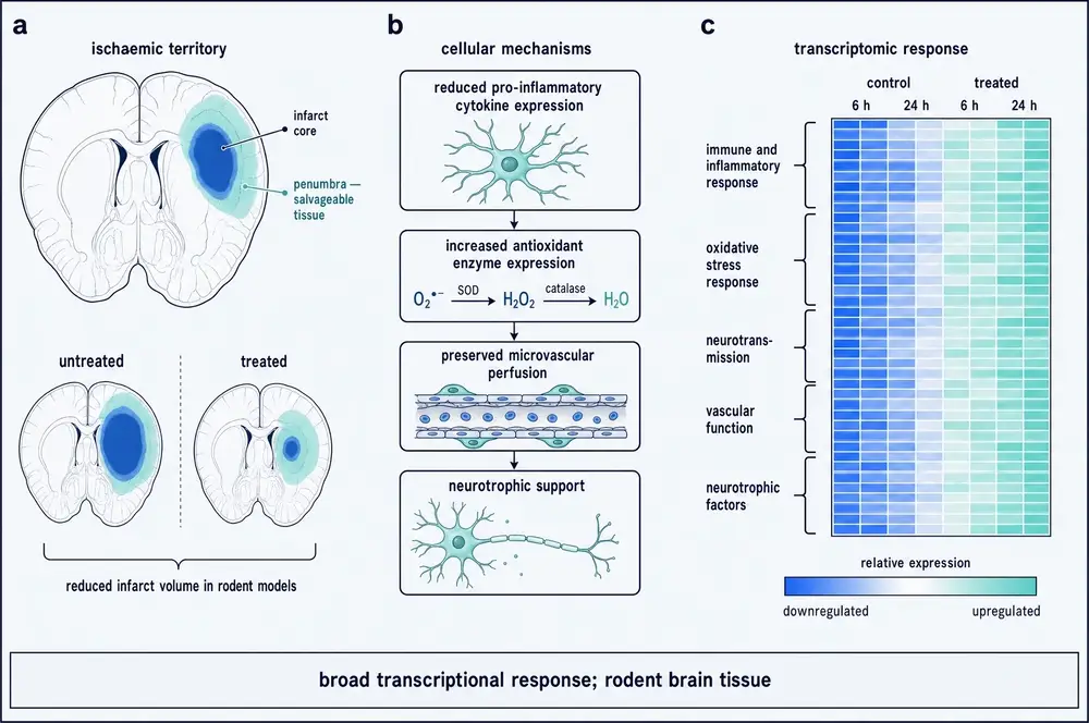
Two classes of gene dominate what changes. At twenty-four hours in cortex, immune and inflammatory genes go down and neurotransmission genes go up. The inflammatory cluster that falls includes Cd14, Hspb1, Socs3, Cd44, Cxcl16 and the MHC class II genes; chemokines Ccl2, Ccl6 and Ccl9 fall roughly threefold where ischemia had raised them as much as fivefold. The neurotransmission cluster that rises includes the dopamine receptors Drd1 and Drd2, the muscarinic receptor Chrm1, glutamate receptors Grm3, Grm5, Grin2c and Grik2, and GABA-A subunit Gabra5. A core of 111 genes moved the same way in both frontal cortex and striatum, and they cluster under the heading "neuroactive ligand-receptor interaction".
08Compensation, and its three boundaries
The headline claim from this programme is arresting. Comparing the genes Semax changed against the genes ischemia changed, in frontal cortex at twenty-four hours, 1,327 of 1,335 overlapping genes moved in the direction opposite to ischemia — 99.4% (Filippenkov et al., 2024b). In the subcortex study the figure was 312 of 313, with a single gene, Mx1, moving with ischemia rather than against it (Filippenkov et al., 2020). The authors describe the peptide as compensating for the effect of ischemia, and on the face of it that is what the numbers say.
Three boundaries have to travel with that figure or it misleads.
First, it is not a statistical interaction test. The comparison is assembled from two separate pairwise contrasts — treated versus untreated ischemia, and untreated ischemia versus sham — and no formal interaction model is reported in any of these papers. That is a real methodological limit on how much weight the 99.4% can carry.
Second, it is regional, and it fails where the damage is worst. The same cohort of animals yielded 1,539 changed genes in frontal cortex and only 343 in striatum. Only eight KEGG pathways overlapped between the two regions. When the group checked three specific genes by quantitative PCR — Hspb1, Cxcl16 and Casp3, all raised by ischemia in both regions — Semax significantly lowered them only in the frontal cortex; in striatum the changes did not reach significance (Filippenkov et al., 2025). The authors' own reading is that striatal cells sitting in the area of severe damage have limited ability to initiate regenerative mechanisms. Put plainly: the compensation happens in tissue that was going to survive, not in tissue that was dying.
Third, it is late. At 4.5 hours in subcortex, Semax produced zero differentially expressed genes out of more than 17,000 assessed. And in frontal cortex at that same early timepoint, the effect was partly anti-compensatory: of 93 genes overlapping with ischemia's response, 38 moved in the same direction as ischemia, not against it.
There is one more wrinkle, and the group raised it themselves. In 2024 they looked at what Semax does to the transcriptome of a healthy rat brain, and found 258 differentially expressed genes in normal frontal cortex at 22.5 hours — mostly decreases in immune-system gene expression (Filippenkov et al., 2024a). They framed the study explicitly as work on "understanding the risks of their use". That finding sits awkwardly beside an earlier result from the same programme, in which Semax produced zero differentially expressed genes in the hippocampus of unstressed rats at six hours (Filippenkov et al., 2021). Different region, different timepoint, different answer — and the question of whether this compound quietly suppresses immune gene expression in an uninjured brain is, on this evidence, open.
09What the proteins say
Messenger RNA is a proxy. In 2021 the group measured protein directly, in rats subjected to the same ninety-minute occlusion, at twenty-four hours, with five to seven animals per group (Sudarkina et al., 2021). They measured four proteins, which they acknowledge as the study's chief limitation.
| Protein | Subcortex — the lesion | Frontoparietal cortex — adjacent tissue |
|---|---|---|
| phospho-JNK | ischemia raised it >2×; Semax lowered it >1.5× | ischemia raised it >2×; Semax lowered it >1.5× |
| phospho-CREB | ischemia lowered it >1.4×; Semax restored it >1.5× | unchanged by ischemia; Semax's rise not significant |
| MMP-9 | ischemia raised it ~1.7×; Semax's fall not significant | ischemia raised it ~2×; Semax lowered it >1.5× |
| c-Fos | no significant change in any group | ischemia raised it ~2×; Semax lowered it >1.5× |
Two things stand out. The first is a genuine cross-check: in the frontoparietal cortex, both MMP-9 and c-Fos moved the same way at transcript and at protein level, which is the only place in this literature where messenger RNA and protein were measured in the same tissue and agreed. The second is that the pattern is regionally split — the peptide restored CREB phosphorylation in the lesion but not in adjacent cortex, and lowered MMP-9 and c-Fos in adjacent cortex but not in the lesion. The authors of a companion paper put the general caution well: the correlation between transcriptome and proteome profiles is non-linear.
10The missing receptor
Here is the fact that most sharply separates what is known about this compound from how it is described: after four decades, no molecular target for Semax has been identified.
That sentence needs care, because it is easy to state too strongly. A binding site has been characterised. Working with tritium-labelled Semax and plasma membranes from the basal nuclei of rat forebrain, Dolotov and colleagues found binding that was time-dependent, specific, reversible and dependent on calcium ions, with a dissociation constant of 2.4 ± 1.0 nM and a site density of 33.5 ± 7.9 fmol/mg protein (Dolotov et al., 2004; Dolotov et al., 2006a). That is a real, saturable, nanomolar site, and the same preparation showed the peptide's half-life in the presence of those membranes exceeded an hour, with dipeptidyl aminopeptidases cleaving it successively to His-Phe-Pro-Gly-Pro and then Pro-Gly-Pro.
What has never happened is the next step. Nobody has identified the protein. No knockdown, no knockout, no antagonist experiment, and no cloning or purification of the site appears anywhere in the corpus assembled for this monograph. A binding constant without a molecular identity tells you the compound sticks to something in forebrain membranes; it does not tell you what, and it does not license any statement about a receptor. The compound is routinely called a "melanocortin derivative", and that is a statement about its structure — the Met-Glu-His-Phe head coincides with a corresponding stretch of α-melanocyte-stimulating hormone — not a statement about its pharmacology.
The one piece of receptor pharmacology in the record is negative. At 1 µM, Semax did not affect cyclic AMP in HEK293 cells expressing the human melanocortin-4 receptor; it antagonised the cAMP response to α-MSH in those cells. That is evidence Semax is not an MC4 agonist. It is not evidence of a mechanism. And the authors of the 2024 vascularisation study state the position without hedging: the involvement of melanocortin receptors in the binding of these peptides has not been demonstrated to date (Stavchansky et al., 2024).
What fills the gap is a hypothesis called "synactone" — the idea that a parent peptide and the pattern of metabolites it generates act together as one regulatory complex, interacting allosterically with various receptor systems. It is stated entirely in modal language: peptides "are able to" provide allosteric interactions, the metabolite patterns "can be" overlapped. It is a reasonable research programme. It is not a mechanism, and it should not be reported as one.
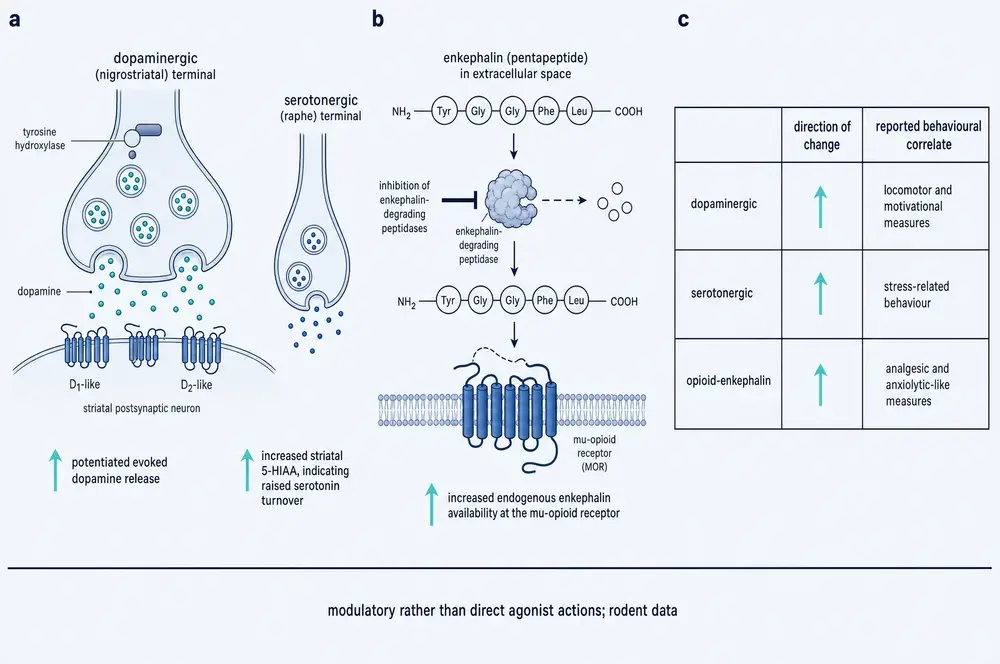
There is, however, one action of this compound that is neither a binding site of unknown identity nor a downstream expression change, but a named enzyme inhibition with a measured potency — and it is easy to miss because it sits outside the neurotrophin literature. Semax inhibits the enzymes that degrade enkephalin in human serum, with an IC₅₀ of 10 µM, which the authors note is more potent than puromycin (Kost et al., 2001). The structure–activity pattern is informative: the pentapeptide fragment retains the inhibition, while tri-, tetra- and hexapeptide fragments do not. Because those enzymes degrade regulatory peptides generally and not only enkephalins, the authors propose this as one mechanism of the compound's biological activity. It is in-vitro human material rather than a living system, and it does not by itself explain the behavioural findings — but it is the most concrete biochemical action attributed to Semax anywhere in this corpus.
Two further findings are worth logging because they narrow the search. A 2023 study reported that ACTH(4–7)PGP and its relatives alter binding of tritiated GABA to rat brain membranes differently at high- and low-affinity sites, which the authors read as allosteric modulation of the GABA receptor system (Vyunova et al., 2023). And a 2025 study found that 1 µM Semax raised the frequency of spontaneous calcium fluctuations in rat hippocampal CA1 neurons but had no effect on proton-induced calcium responses in cerebellar granule cells — which argues specifically against an acid-sensing ion channel mechanism (Kolbaev et al., 2025). Ruling things out is progress.
The most substantial positive lead is also the newest and comes from outside the Moscow programme. In 2025, working in female C57BL/6 mice with spinal cord injury, a group publishing in the British Journal of Pharmacology reported that Semax acts on the µ-opioid receptor gene Oprm1, promoting deubiquitination and functional restoration of the receptor, and modulating lysosomal membrane permeabilisation (Liu et al., 2025). This is the first Semax paper in a major international pharmacology journal, and it proposes an identifiable molecular target. It is a single mouse study and it has not been replicated. It is also the most promising thing to happen to this compound's mechanism in twenty years.
11Pro-Gly-Pro: not inert, and not the active part
The tail deserves a section of its own, because the story about it in the consumer literature is wrong in both directions.
Pro-Gly-Pro is not a synthetic invention. It is a fragment produced by the body's own breakdown of collagen and elastin, cleaved by prolyl endopeptidases from activated neutrophils, microglia and astrocytes, and it circulates in healthy human serum at around 350 pg/mL. It has its own literature as a neutrophil chemoattractant — its structure reproduces a key unit of the CXCL2 chemokine and it binds CXCR2.
So when Semax is degraded and PGP predominates in the tissue, the body is not being left with debris. It is being given more of a signalling molecule it already makes.
The decisive experiment on what the tail contributes was published in 2022. Rats with transient occlusion received Semax at 100 µg/kg, PGP at 37.5 µg/kg, or Pro-Gly-Pro-Leu at 200 µg/kg, all intraperitoneally, eight or more animals per group, with brain examined at twenty-four hours (Stavchansky et al., 2022). The result is clean:
- Semax reversed both the inflammatory-gene rise and the neurotransmitter-gene fall caused by ischemia.
- PGP alone changed none of the six inflammatory genes. Neither did PGPL. Both moved only neurotransmitter genes — PGP raised Gabra5 1.72-fold; PGPL raised Cplx2 2.17-fold, Chrm1 1.85-fold and Gria3 1.70-fold.
- Head to head against PGP, Semax drove interleukin-1β transcript to 0.32 of the PGP value (p = 0.027), interleukin-6 to 0.39 (p = 0.034) and Socs3 to 0.39 (p = 0.040). No neurotransmitter gene differed between them.
The authors' conclusion is that the anti-inflammatory effect is elicited by the ACTH(4–7) fragment, which PGP and PGPL do not contain. The division of labour is therefore reasonably clear: the tail carries the neurotransmitter effects, the head carries the anti-inflammatory ones. Anyone describing Semax as "essentially a PGP delivery system" has it half right and half backwards.
12An ATCUN motif, and what it means
Around 2016 an Italian coordination-chemistry group in Catania noticed something about Semax that the Moscow pharmacologists had not been looking for. Read the first three residues again: methionine, glutamate, histidine. A peptide with a free amino group at the front and a histidine in the third position carries what is called an ATCUN motif — amino-terminal copper and nickel binding site. It is not exotic. Human serum albumin has one, and so do more than four hundred human proteins.
What the motif does is cage a copper ion in a flat square of four nitrogen atoms. The donors are the free N-terminal amine, the two backbone amide nitrogens immediately after it, and one nitrogen from the histidine imidazole ring. Because the two backbone amides must lose their protons to donate, the resulting complex carries a characteristic formula, [CuLH₋₂]²⁻. At physiological pH this is the only copper species Semax forms — 100% of it — which makes the system unusually clean to study and gives it a diagnostic light-absorption band at 522 nm, well separated from the roughly 620 nm band of copper bound to amyloid-β.
The selectivity is extreme. Against zinc, the same peptide binds with a dissociation constant around 1.8 × 10⁻⁵ M — roughly ten orders of magnitude weaker than its reported copper constant. Whatever Semax is doing with metals, it is doing it with copper.
13Silencing the Fenton reaction — and failing to
Copper is useful and dangerous for the same reason: it moves easily between two oxidation states. A loose copper ion in the presence of a reducing agent such as ascorbate cycles between Cu(II) and Cu(I), and each cycle can generate reactive oxygen species, including the hydroxyl radical — the most destructive of them. This is Fenton chemistry, and it is one of the standing hypotheses for how metal dysregulation contributes to neurodegeneration.
A copper ion locked in a rigid square-planar cage cannot easily do this, because Cu(I) prefers a completely different geometry and the cage will not accommodate it. So the prediction is that Semax should shut copper's redox cycling down. In 2025 a group tested it directly (Tomasello et al., 2025). In buffer at pH 7.4 with ascorbate at 150 µM, copper at 20 µM and Semax at 25 µM, they watched ascorbate disappear and hydroxyl radicals appear.
Free copper consumed all the ascorbate within thirty minutes. With Semax present, in the authors' phrase, the copper redox cycling was completely silenced — a small immediate consumption of ascorbate, then nothing. When the Semax–copper complex was pre-formed and amyloid-β added afterwards, there was no ascorbate consumption and no radical formation at all, and adding amyloid produced no change in the absorption spectrum: once copper is bound to Semax, amyloid cannot take it back. Going the other way, adding Semax five minutes into a running amyloid–copper reaction changed both the rate and the level of radical production, which means Semax can pull copper out of a pre-formed amyloid complex.
In cells the effect carried through. In human SH-SY5Y neuroblastoma, copper plus ascorbate drove the fraction of cells positive for a reactive-oxygen probe to 68%, and to 79% with amyloid added; Semax brought both down to about 40%. Viability by MTT assay fell to 59% and 63% under those insults and recovered to 83% and 82% with Semax present.
And then the authors of that same paper wrote the sentence that keeps the whole story honest. Even though Semax forms redox-inert copper complexes and can extract copper from amyloid, the Fenton–Haber–Weiss reaction was not completely inhibited — because a fraction of the copper gets kinetically reduced to Cu(I) before the thermodynamically favoured Semax complex has time to form, and, in their words, the resulting Cu(I) cannot form a complex with Semax. A redox-inert Cu(II) ATCUN motif, they conclude, is not enough to inhibit ROS production. The order in which things are mixed decides the outcome.
14The affinity that shrank two hundredfold
Everything in the section above depends on Semax being a very strong copper binder. The number carrying that weight is a conditional dissociation constant of 1.3 × 10⁻¹⁵ M at pH 7.4 — femtomolar, which would make Semax an extraordinarily avid chelator. It is quoted in the 2022 amyloid paper and twice more in the 2025 reactive-oxygen paper.
It was determined by potentiometric titration of the peptide in isolation. And in 2023, an Estonian group at Tartu asked a different question: not how tightly does this peptide hold copper in a beaker, but can it take copper away from the protein that actually holds most of it in the body?
They mixed human serum albumin at 5 µM with copper at 5 µM and varying amounts of each test peptide, separated the species by size-exclusion chromatography, and measured copper by inductively coupled plasma mass spectrometry, three times per peptide (Noormägi et al., 2023). The Semax sequence came out at 2.89 ± 0.24 × 10⁻¹³ M.
That is 222 times weaker than the potentiometric figure. More to the point, albumin's own constant measured the same way is 9.55 × 10⁻¹⁴ M — so on this measurement albumin binds copper about three times more tightly than the Semax motif does, exactly reversing the published claim that the peptide exceeds albumin by 73-fold. The authors say so directly: in the literature these peptides show affinities exceeding albumin by 44 and 73 times, however in the direct competition experiments they displayed rather similar values to albumin and to the simplest model peptide, glycyl-glycyl-histidine.
Their conclusion is the one sentence in this Part that a reader should carry away: ATCUN motifs cannot compete with excess albumin for the binding of copper ions in the blood and cerebrospinal fluid. Albumin sits at 650 µM in blood and 3 µM in cerebrospinal fluid. The buffer experiments in section 13 used Semax at 25 µM with no albumin present at all.
Two further points sharpen it. The Tartu group argues that competition experiments describe biological conditions more correctly than isolated titrations, and notes that published ATCUN constants vary by more than three orders of magnitude depending on method — this is a field with a measurement problem, not a single rogue result. And the peptide they measured was the amidated form of the Semax sequence; they also found that C-terminal amidation raises copper affinity by roughly threefold. Semax as sold and studied is the free acid, so the honest inference — and this is inference, not a sourced statement — is that real Semax should bind copper no better than 2.89 × 10⁻¹³ M and plausibly somewhat worse.
15Amyloid: a contradiction nobody has resolved
Whether Semax does anything to amyloid-β itself, as opposed to the copper that amyloid binds, is genuinely unsettled — and the two papers that disagree do not cite each other.
In 2017 a group used Semax as a negative control in a peptide microarray designed to find amyloid-binding ligands, and it behaved like one (Galati et al., 2017). Where the known binder KLVFF produced bright spots of labelled amyloid, Semax produced black ones — the amyloid preferred the blocked slide surface to the Semax spots. The authors describe an anti-bonding interaction and a poor ability of Semax to interact with amyloid, and note that this was consistent with the absence of any anti-aggregating activity in their thioflavin-T assay at equimolar peptide, without copper.
In 2022 a different group reported concentration-dependent anti-aggregating activity and inferred a direct interaction between the Semax–copper complex and amyloid oligomers (Sciacca et al., 2022). The inference is explicitly hedged — they call it a hypothesis that in any case needs more in-depth studies to be confirmed.
The two are partly reconcilable, and it is worth doing the work rather than picking a side. The 2017 study tested one condition: 1:1 peptide to amyloid, no copper. The 2022 study agrees that at an equimolar ratio Semax does not significantly perturb any kinetic parameter of amyloid fibril formation. Its effects appear only at a five-fold excess of peptide, or with copper present, or in lipid membranes. So both papers agree on the condition they share. What remains unresolved is the direct-binding question: one says the peptide repels monomeric amyloid, the other infers it stabilises oligomers.
Where the 2022 data are strongest is on membranes, and the numbers are worth stating because they are the most quantitatively solid amyloid result in this literature. Using vesicles made from brain lipid extract in artificial cerebrospinal fluid, with amyloid-β(1–40) at 20 µM, the fraction of entrapped dye released fell from 0.68 with amyloid alone to 0.15 with copper and a five-fold excess of Semax, and the half-time of leakage more than doubled from 688 to about 1,600 minutes. In the same system, total fibre mass in the presence of copper fell from an arbitrary 21.5 to 3.7 and 5.8 — reductions of roughly 83% and 73%.
One caution about all of it. In that same paper copper accelerated amyloid fibrillation in simple buffer and retarded it in brain-lipid vesicles. The authors note the reversal and do not resolve it. Any statement about what copper does to amyloid aggregation has to specify the medium, and by extension so does any statement about what Semax does to that process.
16Stroke in rodents
Two primary experiments carry the animal stroke case, and they are worth setting side by side because they used different models, different doses, different routes, and reached different verdicts.
The first used photochemically induced thrombosis (Shakova et al., 2021). Outbred male Wistar rats, 200–230 g, ninety-six animals, twenty-four per group — a large study by the standards of this literature. Rose bengal was injected intravenously and the prefrontal cortex illuminated through the skull, clotting the vessels beneath. Semax was given intranasally at 25 µg/kg, the first dose two hours after the insult and then daily for six more days. The comparator was Mexidol, a succinate-containing drug used clinically in Russia, at 100 mg/kg intraperitoneally.
The untreated lesion took up 40% of the prefrontal cortex at day one. A single dose of Semax at two hours cut the necrotic area 1.6-fold (p < 0.05). By day twenty-one, after the full course, the damaged region was 2.4-fold smaller than in saline animals, and the number of surviving neurons in the penumbra was 40% higher.
Three things in that same paper complicate the picture, and they are stated in it plainly.
- The six doses after the first added nothing to acute lesion size. In the authors' words, over the following six days of treatment the size of the necrotic area did not differ significantly in either group.
- Semax lost the head-to-head. At day twenty-one the Mexidol-treated damaged area was 1.5-fold smaller than the Semax-treated one (p < 0.05). The authors conclude that Mexidol showed greater protective efficiency.
- The lesion kept growing under a full course of Semax. Between day three and day seven the penumbra expanded 40% centrally and 15% laterally with a 20% rise in neutrophil infiltration, where Mexidol restricted the spread. Semax also failed to move synaptophysin, VE-cadherin, or the inflammatory cell counts at day twenty-one — all of which Mexidol did move.
The second experiment used transient middle-cerebral-artery occlusion: ninety minutes of monofilament occlusion followed by reperfusion, MRI-verified, with Semax at 100 µg/kg intraperitoneally in three doses, and five animals per group (Stavchansky et al., 2024). Here the headline result is a null. Semax did not reach statistical significance on infarct area — the paper reports only a trend towards a decrease. The sibling peptide ACTH(6–9)PGP, tested alongside it, did reach significance, cutting relative infarct area to 54.2 ± 2.3%.
What Semax did move in that study was tissue composition. In the perifocal zone, neuron volume density rose 1.5-fold and blood-vessel volume density 1.25-fold (p < 0.01), and proliferating-cell nuclei rose 1.4-fold. The paper is titled for vascularisation, so its central caveat deserves quoting in substance: there were no significant microscopic signs of neoangiogenesis in the penumbra after either peptide. The vascular effect is protection and recruitment of vessels that were already there — not the growth of new ones. Anyone reading "Semax promotes angiogenesis" is reading past the paper's own finding.
One methodological point applies to the second study and was raised by the same laboratory. In 2023 they reported that in this exact occlusion protocol, the volume of cerebral damage at twenty-four hours was inversely proportional to the duration of isoflurane anaesthesia — eight of ten rats lesioned under long anaesthesia against ten of ten under short, with the lesion spreading to cortex in the latter (Shpetko et al., 2023). They advise that researchers take the background effects of anaesthesia into account when testing neuroprotective drugs. The Semax occlusion study used isoflurane throughout and did not control for it.
17Stress, learning and dopamine
The behavioural literature is where the gap between reputation and evidence is widest, and the reason is not that the studies are bad. It is that they are small, single-dose, almost entirely in male rats, and — crucially — almost entirely in impaired animals.
Stress. Male Wistar rats received Semax at 100 µg/kg intraperitoneally thirty minutes before an hour of restraint under bright light and intermittent noise, then were tested on an elevated zero maze four hours later, nine to ten animals per group (Filippenkov et al., 2021). Semax attenuated one of the three measures the stress had altered. The comparator ACTH(6–9)PGP attenuated all three. And the authors report a problem with their own model that governs how the result can be read: the restraint protocol did not induce any significant changes in the conventional indices of anxiety — time in and entries into the open sectors, locomotor activity, exploration. The effect appears only on non-conventional sector-transition measures, which the authors themselves note may equally indicate hyperactivity, impulsivity or escape motivation.
The same study reports the cleanest specificity result in this literature. In non-stressed rats, Semax produced no behavioural effect at all, and zero differentially expressed genes out of more than seventeen thousand. The authors read this as evidence of safety. It is also evidence that the compound does very little to an undisturbed nervous system — which is worth holding beside the way it is marketed.
Development. Rat pups isolated five hours a day for their first two weeks were given Semax at 50 µg/kg intranasally on days fifteen to twenty-eight, after the stressor had ended (Volodina et al., 2012). At day sixty-five the isolated animals showed a blunted corticosterone response to a forced swim, and Semax restored it. The nulls are extensive: no effect on body weight during the dosing window, no glucose difference at day forty-eight, no corticosterone difference at sixty minutes, and no difference at baseline. One result overshot rather than normalised — treated animals ate faster, longer and more than both the isolated group and the untouched controls, on a measure where isolation had produced no deficit at all.
Learning and memory. The most substantial recent study gave Semax at 50 µg/kg intranasally every other day for a month to APP/PS1 transgenic mice, ten per group, starting at six months (Radchenko et al., 2025). Cortical amyloid plaques were 2.8-fold fewer at 7.5 months. But the behavioural readouts have to be reported with their problems: the transgenic model showed no deficit against wild-type on the novel-object preference index — the very endpoint both peptides are said to improve; performance in the Barnes maze improved on days two and three and then deteriorated on day four; and the day-five probe is described as Semax having reduced time in the target zone and entries into it, which conventionally reads as worse spatial memory, while the authors conclude the peptides corrected performance to wild-type levels. The statistics section states that the behavioural data "were characterized by parametric distribution by the Kruskal–Wallis H test", which is a non-parametric test. No blinding or randomisation is described.
Dopamine. Mice given MPTP, a toxin that destroys nigrostriatal dopamine neurons, received a single 50 µg/kg intranasal dose of Semax either twelve hours before the toxin or one hour after the last dose of it (Kolacheva et al., 2021). Pre-treatment raised striatal dopamine significantly; post-treatment did nothing. The authors describe the effect in their own words as "slightly but reliably" increased, note that the reference drug nomifensine held dopamine at the control level outright, and suggest the design should be altered with multiple injections to enhance the effect. The group size is printed as "n = 5–0", an unrecoverable typographical error. And in intact animals, systemic Semax has been reported not to alter striatal dopamine or its metabolites at all — only serotonin and its metabolite rose.
Depression. No forced-swim, tail-suspension or sucrose-preference study of any Semax family member appeared in the corpus assembled for this monograph, though a 2024 study in European Journal of Pharmacology reports antidepressant-like and antistress effects of Semax and Melanotan II in a chronic unpredictable stress model at 60 nmol/kg (Inozemtseva et al., 2024). The wider melanocortin literature runs against the obvious reading: a 2023 review notes that most studies point to antidepressant and anxiolytic properties of melanocortin receptor antagonists and anxiogenic effects of agonists, and that there are currently no data on melanocortin effects on depressive and anxious behaviour in humans (Markov et al., 2023).
18The human record
This is the section that decides what the compound is. A harvest of 205 verified publication records spanning 1986 to 2026 was screened for studies that actually administered a Semax family compound to human subjects and reported an outcome. Eighteen records qualify. Three of those are title-only in the index, with no recoverable design.
Of the remaining fifteen: none is randomised, blinded and placebo-controlled with a clinical endpoint. The exact shape of the record matters more than any summary of it, so here it is.
Grouped by what they were for, the eighteen break down as five in stroke and cerebrovascular disease, five in optic-nerve, glaucoma and retinal conditions, four in healthy volunteers or general cognition, and four in miscellaneous indications — peptic ulcer, motor neuron disease, psoriasis with metabolic syndrome, and analgesia.
The stroke record. The first report, in 1997, compared thirty treated patients against eighty controls in acute hemispheric ischemic stroke and found some influence on the rate of restoration of neurological function (Gusev et al., 1997). Arms of thirty and eighty are not a randomised design, and none is described. A 2005 evaluation of 187 patients with chronic cerebrovascular insufficiency reported significant clinical improvement and reduced stroke risk with no effect sizes and no p-values (Gusev et al., 2005). A 2018 study of 110 post-stroke rehabilitation patients reported that Semax raised plasma BDNF and accelerated improvement in Barthel score (Gusev et al., 2018); it is the source of the only concrete human regimen found anywhere in this build — two courses of 6,000 µg per day for ten days, twenty days apart — and it is reported here as a study parameter, not as a recommendation.
A larger and much more striking trial is cited at second hand in a 2013 paper: a double-blind, placebo-controlled study of 160 acute carotid ischemic stroke patients reporting mortality reductions of 76.6% and 77.3% (Indharty et al., 2013). That primary report could not be obtained in this build, and the citing paper describes the same study inconsistently — once as double-blind and placebo-controlled, once as three ascending dose arms against a standard-therapy group, which are different designs. A 77% relative mortality reduction in severe stroke would be one of the most important results in neurology. It has no corroboration anywhere in this corpus and cannot be treated as evidence on the strength of a second-hand summary.
The one paper tagged as a randomised controlled trial. It is a 2007 study in motor neuron disease, and three things about it deserve stating together (Serdiuk et al., 2007). Its own abstract calls it an open-label trial in sequential groups of patients — contradicting the index tag. It enrolled twenty-seven patients on intranasal Semax at 12 mg per day in two ten-day courses. And its result was negative: Semax did not influence either the course of chronic partial denervation on electromyography or the dynamics of clinical estimates. It improved only self-reported quality of life. The single most rigorously labelled human study of this compound found that it did not work for what it was being tested for.
The only primary human data with a hard biomarker is not stroke at all but moderate head injury: forty patients, four of whom died in the first three days and were excluded, no placebo, no blinding, no registration (Indharty et al., 2013). Serum Bcl-2 rose from 1.39 to 3.70 ng/mL under intranasal Semax against no significant change on standard therapy. And then the null that matters: there was no significant correlation between Bcl-2 concentration and either the Barthel Index or the Mini-Mental State Examination at any timepoint in either group. The biomarker moved; the two functional outcomes did not follow it.
The two placebo-controlled studies are both single-dose functional-MRI experiments in healthy volunteers. One, with fourteen on Semax and ten on placebo, found a larger rostral subcomponent of the default mode network (Lebedeva et al., 2018). The other, with fifty-two participants comparing Semax, Selank and placebo, found differences in right amygdala connectivity (Panikratova et al., 2020). These are real, controlled human data. They measure brain imaging, not cognition, mood or clinical outcome.
Cognitive enhancement. Cognition is the single largest topic in the literature — 71 of 205 records touch learning, memory or nootropic function. Not one is a controlled human study with a cognitive outcome. The only controlled human data are the two imaging studies above.
19Safety, as far as it goes
Across four decades and 205 indexed records, there is no dedicated safety study, no systematic adverse-event tabulation, no dose-ranging toxicity work, and no long-term follow-up of any Semax family compound. Safety is asserted rather than measured. A 2005 paper states the drug is featured by a minor percentage of side-effects and is well tolerated including in older age groups, without a numerator, a denominator, an event list or a severity grading. The founding group's 1997 review states that in no case did it produce negative side actions or complications.
Against that, the corpus contains exactly one specific adverse observation, and it has never been followed up. In a 1999 series of seventy-three patients with post-hypoxic encephalopathy — fourteen of them in a persistent vegetative state — the authors report that in some cases the EEG showed episodes of paroxysmal activity after Semax injection, and therefore the first injection should be carried out with monitoring of the brain's bioelectric activity (Alekseeva et al., 1999). That is a drug-attributed electrophysiological signal serious enough that the investigators changed their own procedure. Nothing in the subsequent twenty-seven years of literature examined here returns to it.
Three further items belong in the safety column, none of them reassuring and none of them alarming on its own:
- A 2017 screen found that several biologically active peptides including this class produced no toxic effect on mouse embryos during the embryonic and foetal period (Kobylyanskii et al., 2017). Mouse embryos. Not human reproductive safety.
- The 2024 study of gene expression in the healthy rat brain found Semax mainly decreasing immune-system gene expression, and its authors framed the work explicitly as understanding the risks of use (Filippenkov et al., 2024a). What it means to suppress immune gene expression in an uninjured brain is unresolved.
- The acetyl-Pro-Gly-Pro question raised in section 11: if Semax acts partly as a source of PGP, chronic administration implies chronic exposure to a metabolite with a documented association to pathological tissue remodelling in chronic lung disease (Bakaeva et al., 2024).
And one finding from outside the clinical literature entirely. In 2020, a forensic laboratory reported that Selank and Semax were identified in seized pharmaceutical preparations in 2017 and 2018, noted that these peptides have not completed any clinical trials, observed that they are freely available online as lyophilised powder and nasal sprays, and described them as potentially dangerous molecules for which detection methods are needed (Vanhee et al., 2020). That is the only assessment of the supply channel in this corpus, and it is negative.
20What the reviews say, and what they leave out
Three review articles published in 2026 are the most recent literature on this compound, and because recency carries weight they need to be read carefully rather than simply counted.
The gerontology review is the most honest (Mavrych et al., 2026). It states plainly that Semax has no independent Western validation; that the absence of serious adverse events in the published Russian literature is encouraging but insufficient for conclusive safety determinations given publication bias and differing pharmacovigilance standards; that long-term safety surveillance is absent; that dosing protocols rest on preclinical extrapolation and anecdotal clinical experience rather than systematic dose-finding, with cyclical dosing patterns lacking empirical justification; and that the review should be treated as hypothesis-generating rather than practice-defining. It calls for multi-centre randomised placebo-controlled trials of Semax. It also rests, by its own account, on twenty primary sources across nine peptides.
The aesthetic and endocrine review supplies useful regulatory detail (Renke et al., 2026): Semax is officially indicated in Russia for stroke and neurological disease; in the rest of the world it is not approved and is obtained from compounding pharmacies or the internet without registration. Its human claim, however, is unusable as written — that intranasal Semax improved post-stroke neurological deficits and "modulated" inflammatory cytokines better than placebo, with no sample size, no design, no effect size, and not even the direction of the cytokine change.
The orthopaedics review is the one to be careful with (Rahman et al., 2026). Two of the four sentences it devotes to Semax carry no citation at all: that Semax may be useful in cases involving prolonged neuromuscular retraining or peripheral nerve recovery such as brachial plexopathies, and that its reported effects on attention and mental stamina could indirectly support compliance in complex rehabilitation regimens. Nothing in this monograph's corpus supports Semax for peripheral nerve injury, brachial plexopathy or rehabilitation adherence; the leap runs from ischemic-brain rat transcriptomics to human peripheral nerve across species, tissue, injury mechanism and outcome. The same review demands lot-level certificates of analysis, current good manufacturing practice, and third-party sterility and endotoxin testing. Its rigour is applied to sourcing, not to claims.
All three omit the same two things: that Semax's anti-inflammatory transcriptional effect requires the ACTH fragment and is not reproduced by the Pro-Gly-Pro tail, and the acetyl-PGP concern. And all three place Semax in a category with BPC-157, TB-500, GHK-Cu and epitalon — a grouping that is accurate about how it is sold and misleading about where its evidence came from.
On anti-doping the two most recent sources disagree, and the disagreement should be reported rather than resolved. A 2025 survey of the Polish supplement market places Semax among substances of unclear status — absent from the prohibited list, absent from the non-prohibited list, and grouped with substances presumed to meet the criteria by structural or functional similarity (Pokrywka et al., 2025). The 2026 endocrine review states flatly that WADA prohibits Semax under category S2 or as an unapproved substance, framing it as cognitive doping (Renke et al., 2026). Both are review-level statements; neither reproduces the list entry. Anyone whose situation depends on the answer should read the current Prohibited List rather than either paper.
21N-acetyl Semax: two papers, one negative
The commercial case for N-acetyl Semax is chemically literate. Aminopeptidases attack a peptide from its free amino terminus; cap that terminus and you should slow the attack. The laboratory that discovered Semax had already shown the principle works on this exact molecule: substituting alanine, glycine or threonine for the N-terminal methionine produced analogues more stable to aminopeptidase attack than Semax itself (Shevchenko et al., 2011). They extended the work to carboxypeptidases in 2013 (Shevchenko et al., 2013b), and in the same year published a study titled, in translation, "Stability of Semax acetyl to proteolysis in various biological media" (Shevchenko et al., 2013a). That paper is title-only in the index; no abstract, no recoverable result. What it establishes is that the question was asked.
The second paper is the one that matters, and it did not come from Moscow. In 2016 an Italian coordination-chemistry group published a direct comparison of Semax and N-acetyl Semax in the Journal of Inorganic Biochemistry (Magrì et al., 2016). They were asking a metal-binding question, not a nootropic one. Their findings, in order:
- At physiological pH, acetylated Semax forms a distorted CuN₃O chromophore with a weak apical interaction from the methionine sulphur, where unmodified Semax forms CuN₄. Capping the amine removes a nitrogen donor from the coordination sphere — exactly as the ATCUN geometry in section 12 predicts it must.
- The reduced ligand field makes the acetylated complex's formal redox potential more positive than the corresponding Semax species.
- Unmodified Semax's complex is redox-stable and unreactive against ascorbic acid. The acetylated form is not.
- And the biological endpoint: "Semax acetylation did not protect from Cu(II) induced toxicity on a SH-SY5Y neuroblastoma cell line, thus demonstrating the crucial role played by the free NH₂ terminus in the cell protection."
- Zinc binding was unaffected: both peptides formed zinc complexes of comparable strength, and acetylation did not change zinc entry into cells.
That is the entire primary biological literature on N-acetyl Semax. One comparison, in one cell line, against one mechanism — and the modification lost. The paper's own forward-looking proposal for acetylated Semax is as an ionophore in antibody-drug conjugates, to induce copper dysregulation in tumour cells. Explicitly not as a nootropic.
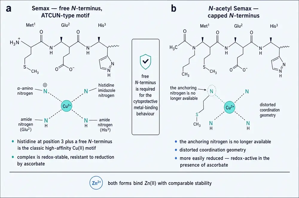
It is worth being precise about the scope of this result, because overstating it would repeat the error the monograph is documenting. This is a copper-mediated cytotoxicity assay. It does not measure cognition, and it does not measure whether acetylation extends the molecule's half-life — which it plausibly does. What it establishes is narrower and harder: for the one mechanism where Semax's activity is best characterised at a molecular level, the free N-terminal amine is required, and acetylation removes it. A modification that buys stability by disabling the binding site has not obviously improved anything.
22N-acetyl Semax amidate: nothing
The amidated form adds a second cap, replacing the C-terminal acid with an amide. The arithmetic checks out: the internal product record gives Semax as C₃₇H₅₁N₉O₁₀S at 813.9 and the amidate as C₃₉H₅₄N₁₀O₁₀S at 855.0, and the difference — two carbons, three hydrogens, one nitrogen, no change in oxygen — is exactly what acetylation plus amidation predicts. Whatever is in the vial, the recorded formula is internally consistent with the name.
What does not exist is any evidence about it. Searches of the indexed literature for "N-acetyl semax amidate", "semax amidate", "NA-Semax" and "amidated Semax" return zero records. Not a pharmacokinetic study, not an animal experiment, not a cell assay, not a human observation. The internal dossier for this product records "0 cited claims" and returns an explicit evidence gap for all twenty-four of its substantive sections, including identity, sequence, origin, mechanism, pharmacokinetics, safety and regulatory status.
One structural detail is worth carrying from Part Three, because it points in an unexpected direction. In the albumin-competition study, C-terminal amidation raised copper affinity roughly threefold (Noormägi et al., 2023). So of the two caps on this molecule, one plausibly helps the copper interaction and the other demonstrably breaks it. Nobody has measured the combination. It is a reasonable thing to want to know and an unreasonable thing to assume.
There is also a market observation in the internal record that says something about how this product is sold. The Semax product entry and the amidate product entry carry the identical count of nine bibliography associations, with overlapping citations, and the one full text retained against the amidate is a plain-Semax rat ischemia paper. No candidate citation attached to the amidate mentions acetylation or amidation at all. The bibliography sold with the modified product is the literature on the unmodified one.
23Adamax, and a lesson about names
Adamax has no molecular formula, no molecular weight, no registry identifier, and no amino-acid sequence in any source consulted for this monograph. The internal record classifies it as a "Semax-related peptide" and otherwise records, field by field: PubChem identifier not locally linked; molecular formula not locally linked; sequence — and this is the record's own wording — "no-local-defined-sequence; do not imply one"; externally verified identifiers, no record. Its stated editorial action is to verify the controlled identity, sequence and aliases before evidence synthesis.
That instruction was not followed, and what happened next is the most instructive thing in this monograph.
An automated pipeline generated a thirty-two-page evidence dossier for Adamax reporting nineteen source documents, 123 cited claims, zero thin flags, and an evidence-tier breakdown that included thirteen human-in-vivo citations. Every one of those 123 citations resolves to a single document: a 2023 paper titled "Enhancing accuracy in brain stroke detection: Multi-layer perceptron with Adadelta, RMSProp and AdaMax optimizers."
AdaMax is a gradient-descent optimisation algorithm. It is a variant of Adam, published in 2015, used to train neural networks. It has no relationship to peptides of any kind. The full candidate bibliography assembled for the compound — forty-seven items — is entirely machine-learning literature: convolutional networks for skin-cancer classification, intrusion detection, potato-blight detection, furniture image classification, the Oblique Mercator projection, wind-speed regression, pig-production data. Not one concerns a molecule.
The same collision poisoned the construction of this monograph. The initial PubMed query for this compound family included the term adamax and returned 252 records; forty-three of them were deep-learning papers. The initial scan of the local library returned 424 candidate documents, of which twenty-three were admitted purely on the strength of the optimiser's name. Two further collisions surfaced later and were subtler: semaxanib, an unrelated kinase inhibitor whose name contains the string; and SEmax and SeMax, which variously denote shielding-effectiveness maximum, standard-error maximum, a pesticide-exposure metric, the sella-to-maxilla cephalometric distance, and the maximum-sensitivity and maximum-senescence parameters of two unrelated mathematical models. Sixteen documents entered the corpus on those and had to be removed one filter at a time.
None of this establishes that Adamax is not a real substance. Something is being shipped in those vials, and the most economical reading of a "Semax-related peptide" sold alongside the acetylated and amidated forms is that it is one of them under a trade name. But that is an inference about marketing, not a fact about chemistry, and this monograph will not assert it. What can be said is that no compound called Adamax has any scientific literature, and that the only public record bearing the name in a research index is an optimisation algorithm.
24The shape of the literature
Step back from what the studies found and look at who did them and where they appeared. The pattern is unusual, and it bears directly on how much confidence any single finding can carry.
None of this makes the work wrong. Concentrated authorship is normal for a compound that never left the institute that made it, and Russian-language publication is a fact about geopolitics rather than about quality. But it has three consequences that a reader should hold.
First, the clinical claim has never been independently replicated. The stroke findings originate with one Moscow group across 1997 to 2018 and are thereafter propagated by narrative reviews citing that same handful of studies. The 32% of records without a top-twenty author are, on inspection, mostly one-off mentions of Semax as a comparator in unrelated pharmacology, plus the small Italian coordination-chemistry cluster — which makes no efficacy claim at all and whose one biological comparison went against the compound's marketed form.
Second, a quarter of the evidence base cannot be read. Fifty-two of the 205 records exist in the index only as an English abstract of a Russian paper. For those, the abstract is the record. This monograph has treated them as abstract-level evidence throughout and said so; a reader who wants to go behind them will need Russian and a library.
Third, the field has changed direction. Of fifteen records from 2024 to 2026, seven are animal or in-vitro mechanism studies, five are reviews, one is a human ophthalmology study in which both arms received Semax, and two are non-Russian chemistry papers. Zero are new clinical trials. The centre of gravity moved from clinical assertion to rat transcriptomics and coordination chemistry, and it has stayed there for a decade.
25The market and the evidence
Set the two records side by side and the asymmetry is the finding.
The 2026 literature describes the market this compound now sits in with unusual candour. A review of unregulated peptide use places Semax in a tier whose defining concern is that online visibility may exceed human evidence and safety characterisation, and notes that "for research use only" labelling may function more as a legal disclaimer than a meaningful safety warning (Hailu et al., 2026). On product quality it is blunt: certificates of analysis and purity claims may not reliably indicate pharmaceutical-grade quality, sterility, endotoxin burden, degradation, storage conditions, batch consistency, or whether the delivered product matches what was tested.
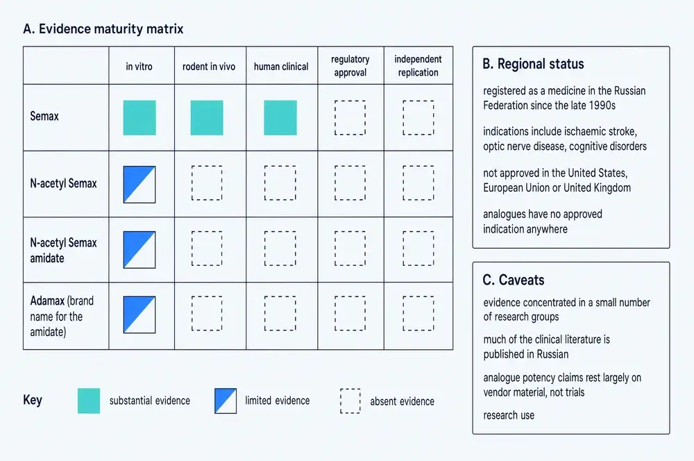
That last clause is the one that connects back to sections 21 to 23. If the delivered product may not match what was tested, and what was tested for three of the four products is nothing, then the certificate accompanying a vial of N-acetyl Semax amidate is attesting to the identity of a substance with no published pharmacology. The document is doing real work — confirming the molecule is what the label says — and no work at all on the question a buyer actually has.
26What would settle it
This monograph has been sceptical throughout, and scepticism is cheap unless it names what would change its mind. Six things would.
A randomised, blinded, placebo-controlled trial with a clinical endpoint. Not imaging, not a biomarker — a clinical outcome in a defined population. Forty years and roughly two hundred papers have not produced one. The 2026 gerontology review calls for exactly this. Until it exists, the human case rests on eighteen mostly uncontrolled studies, one of which was tagged as randomised, describes itself as open-label, and reported a negative result.
A molecular target. No binding assay, no dissociation constant, no knockdown, no antagonist experiment exists for this compound. The 2025 British Journal of Pharmacology paper proposing the µ-opioid receptor gene as a target (Liu et al., 2025) is the first serious candidate in twenty years and needs replication. A compound with a named receptor is a different scientific object from one without.
Resolution of the copper affinity conflict. The femtomolar constant from isolated titration and the picomolar constant from direct albumin competition differ by a factor of 222 and imply opposite conclusions about whether the mechanism can operate in a body. The 2025 Semax paper does not cite the 2023 competition result. Somebody should measure free-acid Semax against albumin directly and publish the number.
An in-vivo test of the copper mechanism. Every copper experiment in this literature is cell-free or cell-culture. Nobody has measured a brain, plasma or cerebrospinal-fluid concentration of Semax and compared it against the 20–25 µM used in the assays.
A dose–response study. Every primary transcriptomic experiment used 100 µg/kg intraperitoneally; every intranasal animal study used 25 or 50 µg/kg. There is no dose-ranging work, in any species, for any endpoint, anywhere in this corpus. Without it, nothing can be said about whether more is better, worse, or the same.
Any experiment at all on the modified forms. A single head-to-head comparison of Semax against N-acetyl Semax and against the amidate — in one animal model, on one endpoint, with a pharmacokinetic arm — would answer more commercial questions than the entire existing literature on the modified compounds, which consists of two papers, one of them title-only, and one negative result in a cell line.
27References
Generated from verified NCBI records rather than from recall. Author lists, journal names, volumes, pages and identifiers are taken from the PubMed record for each citation. This series has twice shipped reference lists drafted from memory in which identifiers pointed at real but unrelated papers; every entry below was re-derived from the harvest, and the author, journal and title line of each was read back before the build was allowed to run.
- Akimov MG, Fomina-Ageeva EV, Dudina PV, Andreeva LA, Myasoyedov NF, Bezuglov VV. ACTH(6-9)PGP Peptide Protects SH-SY5Y Cells from H2O2, tert-Butyl Hydroperoxide, and Cyanide Cytotoxicity via Stimulation of Proliferation and Induction of Prosurvival-Related Genes. Molecules. 2021;26(7).
PMID 33810344 · doi:10.3390/molecules26071878 · PMC8036943 - Alekseeva GV, Bottaev NA, Goroshkova VV. [Use of semax at a follow-up of patients with posthypoxic encephalopathy]. Anesteziologiia i reanimatologiia. 1999:40-3.
PMID 10199046 - Antonova LV, Kamenskiĭ AA, Vlasova TI, Sarycheva NIu, Nezavibat'ko VN. [Catecholaminergic component of the action of a heptapeptide analog of ACTH4-10 on the open-field behavior of rats]. Biulleten' eksperimental'noi biologii i meditsiny. 1986;102(11):569-71.
PMID 3022845 - Asadullah A, Bajamal AH, Parenrengi MA, Turchan A, Utomo B, Sudiana IK, et al.. Effect of ACTH4-10Pro8-Gly9-Pro10 on anti-inflammatory cytokine (IL-4, IL-10, IL-13) expression in acute spinal cord injury models (male Sprague Dawley rats). F1000Research. 2023;12:194.
PMID 41179234 · doi:10.12688/f1000research.127413.2 · PMC12579320 - Ashmarin IP. [Oligopeptides--modulators of memory and pain (structure, properties, probable evolutionary origin)]. Zh Evol Biokhim Fiziol. 1977;13(5):570-8.
PMID 200038 - Asmarin IP, Nezavibat'ko VN, Miasoedov NF, Kamenskiĭ AA, Grivennikov IA, Ponomareva-Stepnaia MA, et al.. [A nootropic adrenocorticotropin analog 4-10-semax (l5 years experience in its design and study)]. Zhurnal vysshei nervnoi deiatelnosti imeni I P Pavlova. 1997;47(2):420-30.
PMID 9173745 - Bakaeva Z, Goncharov M, Frolov F, Krasilnikova I, Sorokina E, Zgodova A, et al.. Regulatory Peptide Pro-Gly-Pro Accelerates Neuroregeneration of Primary Neuroglial Culture after Mechanical Injury in Scratch Test. Int J Mol Sci. 2024;25(20).
PMID 39456669 · doi:10.3390/ijms252010886 · PMC11507231 - Conti F, McCue JJ, DiTuro P, Galpin AJ, Wood TR. Mitigating Traumatic Brain Injury: A Narrative Review of Supplementation and Dietary Protocols. Nutrients. 2024;16(15).
PMID 39125311 · doi:10.3390/nu16152430 · PMC11314487 - Deigin VI, Poluektova EA, Beniashvili AG, Kozin SA, Poluektov YM. Development of Peptide Biopharmaceuticals in Russia. Pharmaceutics. 2022;14(4).
PMID 35456550 · doi:10.3390/pharmaceutics14040716 · PMC9030433 - Dergunova LV, Filippenkov IB, Limborska SA, Myasoedov NF. Neuroprotective Peptides and New Strategies for Ischemic Stroke Drug Discoveries. Genes (Basel). 2023;14(5).
PMID 37239313 · doi:10.3390/genes14050953 · PMC10218113 - Dmitrieva VG, Povarova OV, Skvortsova VI, Limborska SA, Myasoedov NF, Dergunova LV. Semax and Pro-Gly-Pro activate the transcription of neurotrophins and their receptor genes after cerebral ischemia. Cellular and molecular neurobiology. 2010;30(1):71-9.
PMID 19633950 · doi:10.1007/s10571-009-9432-0 · PMC11498467 - Dolotov OV, Zolotarev IuA, Dorokhova EM, Andreeva LA, Alfeeva LIu, Grivennikov IA, et al.. [The binding of Semax, ACTH 4-10 heptapeptide, to plasma membranes of the rat forebrain basal nuclei and its biodegradation]. Bioorganicheskaia khimiia. 2004;30(3):241-6.
PMID 15344653 · doi:10.1023/b:rubi.0000030127.46845.f0 - Dolotov OV, Karpenko EA, Seredenina TS, Inozemtseva LS, Levitskaya NG, Zolotarev YA, et al.. Semax, an analogue of adrenocorticotropin (4-10), binds specifically and increases levels of brain-derived neurotrophic factor protein in rat basal forebrain. Journal of neurochemistry. 2006;97 Suppl 1:82-6.
PMID 16635254 · doi:10.1111/j.1471-4159.2006.03658.x - Dolotov OV, Karpenko EA, Inozemtseva LS, Seredenina TS, Levitskaya NG, Rozyczka J, et al.. Semax, an analog of ACTH(4-10) with cognitive effects, regulates BDNF and trkB expression in the rat hippocampus. Brain research. 2006;1117(1):54-60.
PMID 16996037 · doi:10.1016/j.brainres.2006.07.108 - Dolotov OV, Seredenina TS, Levitskaya NG, Kamensky AA, Andreeva LA, Alfeeva LY, et al.. The heptapeptide SEMAX stimulates BDNF expression in different areas of the rat brain in vivo. Doklady biological sciences : proceedings of the Academy of Sciences of the USSR, Biological sciences sections. 2003;391:292-5.
PMID 14556513 · doi:10.1023/a:1025177812262 - Filippenkov IB, Stavchansky VV, Denisova AE, Yuzhakov VV, Sevan'kaeva LE, Sudarkina OY, et al.. Novel Insights into the Protective Properties of ACTH(4-7)PGP (Semax) Peptide at the Transcriptome Level Following Cerebral Ischaemia-Reperfusion in Rats. Genes. 2020;11(6).
PMID 32580520 · doi:10.3390/genes11060681 · PMC7350263 - Filippenkov IB, Remizova JA, Stavchansky VV, Denisova AE, Gubsky LV, Myasoedov NF, et al.. Synthetic Adrenocorticotropic Peptides Modulate the Expression Pattern of Immune Genes in Rat Brain following the Early Post-Stroke Period. Genes. 2023;14(7).
PMID 37510287 · doi:10.3390/genes14071382 · PMC10379992 - Filippenkov IB, Glazova NY, Sebentsova EA, Stavchansky VV, Andreeva LA, Myasoedov NF, et al.. Changes of Transcriptomic Activity in Rat Brain Cells under the Influence of Synthetic Adrenocorticotropic Hormone-Like Peptides. Biochemistry. Biokhimiia. 2024;89(9):1643-1656.
PMID 39418522 · doi:10.1134/S0006297924090104 - Filippenkov IB, Shpetko YY, Stavchansky VV, Denisova AE, Gubsky LV, Andreeva LA, et al.. ACTH-like Peptides Compensate Rat Brain Gene Expression Profile Disrupted by Ischemia a Day After Experimental Stroke. Biomedicines. 2024;12(12).
PMID 39767736 · doi:10.3390/biomedicines12122830 · PMC11673339 - Filippenkov IB, Shpetko YY, Ales DA, Stavchansky VV, Denisova AE, Yuzhakov VV, et al.. Genes That Associated with Action of ACTH-like Peptides with Neuroprotective Potential in Rat Brain Regions with Different Degrees of Ischemic Damage. International journal of molecular sciences. 2025;26(13).
PMID 40650034 · doi:10.3390/ijms26136256 · PMC12249733 - Filippenkov IB, Stavchansky VV, Glazova NY, Sebentsova EA, Remizova JA, Valieva LV, et al.. Antistress Action of Melanocortin Derivatives Associated with Correction of Gene Expression Patterns in the Hippocampus of Male Rats Following Acute Stress. International journal of molecular sciences. 2021;22(18).
PMID 34576218 · doi:10.3390/ijms221810054 · PMC8469576 - Galati C, Spinella N, Renna L, Milardi D, Attanasio F, Sciacca MFM, et al.. Strategy to discover full-length amyloid-beta peptide ligands using high-efficiency microarray technology. Beilstein journal of nanotechnology. 2017;8:2446-2453.
PMID 29234579 · doi:10.3762/bjnano.8.243 · PMC5704750 - Grivennikov IA, Dolotov OV, Zolotarev YA, Andreeva LA, Myasoedov NF, Leacher L, et al.. Effects of behaviorally active ACTH (4-10) analogue - Semax on rat basal forebrain cholinergic neurons. Restorative neurology and neuroscience. 2008;26(1):35-43.
PMID 18431004 - Gusev EI, Skvortsova VI, Miasoedov NF, Nezavibat'ko VN, Zhuravleva EIu, Vanichkin AV. [Effectiveness of semax in acute period of hemispheric ischemic stroke (a clinical and electrophysiological study)]. Zhurnal nevrologii i psikhiatrii imeni S.S. Korsakova. 1997;97(6):26-34.
PMID 11517472 - Gusev EI, Skvortsova VI, Chukanova EI. [Semax in prevention of disease progress and development of exacerbations in patients with cerebrovascular insufficiency]. Zhurnal nevrologii i psikhiatrii imeni S.S. Korsakova. 2005;105(2):35-40.
PMID 15792140 - Gusev EI, Martynov MY, Kostenko EV, Petrova LV, Bobyreva SN. [The efficacy of semax in the tretament of patients at different stages of ischemic stroke]. Zhurnal nevrologii i psikhiatrii imeni S.S. Korsakova. 2018;118(3. Vyp. 2):61-68.
PMID 29798983 · doi:10.17116/jnevro20181183261-68 - Hailu KT, Abriha FN, Duguma YM, Haddad RR, Liyew T, Kasagga A. Unregulated Peptide Use in the Age of Biohacking: Digital Promotion, Gray-Market Access, and Emerging Public Health Risks. Cureus. 2026;18(6):e110657.
PMID 42437212 · doi:10.7759/cureus.110657 · PMC13355462 - Indharty RS. The increase of serum Bcl-2 concentration in moderate head injury outcome: The role of ACTH4-10Pro(8)-Gly(9)-Pro(10.). Asian J Neurosurg. 2013;8(2):83-9.
PMID 24049550 · doi:10.4103/1793-5482.116381 · PMC3775187 - Inozemtseva LS, Yatsenko KA, Glazova NY, Kamensky AA, Myasoedov NF, Levitskaya NG, et al.. Antidepressant-like and antistress effects of the ACTH(4-10) synthetic analogs Semax and Melanotan II on male rats in a model of chronic unpredictable stress. European journal of pharmacology. 2024;984:177068.
PMID 39442746 · doi:10.1016/j.ejphar.2024.177068 - Ivanikov IO, Brekhova ME, Samonina GE, Myasoedov NF, Ashmarin IP. Therapy of peptic ulcer with semax peptide. Bulletin of experimental biology and medicine. 2002;134(1):73-4.
PMID 12459874 · doi:10.1023/a:1020621124776 - Kaplan AIa, Koshelev VB, Nezavibat'ko VN, Ashmarin IP. [Increased resistance to hypoxia effected by the neuropeptide preparation SEMAX]. Fiziologiia cheloveka. 1992;18(5):104-7.
PMID 1335426 - Khavinson VK, Popovich IG, Linkova NS, Mironova ES, Ilina AR. Peptide Regulation of Gene Expression: A Systematic Review. Molecules. 2021;26(22).
PMID 34834147 · doi:10.3390/molecules26227053 · PMC8619776 - Kobylyanskii AG, Zolotarev YA, Andreeva LA, Grivennikov IA, Myasoedov NF. Studying the Toxic Effects of Some Biologically Active Peptides on the Model of Mouse Embryonic Stem Cells. Bulletin of experimental biology and medicine. 2017;163(6):731-736.
PMID 29063333 · doi:10.1007/s10517-017-3891-y - Kolacheva AA, Ugrumov MV. A Mouse Model of Nigrostriatal Dopaminergic Axonal Degeneration As a Tool for Testing Neuroprotectors. Acta naturae. 2021;13(3):110-113.
PMID 34707903 · doi:10.32607/actanaturae.11433 · PMC8526182 - Kolbaev SN, Sharonova IN, Skrebitsky VG. The Effect of Peptide Semax, an ACTH(4-10) Analogue, on Intracellular Calcium Dynamics in Rat Brain Neurons. Bulletin of experimental biology and medicine. 2025;179(4):416-420.
PMID 41171324 · doi:10.1007/s10517-025-06501-z - Kost NV, Sokolov OIu, Gabaeva MV, Grivennikov IA, Andreeva LA, Miasoedov NF, et al.. [Semax and selank inhibit the enkephalin-degrading enzymes from human serum]]. Bioorganicheskaia khimiia. 2001;27(3):180-3.
PMID 11443939 · doi:10.1023/a:1011373002885 - Lebedeva IS, Panikratova YR, Sokolov OY, Kupriyanov DA, Rumshiskaya AD, Kost NV, et al.. Effects of Semax on the Default Mode Network of the Brain. Bulletin of experimental biology and medicine. 2018;165(5):653-656.
PMID 30225715 · doi:10.1007/s10517-018-4234-3 - Liu R, Chen Y, Huang H, Li X, Lv J, Jiang L, et al.. Semax peptide targets the μ opioid receptor gene Oprm1 to promote deubiquitination and functional recovery after spinal cord injury in female mice. British journal of pharmacology. 2025;182(22):5489-5516.
PMID 40692165 · doi:10.1111/bph.70122 - Magrì A, Tabbì G, Giuffrida A, Pappalardo G, Satriano C, Naletova I, et al.. Influence of the N-terminus acetylation of Semax, a synthetic analog of ACTH(4-10), on copper(II) and zinc(II) coordination and biological properties. Journal of inorganic biochemistry. 2016;164:59-69.
PMID 27586814 · doi:10.1016/j.jinorgbio.2016.08.013 - Markov DD, Dolotov OV, Grivennikov IA. The Melanocortin System: A Promising Target for the Development of New Antidepressant Drugs. Int J Mol Sci. 2023;24(7).
PMID 37047638 · doi:10.3390/ijms24076664 · PMC10094937 - Mavrych V, Shypilova I, Bolgova O. Therapeutic peptides in gerontology: mechanisms and applications for healthy aging. Frontiers in aging. 2026;7:1790247.
PMID 42021992 · doi:10.3389/fragi.2026.1790247 · PMC13095733 - Medvedeva EV, Dmitrieva VG, Povarova OV, Limborska SA, Skvortsova VI, Myasoedov NF, et al.. The peptide semax affects the expression of genes related to the immune and vascular systems in rat brain focal ischemia: genome-wide transcriptional analysis. BMC genomics. 2014;15:228.
PMID 24661604 · doi:10.1186/1471-2164-15-228 · PMC3987924 - Miasoedova NF, Skvortsova VI, Nasonov EL, Zhuravleva EIu, Grivennikov IA, Arsen'eva EL, et al.. [Investigation of mechanisms of neuro-protective effect of semax in acute period of ischemic stroke]. Zhurnal nevrologii i psikhiatrii imeni S.S. Korsakova. 1999;99(5):15-9.
PMID 10358912 - Nazarova GA, Konchugova TV, Iurova OV, Turova EA, Rassulova MA, Sichinava NV, et al.. [The results of the application of the pre-formed physical factors and neuroprotective therapy for the rehabilitative treatment of the patients with diabetic retinopathy]. Voprosy kurortologii, fizioterapii, i lechebnoi fizicheskoi kultury. 2013:29-32.
PMID 24437205 - Noormägi A, Golubeva T, Berntsson E, Wärmländer SKTS, Tõugu V, Palumaa P. Direct Competition of ATCUN Peptides with Human Serum Albumin for Copper(II) Ions Determined by LC-ICP MS. ACS Omega. 2023;8(37):33912-33919.
PMID 37744839 · doi:10.1021/acsomega.3c04649 · PMC10515390 - Panikratova YR, Lebedeva IS, Sokolov OY, Rumshiskaya AD, Kupriyanov DA, Kost NV, et al.. Functional Connectomic Approach to Studying Selank and Semax Effects. Doklady biological sciences : proceedings of the Academy of Sciences of the USSR, Biological sciences sections. 2020;490(1):9-11.
PMID 32342318 · doi:10.1134/S001249662001007X - Pokrywka A, Surała O, Grabowska K, Przybyła M, Granda D, Małecki A, et al.. "Brain doping" substances: prohibited or not in sports?. Biol Sport. 2025;42(4):189-201.
PMID 41048238 · doi:10.5114/biolsport.2025.150047 · PMC12492343 - Polunin GS, Nurieva SM, Baiandin DL, Sheremet NL, Andreeva LA. [Evaluation of therapeutic effect of new Russian drug semax in optic nerve disease]. Vestnik oftalmologii. 2000;116(1):15-8.
PMID 10741256 - Ponomareva-Stepnaia MA, Porunkevich EA, Skuin'sh AA, Nezavibat'ko VN, Ashmarin IP. [Hormonal activity of the ACTH(4-10) analog--a prolonged-action stimulant of learning]. Biull Eksp Biol Med. 1986;101(3):267-8.
PMID 3006824 - Potaman VN, Antonova LV, Dubynin VA, Zaitzev DA, Kamensky AA, Myasoedov NF, et al.. Entry of the synthetic ACTH(4-10) analogue into the rat brain following intravenous injection. Neuroscience letters. 1991;127(1):133-6.
PMID 1652713 · doi:10.1016/0304-3940(91)90912-d - Potaman VN, Alfeeva LY, Kamensky AA, Levitzkaya NG, Nezavibatko VN. N-terminal degradation of ACTH(4-10) and its synthetic analog semax by the rat blood enzymes. Biochemical and biophysical research communications. 1991;176(2):741-6.
PMID 1851003 · doi:10.1016/s0006-291x(05)80247-5 - Potaman VN, Alfeeva LY, Kamensky AA, Nezavibatko VN. Degradation of ACTH/MSH(4-10) and its synthetic analog semax by rat serum enzymes: an inhibitor study. Peptides. 1993;14(3):491-5.
PMID 8392718 · doi:10.1016/0196-9781(93)90137-6 - Radchenko AI, Kuzubova EV, Apostol AA, Mitkevich VA, Andreeva LA, Limborska SA, et al.. The Potential of the Peptide Drug Semax and Its Derivative for Correcting Pathological Impairments in the Animal Model of Alzheimer's Disease. Acta naturae. 2025;17(4):110-120.
PMID 41479572 · doi:10.32607/actanaturae.27808 · PMC12755871 - Rahman OF, Lee SJ, Seeds WA. Therapeutic Peptides in Orthopaedics: Applications, Challenges, and Future Directions. Journal of the American Academy of Orthopaedic Surgeons. Global research & reviews. 2026;10(1).
PMID 41490200 · doi:10.5435/JAAOSGlobal-D-25-00236 · PMC12753158 - Renke G, Chinellato L. Therapeutic Peptides in Aesthetic, Metabolic and Endocrine Conditions: Effects, Safety, Clinical Applications, and Future Perspectives. Int J Mol Sci. 2026;27(9).
PMID 42123471 · doi:10.3390/ijms27093890 · PMC13164565 - Sciacca MFM, Naletova I, Giuffrida ML, Attanasio F. Semax, a Synthetic Regulatory Peptide, Affects Copper-Induced Abeta Aggregation and Amyloid Formation in Artificial Membrane Models. ACS chemical neuroscience. 2022;13(4):486-496.
PMID 35080861 · doi:10.1021/acschemneuro.1c00707 · PMC8855339 - Serdiuk AV, Levitskiĭ GN, Miasoedov NF, Skvortsova VI. [The study of chronic partial denervation and quality of life in patients with motor neuron disease treated with semax]. Zhurnal nevrologii i psikhiatrii imeni S.S. Korsakova. 2007;107(4):29-39.
PMID 18379501 - Shadrina MI, Dolotov OV, Grivennikov IA, Slominsky PA, Andreeva LA, Inozemtseva LS, et al.. Rapid induction of neurotrophin mRNAs in rat glial cell cultures by Semax, an adrenocorticotropic hormone analog. Neuroscience letters. 2001;308(2):115-8.
PMID 11457573 · doi:10.1016/s0304-3940(01)01994-2 - Shakova FM, Kirova YI, Silachev DN, Romanova GA, Morozov SG. Protective Effects of PGC-1α Activators on Ischemic Stroke in a Rat Model of Photochemically Induced Thrombosis. Brain sciences. 2021;11(3).
PMID 33806692 · doi:10.3390/brainsci11030325 · PMC8002020 - Sheludchenko VM, Matyushchenko AG, Alharki L, Baryshev KV, Balkar SS, Makarova MA, et al.. [Effect of a combined physiotherapeutic approach on changes in functional parameters after endovitreal surgery of rhegmatogenous retinal detachment with a favorable anatomical outcome]. Vestnik oftalmologii. 2026;142(3):29-37.
PMID 42366656 · doi:10.17116/oftalma202614203129 - Shevchenko KV, Nagaev IIu, Alfeeva LIu, Andreeva LA, Kamenskiĭ AA, Levitskaia NG, et al.. [Kinetics of Semax penetration into the brain and blood of rats after its intranasal administration]. Bioorganicheskaia khimiia. 2006;32(1):64-70.
PMID 16523722 · doi:10.1134/s1068162006010055 - Shevchenko KV, Khrapko AP, Schwets VI, Myasoyedov NF. The use of phospholipid nanoparticles for increasing Semax resistance in various proteolytic biological media. Doklady biological sciences : proceedings of the Academy of Sciences of the USSR, Biological sciences sections. 2009;429:505-7.
PMID 20170058 · doi:10.1134/s0012496609060076 - Shevchenko KV, V'iunova TV, Nagaev IIu, Andreeva LA, Alfeeva LIu, Miasoedov NF. [Proteolysis of semax analogues with different N-terminal amino acids by aminopeptidases]. Bioorganicheskaia khimiia. 2011;37(4):475-82.
PMID 22096989 · doi:10.1134/s1068162011040133 - Shevchenko KV, Nagaev IY, Andreeva LA, Shevchenko VP, Myasoedov NF. Stability of Semax acetyl to proteolysis in various biological media. Doklady biological sciences : proceedings of the Academy of Sciences of the USSR, Biological sciences sections. 2013;449:110-2.
PMID 23652441 · doi:10.1134/S0012496613020166 - Shevchenko KV, Nagaev IY, Andreeva LA, Alfeeva LY, Myasoedov NF. Study of proteolysis of Semax analogues with different N-terminal amino acids by carboxypeptidases. Doklady biological sciences : proceedings of the Academy of Sciences of the USSR, Biological sciences sections. 2013;450:146-8.
PMID 23821053 · doi:10.1134/S0012496613030101 - Sheĭman IM, Ponomareva-Stepnaia MA, Maksimova LA, Nezovibat'ko VN, Ashmarin IP. [Action of adrenocorticotropic hormone and the tripeptides, Glu-His-L-Phe and Glu-His-D-Phe on imprinting in the beetle, Tenebrio molitor]. Zh Evol Biokhim Fiziol. 1978;14(4):398-400.
PMID 209645 - Shpetko YY, Filippenkov IB, Denisova AE, Stavchansky VV, Gubsky LV, Limborska SA, et al.. Isoflurane Anesthesia's Impact on Gene Expression Patterns of Rat Brains in an Ischemic Stroke Model. Genes (Basel). 2023;14(7).
PMID 37510352 · doi:10.3390/genes14071448 · PMC10379230 - Stavchansky VV, Filippenkov IB, Remizova JA, Denisova AE, Mozgovoy IV, Gubsky LV, et al.. Insight into Glyproline Peptides' Activity through the Modulation of the Inflammatory and Neurosignaling Genetic Response Following Cerebral Ischemia-Reperfusion. Genes. 2022;13(12).
PMID 36553646 · doi:10.3390/genes13122380 · PMC9777888 - Stavchansky VV, Yuzhakov VV, Sevan'kaeva LE, Fomina NK, Koretskaya AE, Denisova AE, et al.. Melanocortin Derivatives Induced Vascularization and Neuroglial Proliferation in the Rat Brain under Conditions of Cerebral Ischemia. Current issues in molecular biology. 2024;46(3):2071-2092.
PMID 38534749 · doi:10.3390/cimb46030133 · PMC10969580 - Sternke-Hoffmann R, Liu C, Wang X, Pupart H, Sun X, Dreiser JG, et al.. Protein-Driven Copper Redox Regulation: Uncovering the Role of Disulphide Bonds and Allosteric Modulation. Angew Chem Int Ed Engl. 2026;65(15):e19673.
PMID 41757714 · doi:10.1002/anie.202519673 · PMC13053925 - Sudarkina OY, Filippenkov IB, Stavchansky VV, Denisova AE, Yuzhakov VV, Sevan'kaeva LE, et al.. Brain Protein Expression Profile Confirms the Protective Effect of the ACTH(4-7)PGP Peptide (Semax) in a Rat Model of Cerebral Ischemia-Reperfusion. International journal of molecular sciences. 2021;22(12).
PMID 34201112 · doi:10.3390/ijms22126179 · PMC8226508 - Tomasello MF, Di Rosa MC, Naletova I, Sciacca MFM, Giuffrida A, Maccarrone G, et al.. Semax, a Copper Chelator Peptide, Decreases the Cu(II)-Catalyzed ROS Production and Cytotoxicity of aβ by Metal Ion Stripping and Redox Silencing. Bioinorganic chemistry and applications. 2025;2025:4226220.
PMID 40496623 · doi:10.1155/bca/4226220 · PMC12151629 - Tsai SJ. Semax, an analogue of adrenocorticotropin (4-10), is a potential agent for the treatment of attention-deficit hyperactivity disorder and Rett syndrome. Medical hypotheses. 2007;68(5):1144-6.
PMID 16996699 · doi:10.1016/j.mehy.2006.07.017 - V'yunova TV, Shevchenko KV, Shevchenko VP, Bobrov MY, Bezuglov VV, Myasoedov NF. Specific binding of semax in different regions of the rat brain. Doklady biological sciences : proceedings of the Academy of Sciences of the USSR, Biological sciences sections. 2006;410:376-7.
PMID 17278839 · doi:10.1134/s0012496606050085 - Vanhee C, Francotte A, Janvier S, Deconinck E. The occurrence of putative cognitive enhancing research peptides in seized pharmaceutical preparations: An incentive for controlling agencies to prepare for future encounters of the kind. Drug testing and analysis. 2020;12(3):371-381.
PMID 31667971 · doi:10.1002/dta.2717 - Volodina MA, Sebentsova EA, Glazova NY, Manchenko DM, Inozemtseva LS, Dolotov OV, et al.. Correction of long-lasting negative effects of neonatal isolation in white rats using semax. Acta naturae. 2012;4(1):86-92.
PMID 22708068 · PMC3372995 - Vyunova TV, Andreeva LA, Shevchenko KV, Glazova NY, Sebentsova EA, Levitskaya NG, et al.. Synthetic corticotropins and the GABA-receptor system: Direct and delayed effects. Chemical biology & drug design. 2023;101(6):1393-1405.
PMID 36828803 · doi:10.1111/cbdd.14221 - Yatsenko KA, Glazova NY, Inozemtseva LS, Andreeva LA, Kamensky AA, Grivennikov IA, et al.. Heptapeptide semax attenuates the effects of chronic unpredictable stress in rats. Doklady biological sciences : proceedings of the Academy of Sciences of the USSR, Biological sciences sections. 2013;453:353-7.
PMID 24385169 · doi:10.1134/S0012496613060161 - Zolotarev YA, Dolotov OV, Inozemtseva LS, Dadayan AK, Dorokhova EM, Andreeva LA, et al.. Degradation of the ACTH(4-10) analog Semax in the presence of rat basal forebrain cell cultures and plasma membranes. Amino acids. 2006;30(4):403-8.
PMID 16773243 · doi:10.1007/s00726-006-0328-8
28How this document was assembled
The corpus was built by a nine-stage pipeline against project 05, the Therapeutic Peptide Research Library. Every file with a document extension in the project’s document stores was opened and its extracted text searched for explicit mentions of a Semax-family compound. That sweep opened 45,807 files across twenty-one stores and returned 424 raw matches.
Three separate homographs contaminate any search for this family, and each was caught only after it had already entered the corpus. Semaxanib is a kinase inhibitor whose name contains the string. AdaMax is a gradient-descent optimisation algorithm, and it is why a paper on potato-blight detection entered a peptide corpus — twenty-three local documents were admitted on its name alone. SEmax and SeMax denote shielding-effectiveness maximum, standard-error maximum, a pesticide-exposure metric, the sella-to-maxilla cephalometric distance, and the parameters of two unrelated mathematical models; these survive case-insensitive matching and were removed only by matching case-sensitively and requiring peptide subject matter nearby. Re-verification removed 32 local documents, and a further sixteen were removed from the fetched set at the reading stage.
Classifying what survived by kind of source is the step that matters. Of 392 verified local assets, only 13 were peer-reviewed scientific full texts. The remaining 379 were consumer web content captured for style training, vendor catalogue material and internal working documents — none of which is evidence about the compound. A raw count of 424 would have overstated the local scientific base by a factor of about thirty.
Because the local library held so little primary science, the pipeline queried NCBI by two routes. A PubMed harvest returned 205 verified records spanning 1977 to 2026, with 47 homograph records rejected to a separate list. Only 21 of those carry a PubMed Central identifier — this literature is overwhelmingly Russian-language and abstract-only in the Western index — so a second route searched PubMed Central’s full text, returning 183 candidates of which 67 were confirmed to name a family compound in their body text and 46 were invisible to the first route. Consolidating local and fetched sets and grading each document by how much of it concerns the compound gives the reading corpus this monograph is written from: 65 verified scientific documents, roughly 1,648 printed-page equivalents, of which 19 are studies whose subject is a Semax-family compound, together with the complete 205-record metadata layer.
| Stage | What it does | Result |
|---|---|---|
| 01b | Targeted scan of the project’s document stores | 45,807 files opened |
| 02 | PubMed E-utilities harvest, complete publication record | 205 records |
| 02b | PubMed Central full-text search | 46 records the first route missed |
| 03 | Open-access full-text retrieval | 21 full texts |
| 04 | Homograph re-verification, classification, inventory | 32 removed |
| 04c–04d | Corpus consolidation and relevance grading | 65 documents |
| 05 | Reference list generation from verified records | 79 citations |
| 08 | Commissioned-plate encoding, 1900 px WebP | 10 plates |
| 09 | Figure renumbering in document order | 26 figures |
| 06 | Assembly, citation and token validation, this document | 1 deliverable |
| 07 | Print rendering | A4, 25 mm margins |
Three traps are recorded here because each produced a real error during this build. First, a PubMed article record carries reference and comment lists full of identifier nodes belonging to other papers; parsing them without scoping each lookup to the article’s own subtree silently assigns a bibliography entry’s identifiers to the article being read, and every lookup in stage 02 is scoped for this reason. Second, raw match counts flatter a corpus badly. Third — and this one is specific to this compound family — an identity check must run before a compliance check. An automated dossier for Adamax passed a research-use-only language filter with zero sentences flagged while being, in its entirety, about a machine-learning optimiser. The filter was reading the prose, and the prose was clean.
29Evidence handling
Findings in this document are labelled by the kind of study that produced them, in the sentence that reports them. Randomised human trials, uncontrolled human series, rodent experiments, cultured-cell measurements, cell-free coordination chemistry, forensic analyses of seized material and narrative reviews are different kinds of claim, and the difference is stated rather than left to the reader to infer. Animal and in-vitro findings are never phrased so as to imply a human outcome, and the species is named every time. Where a claim rests on an abstract because the full text is not accessible — a quarter of this literature exists in the Western index only as an English abstract of a Russian paper — that is said in place.
Recency is weighted, but not blindly. Newer findings take precedence unless a preponderance of evidence contradicts them, and that rule cuts both ways here. The three 2026 reviews are treated as current on regulatory status and on patterns of use, while their pharmacological claims are traced back to the primary studies they rest on, which are mostly from 2001 to 2014. Where a 2023 measurement contradicts a constant that a 2025 paper still quotes without citing it, the conflict is reported as a conflict and the reason the newer result is the more physiologically informative one is given. Recency of publication is not recency of evidence.
The ten commissioned plates in this document were treated as evidence to be checked rather than as illustration. Every value printed on them was verified against the dossier, and the check is recorded in assets/higgsfield/MAPPING.md. It changed the text in three places: it produced the primary source for the hippocampal BDNF and trkB figures in section 06, it surfaced the binding constant in section 10 that an earlier draft had said did not exist, and it supplied the enkephalinase inhibition that section 10 now reports. Where a plate carries something the evidence base does not — the stability ranking in Figure 9, the receptor blockade in Figure 5, the human-evidence rating in Figure 26 — the caption says so in place.
Where the record is silent, silence is reported as silence rather than filled from secondary sources. There is no identified molecular target for this compound, no dose–response study in any species, no dedicated safety study, no human trial that is randomised and blinded with a clinical endpoint, and no primary evidence of any kind for two of the four marketed products. Each of those absences is stated where it belongs rather than omitted, because in this literature the absences are the most decision-relevant facts available.
Continue exploring