
Cartalax A three-letter peptide, a Soviet military laboratory, and the question of what a bioregulator is
Search the world’s largest index of biomedical literature for the word Cartalax and it returns nothing. Not a thin literature — nothing. Search the register of clinical trials and it returns nothing. Search the standard database of drug-like molecules and bioactivity and there is no entry. And yet the compound is real, it is patented, it is sold on four continents, and there are experiments on it. They are filed under a three-letter code that also means “antiepileptic drug”, a development number that also belongs to a fragment of a bacterial protein, and a transliteration almost nobody uses. This monograph is about a molecule that is easy to buy and hard to find — and about the class it belongs to, which does not work the way any other compound in this series works, or claims not to.
Findings are labelled by the kind of study that produced them, in the sentence that reports them. A result in a dish of stem cells is called that. A result in an old rat is called that. Where a number appears, the species, the concentration and the duration travel with it.
Several molecules appear in these pages and they are not interchangeable. Cartalax is the subject: the tripeptide Ala–Glu–Asp, also written AED, also called Kartalaks, and known in its own laboratory as T-31. Epitalon is Ala–Glu–Asp–Gly — the same three residues plus one — and it is a far more famous compound with a far larger literature; almost everything a reader may already believe about “Khavinson peptides” was measured with it. Bronchogen, Cortagen and Cardiogen are the same stem plus leucine, proline and arginine respectively. Sigumir is not a peptide at all but the cartilage-and-bone tissue extract that Cartalax was derived from. Every finding below names the molecule that was actually studied, and where a claim about Cartalax rests on work done with a relative, the sentence says so.
Concentrations and doses appear only as reported experimental parameters. Nothing in this document is a recommendation, and no route or schedule for human use is described or endorsed anywhere in it.
Section 01The absence is the finding
Most monographs in this series begin with a molecule and ask what is known about it. This one has to begin a step earlier, because the ordinary tools for finding out what is known return zero.
On 3 August 2026, a search of PubMed for the term Cartalax in all fields returned no records at all. Not one. A search of PubMed Central, which indexes the full body text of open -access articles rather than only titles and abstracts, found the word twice — once in a review of gene regulation and once in a review of peptide transport, in both cases inside a list of compound names. The Russian transliteration Kartalaks returned a single paper, on jaw surgery in old rats. ClinicalTrials.gov holds no registration for this compound, and none for any compound in its class. ChEMBL, the reference database of bioactive molecules maintained at the European Bioinformatics Institute, has no entry for it: no assay, no target, no measured activity of any kind.
The Radix research dossier generated for this compound before this monograph was commissioned is a document of twenty pages. It contains zero evidence chunks, zero linked source documents, and seventeen claims flagged by its own pipeline as uncited and requiring review. It is, in effect, a form with nothing filled in.
It is worth pausing on what that combination means, because two very different situations produce the same empty search result. One is a compound that nobody has studied — a molecule with a catalogue number and a price and no science behind it at all. The other is a compound that has been studied under names the database does not connect to the name on the label. The distinction matters enormously to a reader trying to decide what to believe, and nothing in the search result itself tells you which one you are looking at.
Cartalax is the second kind, and establishing that took most of the work behind this document. The evidence exists. It is small, it is almost entirely the product of one research group, and it is filed under a three-letter code shared with antiepileptic drugs, a development number shared with a fragment of a bacterial protein, and a Russian transliteration that appears in exactly one indexed paper. A reader who searches the obvious term and finds nothing has not learned that there is nothing; they have learned that the obvious term does not work.
None of this means there is no evidence. It means the evidence is not filed under the name the compound is sold under. The National Library of Medicine maintains a curated Supplementary Concept Record for the molecule — alanyl-glutamyl-aspartic acid — and that record indexes six papers. Six is a small number and it is not zero, and those six, together with about a dozen more that a human curator did not connect to the concept, are what this monograph is built on.
Finding them requires knowing something that no single database states: that Cartalax, Kartalaks, AED, Ala-Glu-Asp and T-31 are all the same molecule. Two of those five strings are among the most heavily overloaded abbreviations in medicine.
Section 02Three letters, eight thousand papers
The compound’s own investigators call it AED. So does epileptology, where it stands for antiepileptic drug and where it appears in several thousand papers about seizure control. So does emergency cardiology, where it means automated external defibrillator. So does analytical chemistry, where it means atomic emission detection. So does dermatology, where it once denoted anhidrotic ectodermal dysplasia. It is also the ISO currency code for the United Arab Emirates dirham, which puts it in health-economics papers about the cost of care in Dubai.
The scale of the problem is worth stating precisely, because it determines what kind of search can work. A PubMed query for the bare string returns 8,214 records. Restricting that set to records that also mention epilepsy, seizures or antiseizure medication accounts for 5,360 of them — about two in three. Defibrillator papers account for a further 1,520. Between them, two fields that have nothing to do with peptide chemistry account for more than five-sixths of every use of the abbreviation in the biomedical literature.
There is no way to separate those from this compound by looking for supporting context, which is the technique that works for most ambiguous names. An antiepileptic-drug paper and a peptide paper both contain the words “drug”, “patients”, “treatment”, “dose” and “trial”. The only method that works is to invert the question: rather than asking what a document must contain to be about this compound, ask what it must not contain. That is how the corpus behind this monograph was assembled, and the Apparatus describes it in detail.
Section 03The molecule
Cartalax is L-alanyl-L-glutamyl-L-aspartic acid: three amino acids joined by two peptide bonds, with a free amino group at one end and a free carboxyl group at the other. All three residues are among the twenty specified by the genetic code, and all three are in the natural L configuration. There is nothing exotic in it at all.
Its molecular formula is C12H19N3O8 and its molecular mass 333.29 daltons. Those values are confirmed against PubChem CID 87815447 and, independently, by recomputing the composition from the three constituent amino acids less one water molecule per peptide bond, which is the arithmetic printed in the figure above. Its CAS registry number is 85806-95-7 and its InChIKey KXEVYGKATAMXJJ-ACZMJKKPSA-N, whose stereochemical layer confirms the all-L assignment.
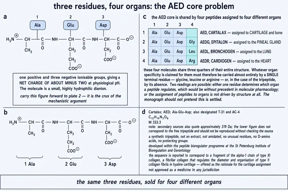
Two properties matter more than the rest. The molecule carries two carboxyl side chains in addition to its C-terminus, so at physiological pH it is triply negatively charged. And its calculated partition coefficient is −4.9, with a polar surface area of 196 Å2 and six hydrogen-bond donors. This is, by a wide margin, one of the most hydrophilic molecules in this monograph series. A compound with those numbers does not cross a lipid membrane by diffusion. If it reaches the inside of a cell — which is where its proposed mechanism requires it to be — something must carry it there. Section 12 takes that question up, and it turns out to be the most interesting thing about this particular peptide.
A small defect in the public record, noted here so it does not propagate. PubChem’s synonym list for CID 87815447 includes the string H-Asp-Glu-Asp-OH alongside the correct H-Ala-Glu-Asp-OH. Asp-Glu-Asp is a different molecule with a different formula and a mass some 44 daltons higher. It appears to be a depositor error. Several vendor descriptions of this compound, meanwhile, call it a tetrapeptide; it is a tripeptide.
Section 04One stem, five compounds, five organs
Cartalax is not an isolated invention. It belongs to a designed series in which each member is built by adding residues to a shared stem, and each member is assigned to a different organ.
The claim embedded in that table is a strong one. Adding a glycine to Ala-Glu-Asp is said to convert a cartilage agent into a pineal agent; adding a leucine makes it a lung agent; a proline, a brain agent; an arginine, a heart agent. Nothing in ordinary molecular pharmacology would lead one to expect a single terminal residue to redirect a molecule between organs in that way, and Section 18 examines what the evidence for it actually is. It is thinner than the confidence with which it is asserted, and it is not nothing.
The naming scheme also has a mundane consequence that shapes this entire document. Because the shorter designation is a strict prefix of the longer ones, a literature search for the subject retrieves its relatives. Of the 107 PubMed records that match the phrase Ala-Glu-Asp, seventy-eight are about a sibling — overwhelmingly about Epitalon — and five are about Cartalax. The remaining twenty-four are about neither: they are papers reporting the primary structure of proteins that happen to contain those three residues in that order, which is a common enough motif to turn up in cytochrome c oxidase, in a ferredoxin, in the human gastrin precursor, and in a hydrolysate of chicken protein. Section 24 returns to that last one, because it is not a coincidence worth discarding.
The cartilage rationale that circulates, and why it fails
There is a structural story in circulation about why this particular tripeptide was assigned to cartilage: that Ala-Glu-Asp corresponds to a fragment of the α1 chain of type XI collagen, the fibrillar collagen that governs the diameter and organisation of type II collagen fibrils in hyaline cartilage. It is an appealing account, and if it were right it would be the only structure-based reason anyone has offered for any assignment in this family.
It does not survive a check that takes about a minute. The human α1(XI) chain, UniProt P12107, is 1,806 residues long and contains the motif Ala-Glu-Asp zero times. Nor does α2(XI). Nor does α1(II), the principal collagen of hyaline cartilage, in human or in cow. Nor does α1(XII). Of the twenty-five reviewed human collagen entries, only two contain the motif at all — a basement-membrane collagen and COL21A1 — and neither is the chain named, nor a cartilage fibrillar collagen. No indexed paper links this compound or its sequence to type XI collagen.
There is also a reason of principle why it could not. The triple-helical domain of a fibrillar collagen is a Gly-X-Y repeat: every third residue is glycine. A run of three consecutive non-glycine residues cannot occur inside it at all. The claim is not merely unsupported; it is incompatible with the architecture of the molecule it invokes.
Section 05What is a bioregulator?
The word is not a pharmacological classification. It does not appear in any regulatory nomenclature, it has no accepted definition outside the literature that coined it, and a reader who assumes it means something like “hormone” or “signalling peptide” will misread everything that follows. It is a term of art belonging to one research tradition, and understanding what that tradition means by it is a precondition for evaluating any claim made about Cartalax.
The tradition begins with a proposition about how the body coordinates itself. On the standard account, long-range regulation is done by hormones released into the blood by endocrine glands and read by receptors on target cells. The Leningrad group proposed an additional layer beneath that one: every tissue, they argued, produces its own small peptides which act locally on the cells of that same tissue, carrying instructions not to a surface receptor but to the genome itself. In 1983 Vyacheslav Morozov and Vladimir Khavinson gave these hypothetical messengers a name — cytomedines (Khavinson & Kuznik, 2013), from the Greek kytos, cell, and the Latin mediator, go-between. The claim attached to the name was tissue specificity: a cytomedine, they wrote, exerts a normalising effect on the tissue it was extracted from, and on no other.
The mechanism is not receptor binding. These peptides are said to enter the cell and the nucleus and to act on DNA, chromatin and transcription directly. There is no named receptor for any of them, no binding constant, no displacement curve.
The concentrations are very low. The class is claimed to act in the nanomolar range and below — in one cartilage experiment at 0.05 ng/mL, which for a 333-dalton molecule is about 150 picomolar. A claim of activity at that concentration is a strong claim and demands a correspondingly strong demonstration.
There are two generations, and they are different kinds of thing. First the tissue extracts — complex mixtures of hundreds of peptides. Then, decades later, individual short peptides of two to four residues, arrived at from those extracts. Cartalax belongs to the second generation; its counterpart Sigumir to the first.
It is worth being explicit about what is and is not unusual here. That small peptides can influence gene expression is not, in itself, an extraordinary claim; that they might reach chromatin and affect it is not physically impossible. What is unusual is the combination: a molecule with no receptor, no measured target, no pharmacokinetics and no dose-response curve, credited with organ-specific effects at picomolar concentrations, on the strength of a literature produced almost entirely by the people who sell it. Each of those qualifications is examined in the sections that follow, and none of them is offered here as a verdict.
Section 06A programme that began in 1973
The work began at the S. M. Kirov Military Medical Academy in Leningrad. Vladimir Khatskelevich Khavinson, born 27 November 1946, graduated from the Academy in 1971; the first experimental papers he published with Vyacheslav Grigorievich Morozov appeared in 1973, and the group’s own retrospective reviews are self-consistent about the starting date. A 1994 paper is titled “Twenty years of study”; a 2009 paper is titled “35-year experience”. Both point to 1973–74. In 1974 the group developed the calf-thymus extraction procedure that produced the preparation later registered as Thymalin.
Both men were career military medical officers holding the rank of colonel, and the institution was a military academy. That much is documented and uncontroversial. A more colourful story is widely repeated in commercial material: that the programme was a classified Ministry of Defence project commissioned to protect submariners from chronic radiation and soldiers from battlefield laser injury, and that it was declassified only after the Soviet collapse in 1991. That version is worth examining, because it is the version most readers will have encountered.
It cannot be sourced. It does not appear in Khavinson’s own peer-reviewed reviews, which describe the origins of the work in ordinary scientific terms and never mention classification, secrecy, or a declassification date. The nearest thing to a scholarly source is a set of essays by Anthony Rimmington, a genuine specialist on Soviet biological research programmes whose book-length work is properly documented — but the essays in question were published on a blogging platform without a single footnote, archival reference or interview attribution, and they make no claim about whether the peptides work. Every other instance traces back to a supplement retailer.
There is also a straightforward internal difficulty with the secrecy account. Thymalin was registered as a medicine by the USSR Ministry of Health on 10 November 1982, and the group was publishing in the open Soviet literature from 1973 onward, including a 1987 review of cytomedines in the Military Medical Journal (Iakovlev et al., 1987). A programme cannot be comprehensively secret and simultaneously producing registered pharmaceuticals and journal articles under its investigators’ names. The honest formulation is that the setting was military and is documented, and that the secrecy narrative is not.
The institutional history after that is straightforward. The St Petersburg Institute of Bioregulation and Gerontology was founded in 1992 with Khavinson as director, a post he held until his death on 5 or 6 January 2024, aged 77. Morozov served as its research director. Khavinson also led a peptide research group at the Pavlov Institute of Physiology of the Russian Academy of Sciences and held the chair of gerontology and geriatrics at the Mechnikov North-Western State Medical University.
Section 07The first generation: extracts
Six preparations from this programme were registered as medicines by the Soviet and then Russian health ministries: Thymalin from thymus (1982), Thymogen and Epithalamin from thymus and pineal gland (both 1990), Cortexin from cerebral cortex (1999), and Prostatilen and Retinalamin from prostate and retina. Each is an acid extract of an animal organ — in practice calf tissue — purified of protein and containing a mixture of small peptides.
What is actually in one of these preparations has been published for exactly one of them. In 2017 the group reported a mass-spectrometric and chromatographic breakdown of the pineal complex (Khavinson et al., 2017).
The composition is worth dwelling on, because it makes the central difficulty of the first generation visible. An extract that is half tripeptides, a quarter dipeptides and a fifth tetrapeptides by count is a mixture of hundreds of distinct molecules. An effect observed with the mixture cannot be attributed to any one of them without an experiment that separates them. That is precisely the experiment the second generation was supposed to be.
Sigumir is the cartilage-and-bone preparation of this family, and it is the natural counterpart of Cartalax. It is not one of the six registered medicines. In Russia it is sold as a БАД — a dietary supplement — and is classed by the institute itself as a “cytomax” parapharmaceutical. Its published literature consists of three papers, all in the same Russian-language journal, of which only one reports original clinical observation. A second cartilage-and-bone extract, Chondrolux, appears in the same literature with its own studies in wound healing and bone repair; the published record does not establish that the two are the same preparation, and results are not transferable between them.
Section 08The second generation, and how it was arrived at
The distinction that matters most in this entire monograph is whether the short peptides were isolated from the extracts — separated, sequenced and then resynthesised — or designed from them, composed out of the residues that a bulk amino-acid analysis found to be abundant. These are different epistemic objects. An isolated peptide is known to exist in the tissue. A designed peptide is a hypothesis about what might be there, because a bulk composition is compatible with an enormous number of sequences.
Khavinson’s own writings answer this question twice, and they do not agree with each other.
The 2013 review with Boris Kuznik states the method plainly: in 1999, Khavinson “suggested the method of creating peptide bioregulators synthesized based on the analysis of the amino acid composition of peptide extracts isolated from animal tissues and consisted of two to four amino acids”. His 2002 review in Neuroendocrinology Letters says the same thing in different words: “based on the data about the amino acid compositions of the peptide preparations, novel principles of the design of biologically active short peptides … has been developed”. That is design from composition.
His 2020 review in Klinicheskaya Meditsina, however, describes the second generation as arising from “isolation of short di-, tri-, tetrapeptides from complex preparations, identification of their primary structures and subsequent synthesis from amino acids”. That is isolation and sequencing — a materially stronger claim, and one the earlier statements do not support.
One compound in the family was genuinely isolated. Thymogen (Glu-Trp) was separated from Thymalin, sequenced, and then synthesised. The 2013 review says so specifically and distinguishes it from the others.
Epitalon was designed. The same review states that Ala-Glu-Asp-Gly “was synthesized based on the amino acid composition of epithalamine”. A 2017 paper adds an important later chapter: the tetrapeptide was subsequently detected in the pineal complex by selective reaction monitoring — eighteen years after it was designed. That is a real result and it partially vindicates the design method. The same paper’s closing sentence, that the extract’s effects “are determined by the effect of its component AEDG”, does not follow from detecting a component.
For Cartalax, neither statement exists. No published paper documents the derivation of Ala-Glu-Asp specifically, and no publication reports detecting it in cartilage tissue. Its origin follows from the general method, which is to say it was designed.
Section 09Sigumir and Cartalax
The pairing of the tripeptide with its parent extract is stated explicitly in the group’s own 2023 review, which names in one sentence “Sigumir, a polypeptide complex of cartilage and bone tissues of young animals, and the AED tripeptide (Kartalax)” (Myakisheva et al., 2023). It is corroborated structurally by the compound’s patent, which claims the sequence specifically for bone and cartilage metabolism.
A 2023 review from the same institute adds a detail that matters: the cartilage polypeptide complex “includes short peptides with a molecular weight from 75 to 846 Da, including the tripeptide AED (Ala-Glu-Asp)”. The supporting reference is a 2020 Russian-language paper on identifying short peptides within organ complexes. If that identification is sound, it places Cartalax in the same position Epitalon reached in 2017: designed first, found later. This monograph reports the claim and notes that the underlying identification has not been published in a form that can be independently examined.
Section 10The patent, and what is sold
Cartalax is patented. Eurasian patent EA 010574 B1, “Peptide normalizing metabolism in bone and cartilaginous tissues, pharmaceutical composition based thereon and method for use thereof”, was filed on 29 January 2007 and granted on 30 October 2008 to SIA Peptides LLC, with Khavinson, Grigoriev, Malinin and Ryzhak named as inventors. Its claims name H-Ala-Glu-Asp-OH explicitly. A corresponding Russian patent, RF 2299741, was granted the same year. No United States or European patent claiming this sequence has been located; the protection appears to be Eurasian and Russian only.
The commercial picture is worth stating plainly, and as commerce rather than as evidence. In Russia, Cartalax is sold in oral capsules as a dietary supplement — explicitly not as a medicine — with claims relating to spinal osteochondrosis, osteoarthrosis, osteoporosis and recovery from fracture. Outside Russia it is sold in a different physical form entirely: 20 milligram lyophilised vials for reconstitution, labelled for research use only and not for human or veterinary use, at roughly $375 for a box of ten. None of the claims made in either market traces to a study of this compound in cartilage, a point made not by a critic but by one of the sellers, whose own archived product page states: “There is no research specific to cartilage and Cartalax that would have earned the peptide its name.”
That sentence was written in 2023 or 2024. It was very nearly true then and it is no longer quite true now, because one cartilage experiment has since been published. Section 17 reports it.
Section 11A peptide that is said to act on DNA
Every other compound in this monograph series works, or is proposed to work, in the same general way: it binds a receptor. The receptor has a name, a gene, a structure and a measured affinity for the ligand; the ligand has a dose-response curve; and the whole thing can be falsified by deleting the receptor and showing that the effect disappears. Sermorelin has GHRH-R. Semaglutide has GLP-1R. GHRP-6 spent twelve years without a named receptor and that was treated, correctly, as a scandal in need of resolution.
The bioregulator claim is different in kind. In the group’s own words, from their 2021 systematic review (Khavinson et al., 2021): “Short peptides, consisting of 2–7 amino acid residues, can penetrate into the nuclei and nucleoli of cells and interact with the nucleosome, the histone proteins, and both single- and double-stranded DNA.” There is no receptor in that sentence. The peptide is proposed to reach the genome and to act on transcription by binding DNA and chromatin directly, in a sequence-dependent way — recognising promoter motifs, and even discriminating the methylation state of the sites it touches.
The difference is not academic bookkeeping. A receptor is what makes a drug claim checkable. Once a target is named, a whole apparatus follows almost automatically: you can measure how tightly the ligand binds, watch a labelled competitor displace it, delete the receptor and see whether the effect survives, and compare potency against every other molecule that touches the same site. Each of those is a way the claim could fail, and a claim that has survived them is worth something precisely because it might not have.
Remove the receptor and that apparatus has nothing to attach to. The proposition that a peptide reaches chromatin and changes transcription is not unfalsifiable in principle — Section 30 sets out three experiments that would settle it, none of them expensive. But it has been supported, so far, almost entirely by observations of the downstream consequence: gene expression changed, therefore the peptide must have acted on the gene. That inference has many other explanations, and the class’s literature has not systematically ruled them out.
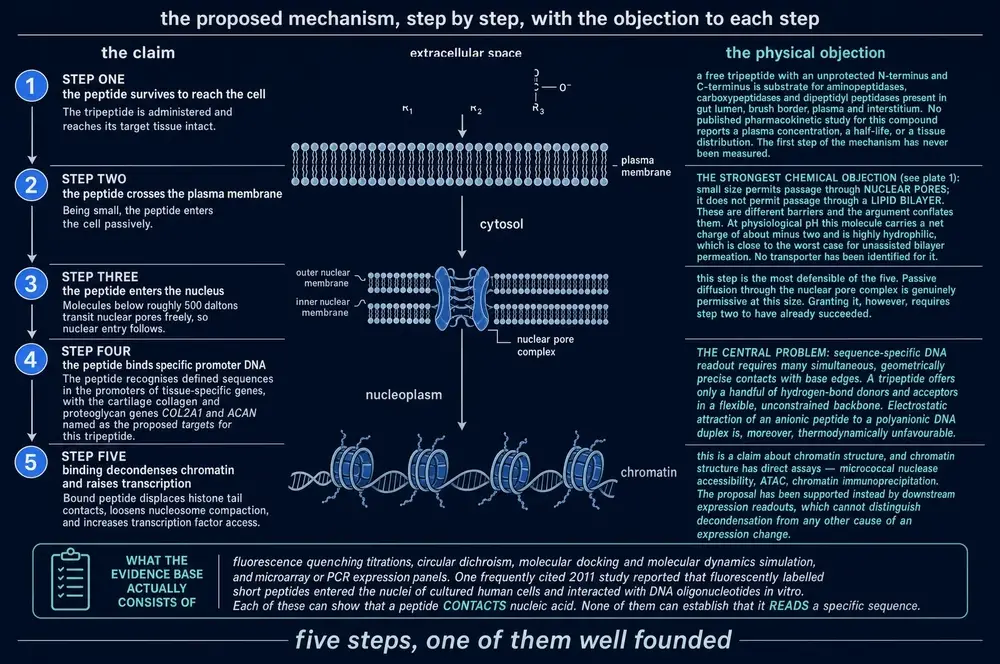
This is not an absurd proposition. Small molecules that bind DNA grooves are ordinary chemistry, and the idea that peptide–nucleic acid interactions might have been an early form of regulation is a respectable, if speculative, evolutionary argument that the group itself makes. But it is a proposition that carries a heavy evidential burden, because it dispenses with every measurement that would normally establish that a drug engages a target. The rest of this Part asks what has been put in their place.
Section 12Getting in: the size window
A triply charged, extremely hydrophilic tripeptide does not cross a plasma membrane by diffusion. If the mechanism above is right, something carries it. The class’s own answer — set out at length in a 2022 review from the institute (Khavinson et al., 2022) — is the peptide transporters.
The proton-dependent oligopeptide transporter family, SLC15, comprises PEPT1, PEPT2, PHT1 and PHT2. PEPT1 sits in the brush border of the small intestine and is a low-affinity, high-capacity carrier; PEPT2 sits in the kidney and is high-affinity. Their defining property, and the reason they matter here, is that they carry di- and tripeptides largely without regard to sequence: they are the route by which the products of protein digestion, and incidentally the β-lactam antibiotics, are absorbed. The review also considers the LAT amino-acid transporters as a possible additional route.
And here the family divides, in a way that the class’s own literature does not draw attention to. The substrate range of the POT transporters is di- and tripeptides. It is not a soft preference: the standard reference review of the family states that neither free amino acids nor peptides of four or more residues are accepted as substrates, and transport experiments have found that tetrapeptides do not even compete for the binding site (Daniel, 2004; Terada et al., 2000).
Cartalax is a tripeptide. Epitalon, Bronchogen, Cortagen and Cardiogen are tetrapeptides. On the class’s own proposed route of entry, the subject of this monograph fits and its four better-known relatives do not.
Two cautions belong immediately alongside that observation, because it is easy to over-read. It is a structural argument, not an experimental result: no published study has shown that Cartalax is transported by PEPT1 or PEPT2, or by anything else. And crossing into an enterocyte is not the same as reaching the bloodstream — the cytosol of an intestinal cell is rich in peptidases whose function is precisely to destroy absorbed di- and tripeptides (Brodin et al., 2002), and the peptidomimetic drugs that survive that gauntlet survive because they were deliberately engineered to resist hydrolysis. An unmodified all-L tripeptide is the substrate those enzymes exist for. What can honestly be said is that the transport hypothesis is testable for this compound in a way it is not for its famous relatives, and that nobody has tested it.
Section 13Binding DNA: what was actually measured
The mechanistic literature of this class is real experimental work, and it is also, almost entirely, work on other molecules.
Taking the strands in order. The melting experiment is a genuine physical measurement: binding of Ala-Glu-Asp-Gly to an alternating poly(dA-dT) duplex lowered its melting temperature from 69.5 °C to 28 °C, with the enthalpy of melting falling from 976 to 445 kJ per mole of base pair (Khavinson et al., 2008). A shift of forty degrees is not a subtle effect. It was measured on a synthetic homopolymer rather than chromatin, and it was measured on the tetrapeptide.
The endonuclease work showed that short peptides alter the way wheat endonucleases hydrolyse phage DNA, and — the striking part — that the peptides discriminate the methylation state of the sites involved (Khavinson et al., 2011). It infers binding from an enzyme’s behaviour rather than measuring it, and it was done in a plant system.
The histone work found that six peptides of the family — AEDG, EDR, AEDL, KEDG, AEDR and KEDW — quench the fluorescence of labelled wheat histones H1, H2b, H3 and H4 (Fedoreyeva et al., 2013), in a manner depending on which histone, which peptide and which oligonucleotide is present. Ala-Glu-Asp is not among the six tested.
The promoter-methylation study is the most consequential of them, because it is the class’s best evidence for tissue specificity: the peptides KEDW and AEDL changed the methylation patterns of specific gene promoters in pancreatic and bronchial cells respectively, in correlation with changes in expression of those genes (Ashapkin et al., 2015). Neither peptide is the subject of this monograph.
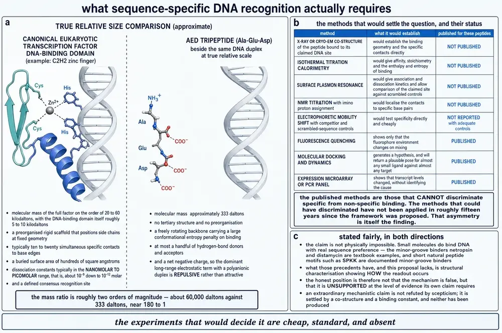
What exists for Cartalax specifically is one molecular docking study, from 2014 (Khavinson et al., 2014), which modelled AED and EDL into the minor groove of the decamer d(ATATATATAT)2 and reported that the complexes formed there were the most energetically favourable. Docking establishes that a pose is plausible. It does not establish that binding occurs, still less that it causes anything. The paper’s own conclusion — that the interaction of these peptides with DNA “is the cause of gene expression” of ageing markers in renal cells — is a considerable distance beyond what a docking calculation can support.
For the whole family there is no crystal structure, no cryo-electron microscopy, and no binding constant measured by isothermal titration calorimetry, surface plasmon resonance or any other physical method.
Section 14The peptide bond — the best control in the class
There is one experiment in this literature that a sceptical pharmacologist would recognise as doing real work, and it deserves to be singled out.
The obvious null hypothesis about a tripeptide of common amino acids applied to cultured cells at nanogram concentrations is that any effect is nutritional or trivially chemical — that the cells are simply receiving alanine, glutamate and aspartate. In 2015 the group tested exactly that (Khavinson et al., 2015). They compared the dipeptide Lys-Glu against an equimolar mixture of free lysine and free glutamate in organotypic spleen-cell culture. The peptide stimulated proliferation. The mixture of its own constituent amino acids inhibited it. Not a smaller effect in the same direction — the opposite direction.
That result does not prove the DNA-binding mechanism, and it was obtained with a different peptide. What it does is close off the most economical alternative explanation for the whole class, and it does so with a control that costs almost nothing to run and that a great many nutraceutical literatures never bother with. It is a point in the tradition’s favour and it should be counted as one.
Section 15What belongs to Cartalax, and what is inherited
The single most common error in commercial and secondary writing about this compound is to describe the mechanism of the class and then attach it to the molecule being sold. The mechanism was established, to whatever degree it has been established, on the tetrapeptides. It is worth setting out the ledger explicitly.
A molecular docking model of minor-groove binding (2014). Changes in gene and protein expression in cultured human and rat cells. Effects in three live-animal models. That is the complete list of target-engagement and activity evidence naming this molecule.
DNA melting-temperature depression — measured on AEDG. Histone-tail binding — measured on AEDG, EDR, AEDL, KEDG, AEDR and KEDW. Promoter-methylation change — measured on KEDW and AEDL. Telomerase activation and telomere elongation, the claim most often attached to this family — measured on AEDG, and the only finding in the entire class that has been independently replicated outside the originating institute. None of these was measured on Ala-Glu-Asp.
The distinction is not pedantry. Epitalon differs from Cartalax by a single glycine, and the class’s own central claim is that a single terminal residue changes which organ a peptide acts on. A tradition that asserts that one-residue changes matter enormously cannot also treat one-residue relatives as interchangeable sources of evidence.
Section 16Why the usual questions have no answers
A reader accustomed to receptor pharmacology will notice that a number of standard questions about this compound simply cannot be asked of the literature, and it is worth naming them together rather than letting each go unremarked in its own section.
There is no dose-response curve. Not a shallow one or a poorly-sampled one — no study of this compound reports more than two concentrations. The chondrogenic experiment reports one concentration for the peptide and one for the extract. The cartilage explant work reports a single figure of 0.05 ng/mL. Without a curve there is no way to distinguish a pharmacological effect from an artefact, no way to estimate potency, and no way to detect the inverted U-shaped responses that small-peptide literatures are prone to.
There are no pharmacokinetics. No absorption study, no plasma concentration–time curve, no half-life, no bioavailability determination, by any route, in any species, for this compound or for any other member of the class. This is not an oversight peculiar to Cartalax; it is a gap that runs through the entire tradition.
There is no toxicology and no safety record. The literature asserts an absence of side effects; it does not report a study designed to find them.
And the route that is sold was never the route that was studied. Counting route words in the group’s own 2014 clinical review of peptide bioregulators gives seventeen mentions of intramuscular administration, eight of injection and five of intranasal delivery — and none of any oral form. The human geroprotection results most often invoked for this family came from intramuscular injection of a pineal extract. The consumer product is a capsule.
None of these absences shows that the compound does nothing. They show that the questions which would settle the matter have not been put.
Section 17Cartilage: the tissue it is named for
Cartalax is named, patented and sold for cartilage. Until 2023 there was no published experiment on this compound in cartilage of any kind — a point that one of its own sellers made in print. There is now one.
Myakisheva and colleagues, working at the St Petersburg institute (Myakisheva et al., 2023), cultured human mesenchymal stem cells through replicative ageing and drove them toward a chondrocyte fate, measuring four markers of chondrogenic differentiation as both gene expression and protein synthesis: the master transcription factor SOX9, the proteoglycan aggrecan, type II collagen, and cartilage oligomeric matrix protein. The AED peptide at 200 ng/mL raised all four. The cartilage polypeptide complex produced the same effect at 2,000 ng/mL.
Two features of that experimental design deserve to be made explicit, because they bound what the result can mean. The cells are mesenchymal stem cells being pushed toward a cartilage fate, not chondrocytes maintaining existing cartilage; the readout is therefore about differentiation rather than repair. And the ageing is replicative — cells worn out by repeated division in a flask — which is a model of one component of tissue ageing and not of osteoarthritis, a disease involving mechanical loading, inflammation, subchondral bone and synovium, none of which is present in a culture dish.
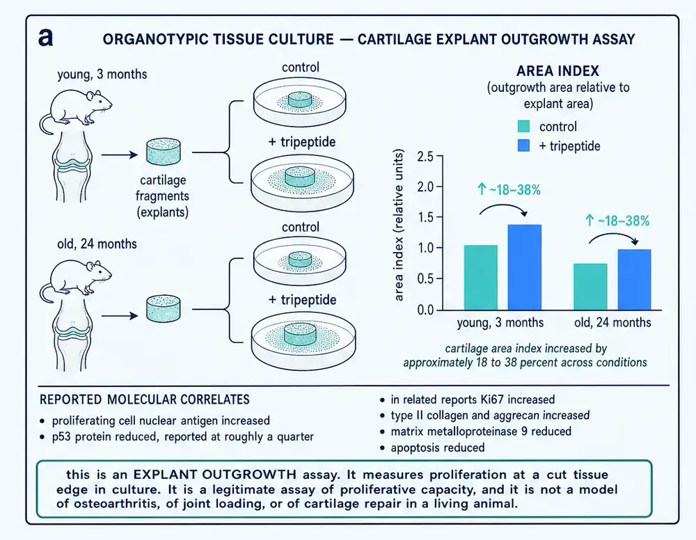
There is a second, older cartilage experiment from the same school, and it is a different kind of assay again: fragments of cartilage from rats aged three months and twenty-four months were placed in organotypic culture and the outgrowth zone measured against the explant area, with short peptides added at 0.05 ng/mL (Smirnov et al., 2011). That design measures proliferative capacity at a cut tissue edge. It is a legitimate assay and it is not a model of joint disease either. Both experiments are cell culture; neither is an animal with a damaged joint.
A tenfold potency advantage for the single peptide over the mixture is the strongest quantitative result this compound has, and it is exactly the result the second-generation programme was designed to produce. It should be read with its limits attached: one experiment, one laboratory, two concentrations, and a report that gives directions of change without effect sizes, variance or replicate counts. It is published in a Russian-language journal whose editor-in-chief is a named inventor on this compound’s patent and a co-author of the paper.
Section 18The senescent chondrocyte
The mechanism proposed to connect cartilage ageing to osteoarthritis is the senescence-associated secretory phenotype. A chondrocyte that has stopped dividing does not simply become inert: it raises the cell-cycle inhibitors p16 and p21, raises p53, secretes tumour necrosis factor α and interleukin 1α, and loses sirtuin 1. The result is a cell that no longer maintains the matrix around it and actively degrades it.
The same group reports that the AED tripeptide and the cartilage polypeptide complex both normalise this profile (Myakisheva et al., 2023). The word “normalise” is theirs, and it is doing a great deal of work: the source reports directions of change without magnitudes for any of the six molecules, so this document reports directions too. What can be said is that the pattern is coherent — the same senescence markers move in the same directions in this compound’s fibroblast and renal-cell studies (Lin'kova et al., 2016; Fridman et al., 2020), which is the kind of internal consistency that would be expected of a real effect and is also the kind that a single laboratory using one set of antibodies would produce either way.
Section 19Bone: the ageing jaw
The most direct live-animal test of this compound against a structural endpoint is a 2016 preclinical study of reparative osteogenesis in old rats (Slugina et al., 2016), and it is the one paper in the world that uses the name Kartalaks in its abstract. Standardised bone cavities were created in the mandible; the peptide was given, in one arm beginning thirty days before surgery; healing was assessed histologically and morphometrically.
The shape of the result is more interesting than a summary would suggest. At thirty days there was no difference from control. Between sixty and one hundred and twenty days the treated animals formed a regenerate containing a smaller proportion of connective tissue and cartilage and showing more advanced remodelling of new bone. Animals that received the peptide only after surgery did less well than those pre-treated.
A null early result reported alongside a positive late one is a good sign about the reporting, not a weakness in it. What the study does not provide is any measurement of the peptide reaching the bone, any dose comparison, or a blinded assessment of the histology.
An earlier and more awkward bone experiment exists. In 2007, Povoroznyuk, Khavinson, Ryzhak and colleagues (Povorozniuk et al., 2007) compared the cartilage tissue extract against the synthetic peptide — identified in that paper by its development code, T-31 — in ovariectomised rats, the standard rodent model of post-menopausal osteoporosis. Both were osteoprotective, both prophylactically and correctively. But the paper records “significantly higher efficacy of the preparation based on cartilaginous tissue extract”.
Section 20When the extract and the peptide were compared
That 2007 result is not isolated. There are two published experiments in which the natural complex and the designed peptide were tested against each other in the same system, and the extract won both times.
The second is a 2015 organotypic study of kidney explants from young and old rats (Chalisova et al., 2015). A polypeptide complex from calf kidney raised the proliferation marker Ki-67 and lowered p53. The short peptides T-31 (AED) and T-35 (EDL) did the same — “but to a lesser degree than the polypeptide complex”.
This matters more than any single positive finding, because the entire rationale for the second generation is that a defined synthetic peptide carries the extract’s activity in a purer and more controllable form. Set against the chondrogenic experiment, where the peptide was effective at one tenth the concentration of the complex, the picture is genuinely mixed. Mixed is the finding, and it is not the finding that any commercial description of this compound reports.
Section 21Kidney: where the siblings come apart
The largest body of work on this compound is not in cartilage at all. It is in kidney, and it is the most useful part of the record because it repeatedly tests several family members side by side.
In cultured renal cells undergoing replicative ageing, AED and EDL raised proliferation and lowered the senescence markers p16, p21 and p53 while raising sirtuin 6 (Khavinson et al., 2014). In rats with cisplatin-induced acute renal failure, AED reduced urinary protein excretion and electrolyte concentration, while EDL produced the most potent nephroprotective effect of the peptides tested (Zamorskii et al., 2015). In old rats, AED and EDL raised urine output by a fifth to two fifths and increased distal sodium transport; AED reduced urinary protein concentration and excretion by between 1.5 and 2.8 times (Zamorskii et al., 2018).
The instructive part is what Ala-Glu-Asp-Gly did in the same animals. It did not behave like its one-residue-shorter relative at all: it reduced glomerular filtration rate by 21 per cent, cut urinary protein concentration by 3.1 times, and lowered absolute sodium reabsorption. Two peptides differing by a single glycine, given to the same strain in the same experiment, produced different renal profiles.
Section 22Head to head
The comparisons of that kind are the most valuable rows in the entire corpus, because they are the only design capable of supporting the tissue-specificity claim at all. A single-peptide study can show that something happened. Only a side-by-side study can show that this peptide does something a near-identical one does not.
Two of those rows deserve particular attention because they are negative.
In 2019, a collaboration between the institute and a group at Chieti-Pescara tested AED, KED, KE and AEDG on the neuronal differentiation of human periodontal ligament stem cells, measuring GAP43 and nestin (Caputi et al., 2019). The markers rose in cells exposed to the combination of all four peptides, and in cells exposed to KED alone. AED alone did not produce the effect. A subsequent review from the same institute nonetheless lists AED among the activators of neuronal differentiation. The primary paper does not support that.
Earlier still, the first published experiment ever to name this compound by its development code — a 2011 study of ageing thymocytes (Lin'kova et al., 2011) — compared T-31 against two other peptides and reported that only AB-9 exhibited a complex geroprotective effect. The compound that would later be named Cartalax did nothing measurable in that system.
Both results are reported here at the same weight as the positive ones, because a class whose central claim is tissue specificity ought to predict exactly this: a cartilage-and-bone peptide should fail in thymus and should not necessarily work in neurons. Read that way, the null results are not embarrassments. They are the closest thing in this literature to a successful prediction.
Section 23Gene expression, and the numbers behind it
The one study of this compound published in a mainstream international journal is a 2020 paper in Molecular Biology Reports, from Ashapkin, Khavinson, Shilovsky, Linkova and Vanyushin (Ashapkin et al., 2020). Human embryonic bone-marrow mesenchymal stem cells were aged in culture by two different methods — serial passage and prolonged stationary phase — and the expression of five genes was measured after exposure to AED, KED and KE at nanomolar concentrations.
Insulin-like growth factor 1 expression rose 3.5 to 5.6-fold with the peptides, in both ageing models. NF-κB expression rose with all three peptides in both models. The divergences are as informative as the agreements: KED inhibited FOXO1 expression by 1.6 to 2.3-fold while KE roughly doubled it in the stationary model and left it unchanged in the passage model; KED suppressed tankyrase-2 in one ageing model and stimulated it in the other. Telomerase reverse transcriptase expression was eightfold higher under stationary ageing than serial passage, independent of peptide.
A 3.5 to 5.6-fold change in a growth-factor transcript at nanomolar peptide concentration is a substantial effect if it is real. The paper is the strongest single piece of evidence this compound has, and it is worth noting both that it passed peer review at a Springer journal and that its senior author, Boris Vanyushin, is a serious plant molecular biologist whose independent work on DNA methylation long predates his involvement with this programme.
Section 24The same molecule, in a bowl of soup
One more paper names this exact molecule, and it comes from a field with no connection to any of the above.
In 1999, a group at Kyoto hydrolysed chicken protein with bromelain and fractionated the digest (Maehashi et al., 1999), isolating eleven peptides and testing each for taste. Ala-Glu-Asp was among them. On its own it tasted of nothing. Combined with inosine monophosphate at 0.02 per cent, it was one of six peptides that enhanced the umami of the mixture.
This is not a false positive to be discarded. It is the same molecule, and it establishes something that no paper in the bioregulator literature does: the sequence Ala-Glu-Asp occurs in ordinary food protein and is released from it by ordinary proteolysis. Anyone who has eaten chicken has been exposed to Cartalax. That observation cuts in two directions at once. It makes any claim of dramatic biological potency at picomolar concentration harder to sustain, since the sequence is not rare and the body encounters it routinely. And it removes at a stroke the most obvious worry about a synthetic peptide of unknown provenance, which is novel toxicity: this is not a novel structure.
Section 25What the evidence supports
It is worth stating the positive case as strongly as it can honestly be stated, because the temptation with a compound like this is to let the absences do all the work.
Cartalax is a chemically real, well-defined, easily synthesised molecule with a granted patent naming its sequence for a specific tissue. Across five independent cell types — skin fibroblasts, renal cells, bone-marrow mesenchymal stem cells, periodontal ligament stem cells and chondrocytes — and in three live-animal models, it has been reported to move a coherent set of markers in a consistent direction: senescence markers down, proliferation markers up, matrix synthesis up, matrix degradation down. The gene-expression work was published in a mainstream international journal and reports effect sizes of 3.5 to 5.6-fold on a growth-factor transcript at nanomolar concentration. The class has run the one control that matters most — peptide against its own constituent amino acids — and the peptide behaved differently from the mixture. And no paper from this group, across five decades and several hundred publications, has ever been retracted.
None of that is nothing. A reader who dismisses this literature because it is Russian, or because the compound is sold by supplement vendors, is reasoning from the wrong premises.
Section 26What it does not support
The negative case is longer, and it is mostly a list of experiments that have not been done rather than experiments that failed.
Four of those rows deserve individual comment.
There is no molecular target. The mechanism that gives this class its identity — direct, sequence-specific interaction with DNA and chromatin — has never been demonstrated for this compound by any method capable of distinguishing specific from non-specific binding. What exists is a docking calculation. The methods that would settle it are standard and inexpensive: a co-structure, isothermal titration calorimetry, surface plasmon resonance, an electrophoretic mobility shift with a scrambled-sequence control. None has been published, in roughly fifteen years. The asymmetry between the discriminating experiments and the non-discriminating ones is itself informative.
There is no independent replication. Not one publication on this compound lacks an author from the originating institute. The Italian collaboration that produced the stem-cell differentiation paper is sometimes cited as external validation; it is not, because Khavinson and two institute colleagues are co-authors. The contrast with the compound’s nearest relative is instructive: Epitalon has now been studied by a group at Brunel University London with no connection to the institute (Al-Dulaimi et al., 2025), and by a group in South Korea (Ullah et al., 2025), and both reported effects. Cartalax has not been touched by anyone outside.
The anti-senescence result cuts both ways, and only one way is marketed. The finding reported most consistently for this compound is that it lowers p16, p21 and p53. Read as rejuvenation of an aged chondrocyte, that is appealing. But those three proteins are the core of the cell’s tumour-suppressor and senescence-enforcement machinery, and an agent that lowers all three is not self-evidently benign; the identical result, reported in an oncology paper, would be described as a liability. The magnitudes are modest and confined to cell culture, so the concern is theoretical rather than demonstrated — but it belongs in the record, and it appears nowhere in the material that sells the compound.
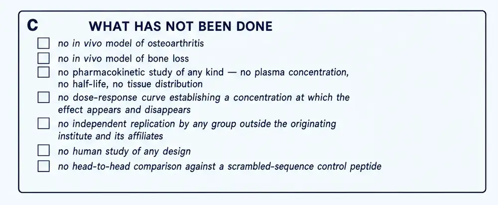
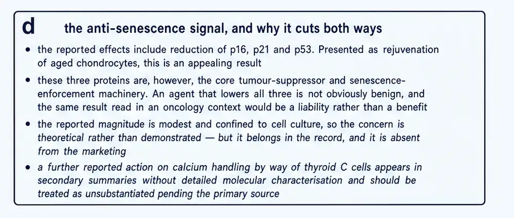
And the evidence stops at the edge of a dish. The two live-animal studies are real and are reported in Sections 19 and 20, but there is no model of osteoarthritis, no model of bone loss beyond the ovariectomy study in which the extract outperformed the peptide, no pharmacokinetics, and no human study of any design.
Section 27Where this literature comes from
Almost all of it has one origin, and the concentration is severe enough to change how much weight any single result can carry.
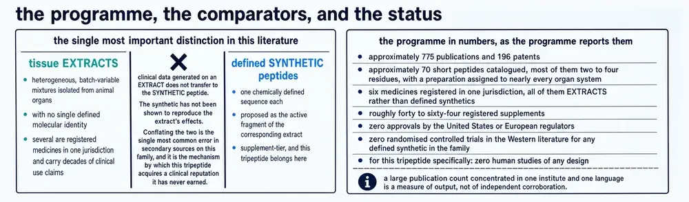
The founder published 494 papers indexed in PubMed. Of those, 321 are in Russian. One hundred and two appeared in a single journal, Bulletin of Experimental Biology and Medicine, and seventy-six in another, Advances in Gerontology — which is published by the St Petersburg Institute of Bioregulation and Gerontology itself, the institute that develops and commercialises these preparations. Its current editor-in-chief is G. A. Ryzhak, the institute’s deputy director for scientific work, a named inventor on this compound’s patent, and a co-author of all three of the 2023 papers that constitute this compound’s entire cartilage evidence.
Two things follow from that, and they pull in opposite directions. Internal consistency across this corpus is worth much less than it looks: agreement between two papers from one group, in a journal that group publishes, is one observation rather than two. But it is also true that no paper by this group has been retracted, and that a null result on a flagship claim — a 2003 study finding no effect of Epitalon on melatonin secretion in rats (Djeridane et al., 2003) — was published in a Western journal with the founder’s own name on it, which is not the behaviour of a group suppressing inconvenient findings.
The right conclusion is neither dismissal nor acceptance. It is that this evidence has not yet been tested by anyone with an incentive to find it wrong.
Section 28Proportion
It helps to set the compound beside things that have been tested properly in the same clinical territory.
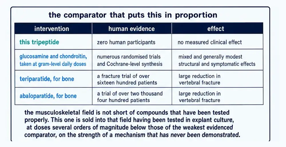
Glucosamine and chondroitin, the most-studied supplements in osteoarthritis, have been through numerous randomised trials and Cochrane-level synthesis, at gram-level daily doses, for mixed and generally modest effects. Teriparatide was tested for fracture prevention in a trial of roughly 1,600 patients and abaloparatide in one of roughly 2,500, both showing large reductions in vertebral fracture (Neer et al., 2001; Miller et al., 2016). Cartalax has been tested in zero human participants.
That is not an argument that it does not work. It is an argument about where it sits: this is a preclinical candidate with an interesting and untested mechanistic hypothesis, sold into a field that is not short of properly evidenced comparators.
Section 29Human use: what is claimed, what is documented
One sentence in the 2023 review states that Sigumir and the AED tripeptide “have shown high efficacy in animal models of OA and oral administration in patients with OA of older age groups”. It is the only assertion of human use of this compound anywhere in the indexed literature, and it is a review sentence: no underlying report is cited that can be retrieved, and no study of Cartalax in human beings can be located in any database.
What does exist, and is frequently confused with it, is a 2012 report of Sigumir — the extract, not the peptide — used in sixty-two elderly patients with temporomandibular joint disorders (Iordanishvili et al., 2012). That study had no randomisation, no blinding and no control arm, and the preparation was one component of a package that also included dental prosthetics, physiotherapy and pharmacotherapy for concurrent disease. It is not evidence about Cartalax, and its design would not support strong conclusions even about Sigumir.
The distinction between the extract and the synthetic peptide is the single most common error made about this family, and it is the mechanism by which this tripeptide acquires a clinical reputation it has not earned. Clinical observations made with a tissue extract do not transfer to a synthetic peptide designed from that extract’s amino-acid composition, particularly when the two direct comparisons that exist both favour the extract.
Section 30What would settle it
Three experiments would move this compound from a claim to a finding, and none of them is expensive or technically difficult.
1. A scrambled-sequence control. Test Ala-Glu-Asp against Asp-Glu-Ala and against a charge-matched tripeptide of unrelated sequence, in the chondrogenic differentiation assay, at matched concentrations. If the effect is specific to the sequence, the controls will be inert. If it is a property of small anionic tripeptides generally, they will not. This is the experiment that the entire tissue-specificity claim rests on and it has never been run.
2. A dose–response curve. Five concentrations spanning four log units in one assay. Without it there is no potency, no way to compare against anything, and no way to distinguish a pharmacological effect from an artefact of a single chosen concentration.
3. A binding measurement. Isothermal titration calorimetry or surface plasmon resonance against the claimed DNA target, with a scrambled-sequence duplex as the comparator. A dissociation constant, or its absence, would settle the mechanism in an afternoon.
Until those are done, the honest position on Cartalax is the one this document has tried to hold throughout: a chemically well-defined molecule, an appealing and not-impossible hypothesis, a small internally consistent body of reported effects, and an evidence base that stops well short of what its own claims require.
This document describes published research. It does not recommend human use of Cartalax or of any other compound named in it, and specifies no dose, route or schedule for any person. Concentrations, routes and durations appear only as reported parameters of the experiments that used them, attached to the species and the system in which they were used. Nothing here is medical advice.
Cartalax holds no marketing authorisation in any jurisdiction. In Russia it is sold as a dietary supplement; elsewhere it is sold labelled for research use only and not for human or veterinary consumption. No pharmacokinetic, toxicological or human safety study of this compound has been published by any route in any species.
Section 31References
- Al-Dulaimi S, Thomas R, Matta S, Roberts T. Epitalon increases telomere length in human cell lines through telomerase upregulation or ALT activity. Biogerontology 2025;26(5):178. PMID 40908429 · doi · PMC12411320
- Al-Dulaimi S, Thomas R, Matta S, Roberts T. Correction: Epitalon increases telomere length in human cell lines through telomerase upregulation or ALT activity. Biogerontology 2025;27(1):1. PMID 41240216 · doi · PMC12619744
- Araj SK, Brzezik J, Mądra-Gackowska K, Szeleszczuk Ł. Overview of Epitalon-Highly Bioactive Pineal Tetrapeptide with Promising Properties. Int J Mol Sci 2025;26(6). PMID 40141333 · doi · PMC11943447
- Ashapkin V, Khavinson V, Shilovsky G, Linkova N, Vanuyshin B. Gene expression in human mesenchymal stem cell aging cultures: modulation by short peptides. Mol Biol Rep 2020;47(6):4323-4329. PMID 32399807 · doi
- Ashapkin VV, Linkova NS, Khavinson VKh, Vanyushin BF. Epigenetic mechanisms of peptidergic regulation of gene expression during aging of human cells. Biochemistry (Mosc) 2015;80(3):310-22. PMID 25761685 · doi
- Bergeon JA, Toth I. Enhancement of oral drug absorption-effect of lipid conjugation on the enzymatic stability and intestinal permeability of l-Glu-l-Trp-NH(2). Bioorg Med Chem 2007;15(22):7048-57. PMID 17845857 · doi
- Brodin B, Nielsen CU, Steffansen B, Frøkjaer S. Transport of peptidomimetic drugs by the intestinal Di/tri-peptide transporter, PepT1. Pharmacol Toxicol 2002;90(6):285-96. PMID 12403049 · doi
- Caputi S, Trubiani O, Sinjari B, Trofimova S, Diomede F, Linkova N et al.. Effect of short peptides on neuronal differentiation of stem cells. Int J Immunopathol Pharmacol 2019;33:2058738419828613. PMID 30791821 · doi · PMC6376556
- Chalisova NI, Lin'kova NS, Nichik TE, Ryzhak AP, Dudkov AV, Ryzhak GA. Peptide Regulation of Cells Renewal Processes in Kidney Tissue Cultures from Young and Old Animals. Bull Exp Biol Med 2015;159(1):124-7. PMID 26033601 · doi
- Daniel H. Molecular and integrative physiology of intestinal peptide transport. Annu Rev Physiol 2004;66:361-84. PMID 14977407 · doi
- Djeridane Y, Khavinson VKh, Anisimov VN, Touitou Y. Effect of a synthetic pineal tetrapeptide (Ala-Glu-Asp-GLy) on melatonin secretion by the pineal gland of young and old rats. J Endocrinol Invest 2003;26(3):211-5. PMID 12809170 · doi
- Fedoreyeva LI, Smirnova TA, Kolomijtseva GY, Khavinson VKh, Vanyushin BF. Interaction of short peptides with FITC-labeled wheat histones and their complexes with deoxyribooligonucleotides. Biochemistry (Mosc) 2013;78(2):166-75. PMID 23581987 · doi
- Fridman NV, Linkova NS, Kozhevnikova EO, Gutop EO, Khavinson VK. Comparison of the Effects of KE and AED Peptides on Functional Activity of Human Skin Fibroblasts during Their Replicative Aging. Bull Exp Biol Med 2020;170(1):154-157. PMID 33231794 · doi
- Iakovlev GM, Morozov VG, Khavinson VKh. [Modern concepts of cytomedins and the problems of bioregulating therapy]. Voen Med Zh 1987;(6):37-40. PMID 3310391
- Ilina A, Khavinson V, Linkova N, Petukhov M. Neuroepigenetic Mechanisms of Action of Ultrashort Peptides in Alzheimer's Disease. Int J Mol Sci 2022;23(8). PMID 35457077 · doi · PMC9032300
- Iordanishvili AK, Samsonov VV, Soldatova LN, Polens AA, Ryzhak GA. [Application of bioregulating therapy in complex treatment of temporomandibular joint diseases in people of elderly and senile age]. Adv Gerontol 2012;25(1):181-6. PMID 22708467
- Khavinson V, Linkova N, Kozhevnikova E, Dyatlova A, Petukhov M. Transport of Biologically Active Ultrashort Peptides Using POT and LAT Carriers. Int J Mol Sci 2022;23(14). PMID 35887081 · doi · PMC9323678
- Khavinson V, Diomede F, Mironova E, Linkova N, Trofimova S, Trubiani O et al.. AEDG Peptide (Epitalon) Stimulates Gene Expression and Protein Synthesis during Neurogenesis: Possible Epigenetic Mechanism. Molecules 2020;25(3). PMID 32019204 · doi · PMC7037223
- Khavinson V, Linkova N, Kozhevnikova E, Trofimova S. EDR Peptide: Possible Mechanism of Gene Expression and Protein Synthesis Regulation Involved in the Pathogenesis of Alzheimer's Disease. Molecules 2020;26(1). PMID 33396470 · doi · PMC7795577
- Khavinson VK, Kopylov AT, Vaskovsky BV, Ryzhak GA, Lin'kova NS. Identification of Peptide AEDG in the Polypeptide Complex of the Pineal Gland. Bull Exp Biol Med 2017;164(1):41-43. PMID 29124531 · doi
- Khavinson VK, Popovich IG, Linkova NS, Mironova ES, Ilina AR. Peptide Regulation of Gene Expression: A Systematic Review. Molecules 2021;26(22). PMID 34834147 · doi · PMC8619776
- Khavinson VKh. Peptides and Ageing. Neuro Endocrinol Lett 2002;23 Suppl 3:11-144. PMID 12374906
- Khavinson VKh, Anisimov VN. [35-year experience in research of peptide regulation of aging]. Adv Gerontol 2009;22(1):11-23. PMID 19827673
- Khavinson VKh, Ryzhak GA, Mikhaĭlova ON. [Saint Petersburg Institute of Bioregulation and Gerontology: achievements and prospects (towards the 20th anniversary)]. Adv Gerontol 2013;26(1):11-9. PMID 24003725
- Khavinson VKh, Tarnovskaia SI, Lin'kova NS, Poliakova VO, Durnova AO, Nichik TE et al.. [Tripeptides slow down aging process in renal cell culture]. Adv Gerontol 2014;27(4):651-6. PMID 25946838
- Khavinson VKh, Solovyov AY, Shataeva LK. Melting of DNA double strand after binding to geroprotective tetrapeptide. Bull Exp Biol Med 2008;146(5):624-6. PMID 19526107 · doi
- Khavinson VKh, Fedoreyeva LI, Vanyushin BF. Site-specific binding of short peptides with DNA modulated eukaryotic endonuclease activity. Bull Exp Biol Med 2011;151(1):66-70. PMID 22442805 · doi
- Khavinson VKh, Tarnovskaya SI, Lin'kova NS, Chervyakova NA, Nichik TE, Elashkina EV et al.. Role of peptide bond in the realization of biological activity of short peptides. Bull Exp Biol Med 2015;158(4):551-4. PMID 25705040 · doi
- Khavinson VKh, Lin'kova NS, Polyakova VO, Durnova AO, Nichik TE, Kvetnoi IM. Peptides regulate expression of signaling molecules in kidney cell cultures during in vitro aging. Bull Exp Biol Med 2014;157(2):261-4. PMID 24958378 · doi
- Korkushko OV, Khavinson VKh, Shatilo VB, Antonyuk-Shcheglova IA. Geroprotective effect of epithalamine (pineal gland peptide preparation) in elderly subjects with accelerated aging. Bull Exp Biol Med 2006;142(3):356-9. PMID 17426848 · doi
- Korkushko OV, Khavinson VKh, Shatilo VB, Antonyk-Sheglova IA. Peptide geroprotector from the pituitary gland inhibits rapid aging of elderly people: results of 15-year follow-up. Bull Exp Biol Med 2011;151(3):366-9. PMID 22451889 · doi
- Kurilov IN, Ryzhak GA. Effect of peptide bioregulator on healing of excision wounds in old animals. Bull Exp Biol Med 2009;148(6):915-9. PMID 21116506 · doi
- Kurilov IN, Ryzhak GA. [Morphological characteristics of bone tissue regeneration process in young and old animals]. Adv Gerontol 2009;22(4):622-5. PMID 20405729
- Lin'kova NS, Polyakova VO, Trofimov AV, Kvetnoy IM, Khavinson VKh. Peptidergic regulation of thymocyte differentiation, proliferation, and apoptosis during aging of the thymus. Bull Exp Biol Med 2011;151(2):239-42. PMID 22238759 · doi
- Lin'kova NS, Drobintseva AO, Orlova OA, Kuznetsova EP, Polyakova VO, Kvetnoy IM et al.. Peptide Regulation of Skin Fibroblast Functions during Their Aging In Vitro. Bull Exp Biol Med 2016;161(1):175-8. PMID 27259496 · doi
- Linkova N, Khavinson V, Diatlova A, Myakisheva S, Ryzhak G. Peptide Regulation of Chondrogenic Stem Cell Differentiation. Int J Mol Sci 2023;24(9). PMID 37176122 · doi · PMC10179481
- Maehashi K, Matsuzaki M, Yamamoto Y, Udaka S. Isolation of peptides from an enzymatic hydrolysate of food proteins and characterization of their taste properties. Biosci Biotechnol Biochem 1999;63(3):555-9. PMID 10227142 · doi
- Miller PD, Hattersley G, Riis BJ, Williams GC, Lau E, Russo LA et al.. Effect of Abaloparatide vs Placebo on New Vertebral Fractures in Postmenopausal Women With Osteoporosis: A Randomized Clinical Trial. JAMA 2016;316(7):722-33. PMID 27533157 · doi
- Myakisheva SN, Linkova NS, Kozhevnikova EO, Polyakova VO, Ryzhak GA. [Peptides prevent the forming of secretory phenotype of chondrocytes associated with the aging.]. Adv Gerontol 2023;36(2):234-238. PMID 37356100
- Myakisheva SN, Linkova NS, Kozhevnikova EO, Ryzhak GA. [Chondrocytes secretory phenotype associated with aging: role in the pathogenesis of osteoarthritis and prospects for peptide bioregulation.]. Adv Gerontol 2023;36(3):313-323. PMID 37782637
- Myakisheva SN, Linkova NS, Diatlova AS, Polyakova VO, Ryzhak GA. [The influence of peptides on the chondrogenic differentiation of human mesenchymal stem cells during replicative aging.]. Adv Gerontol 2023;36(3):383-390. PMID 37782646
- Neer RM, Arnaud CD, Zanchetta JR, Prince R, Gaich GA, Reginster JY et al.. Effect of parathyroid hormone (1-34) on fractures and bone mineral density in postmenopausal women with osteoporosis. N Engl J Med 2001;344(19):1434-41. PMID 11346808 · doi
- Pinelis IS, Pinelis YI, Kuznik BI, Iordanishvili AK, Vasiliev MA. [Age features of bioregulatory therapy of dental diseases.]. Adv Gerontol 2020;33(1):137-152. PMID 32362097
- Povorozniuk VV, Khavinson VKh, Makogonchuk AV, Ryzhak GA, Kreslov EA, Gopkalova IV. [Effect of peptide regulators on the structural and functional status of bone tissue in ageing rats]. Adv Gerontol 2007;20(2):134-7. PMID 18306703
- Ryzhak AP, Chalisova NI, Lin'kova NS, Khalimov RI, Ryzhak GA, Zhekalov AN. [POLYPEPTIDES INFLUENCE ON TISSUE CELL CULTURES REGENERATION OF VARIOUS AGE RATS]. Adv Gerontol 2015;28(1):97-103. PMID 26390619
- Slugina AS, Iordanishvili AK, Serikov AA, Samsonov VV, Ryzhak GA. [OPTIMIZATION OF REPARATIVE OSTEOGENESIS JAWS ON AGING (PRECLINICAL STUDIES)]. Adv Gerontol 2016;29(1):128-133. PMID 28423258
- Smirnov AV, Chalisova NI, Ryzhak GA, Kontsevaia EA, Voĭtsekhovskaia MA. [The geroprotective effect of the amino acids and tripeptides in the cartilage tissue culture in rats]. Adv Gerontol 2011;24(1):139-42. PMID 21809635
- Terada T, Sawada K, Irie M, Saito H, Hashimoto Y, Inui K. Structural requirements for determining the substrate affinity of peptide transporters PEPT1 and PEPT2. Pflugers Arch 2000;440(5):679-84. PMID 11007306 · doi
- Ullah S, Haider Z, Perera CD, Lee SH, Idrees M, Park S et al.. Epitalon-activated telomerase enhance bovine oocyte maturation rate and post-thawed embryo development. Life Sci 2025;362:123381. PMID 39788414 · doi
- Zamorskii II, Shchudrova TS, Lin'kova NS, Nichik TE, Khavinson VKh. Peptides Restore Functional State of the Kidneys During Cisplatin-Induced Acute Renal Failure. Bull Exp Biol Med 2015;159(6):736-9. PMID 26515176 · doi
- Zamorskii II, Shchudrova TS, Zeleniuk VG, Linkova NS, Nichik TE, Khavinson VK. [The influence of peptides on the morphofunctional state of old rats kidneys.]. Adv Gerontol 2018;31(4):498-504. PMID 30607912
- Khavinson VKh, Grigoriev EI, Malinin VV, Ryzhak GA. Peptide normalizing metabolism in bone and cartilaginous tissues, pharmaceutical composition based thereon and method for use thereof. Eurasian Patent EA 010574 B1. Filed 29 January 2007, granted 30 October 2008; assignee SIA Peptides LLC. The claims name H-Ala-Glu-Asp-OH. A corresponding Russian patent, RF 2299741, was granted in 2007. link
- National Center for Biotechnology Information. PubChem Compound Summary for CID 87815447, Cartalax. C12H19N3O8; 333.29 g/mol; CAS 85806-95-7; InChIKey KXEVYGKATAMXJJ-ACZMJKKPSA-N. Retrieved 3 August 2026. The synonym list for this record also carries “H-Asp-Glu-Asp-OH”, which is a different molecule. link
- UniProt Consortium. Collagen alpha-1(XI) chain P12107, collagen alpha-1(II) chain P02458, and 23 further reviewed human collagen entries. Searched for the motif Ala-Glu-Asp on 3 August 2026. The motif is absent from every cartilage collagen examined, in human and in cow, which refutes the type XI collagen rationale printed on one of the commissioned plates. link
- US National Library of Medicine. ClinicalTrials.gov, searched for Cartalax, Kartalaks, Ala-Glu-Asp, Sigumir, Epitalon and Thymalin. Zero registered studies for any compound in this class, searched 3 August 2026; and zero for peptide bioregulators in osteoarthritis. link
- US National Library of Medicine. MeSH Supplementary Concept Record: alanyl-glutamyl-aspartic acid. The only curated statement in the index that a paper is about this molecule. Six records on 3 August 2026. link
- Khavinson VKh, Kuznik BI. Peptide bioregulators: a new class of geroprotectors. Advances in Gerontology 2013;3(3):215–235. The source for the 1999 design-from-amino-acid-composition method, for the Soviet and Russian drug registrations, and for the statement that Thymogen alone was isolated from its extract. Not indexed in PubMed in this English translation. link
- Khavinson VKh. Lekarstvennye peptidnye preparaty: proshloe, nastoyashchee, budushchee. Klinicheskaya Meditsina 2020;98(3):165–177. In Russian; read in full for this monograph. Describes the second generation as ISOLATED from the complexes with their primary structures established, which does not agree with the 2002 and 2013 accounts of the same method. link
- Zhurkovich IK, Kovrova NG, Ryzhak GA, Mironova ES, Khavinson VK. Identification of short peptides as part of polypeptide complexes isolated from animal organs. Biology Bulletin Reviews 2020;140:140–148. In Russian. Cited as the source of the claim that the cartilage polypeptide complex contains the AED tripeptide; not retrievable in a form that could be examined for this monograph. link
- Ryzhak GA, Popovich IG, Khavinson VKh. Prospects for using peptide bioregulators for prevention and treatment of age-associated diseases of the musculoskeletal system. Patogenez 2019;17(2):13–24. In Russian. link
- Alzheimer's Drug Discovery Foundation. Cognitive Vitality Report: Epithalamin and Epithalon. An independent, non-commercial assessment of this family’s evidence base. Its central finding — that every preclinical and clinical study had been conducted by one group with no independent confirmation — remains accurate for this compound, though it is no longer accurate for Epitalon. link
- Uspekhi Gerontologii / Advances in Gerontology. Journal masthead and publisher statement. The journal is published by the St Petersburg Institute of Bioregulation and Gerontology, which develops and commercialises these preparations. Its editor-in-chief is the institute’s deputy director for scientific work, a named inventor on this compound’s patent and a co-author of its cartilage papers. Retrieved 3 August 2026. link
- Peptide Sciences. Cartalax 20 mg (Bioregulator), archived product page. Vendor-authored material, captured 16 January 2024 and held in this project’s source archive. Cited once, as commerce rather than as evidence, for its own statement that there is no research specific to cartilage and Cartalax that would have earned the peptide its name. link
Section 32How this document was assembled
This monograph was compiled from the South Beach Longevity Therapeutic Peptide Research Library (project 05) together with a purpose-built harvest of the external literature. The compound’s identity problem shaped every stage, so the method is described in more detail than usual.
The identity gate, and why it runs backwards
Three separate collisions had to be handled before a single document could be admitted, and each is a different kind of problem requiring a different remedy.
The abbreviation is a first-rank medical homograph. A PubMed search for the bare string AED returns 8,214 records, of which 5,360 also mention epilepsy or antiseizure medication and 1,520 mention defibrillation. The string is additionally the ISO currency code for the United Arab Emirates dirham. Contextual corroboration cannot separate these from the subject, because the vocabulary of a drug trial is shared. The gate is therefore inverted: a disqualify list naming the senses the abbreviation is not runs before any admission logic.
The designation is a strict prefix of four sibling designations. Cartalax is AED; Epitalon is AEDG, Bronchogen AEDL, Cortagen AEDP and Cardiogen AEDR. The spelled-out sequence is the more dangerous form, because Ala-Glu-Asp is contained in Epitalon’s Ala-Glu-Asp-Gly and PubMed tokenises on hyphens. Of the 107 records matching the shorter phrase, 78 belong to a sibling and 5 to the subject. The sibling designations are therefore stripped from the text before identity is tested, in both coded and spelled form and across the six dash characters the corpus actually uses.
The sequence is an ordinary protein motif. Three common residues in a row occur inside real proteins, and six admitted records were protein chemistry — a cytochrome c oxidase subunit, a ferredoxin, the human gastrin precursor, heparin cofactor II and a food-protein hydrolysate. Any run of four or more hyphenated residues, and any run opened by a protecting group such as Ac-, is stripped before testing. One of those six, the 1999 umami paper, was then re-admitted deliberately: it is the same free tripeptide, and Section 24 reports it.
A fourth collision was found only by reading. The compound’s development code T-31 is also the name of a twenty-four-residue tryptic peptide of the Escherichia coli RecA protein, in a 1986 paper that uses the phrase “peptide T-31” repeatedly. The noun that was supposed to disambiguate the code is the noun the other field uses, so the code was demoted to require subject-matter corroboration.
The gate is exercised by a break-test of thirty-seven cases covering every collision above in both directions; the build refuses to proceed if any case decides the wrong way.
The two corpora
Because this compound has almost no literature under its own name, the document is built on two bodies of text which are reported separately and are never conflated. The subject corpus is the set of documents that report work on this tripeptide. The class corpus is the set that concerns the peptide-bioregulator class, the stem siblings or the natural extracts: it is where the mechanism, the discovery history and the natural counterpart live, and it is not evidence about Cartalax. Conflating the two is the single error this compound most invites, because the class literature is two orders of magnitude larger and is written by the same people in the same words.
| Stage | What it did | Output |
|---|---|---|
| 01a | Probed the curated project-05 library for the compound and its class | 0 chunks of 75,264 name it |
| 01z | Measured the PubMed and PMC surface of every candidate designation | 33 counted searches |
| 01y | Classified all 107 “Ala-Glu-Asp” records to size the prefix problem | 78 sibling, 5 subject |
| 00 | Break-tested the identity gate | 37 of 37 correct |
| 02 | Harvested PubMed on an eight-arm query, date-partitioned | 4,645 records, 4,104 kept |
| 02b | Swept PMC body text, which PubMed does not index | 569 matches, 543 new |
| 02d | Classified the harvest by the identity gate | 16 subject records |
| 03 | Fetched the union as JATS XML and rendered to text | 1,796 documents |
| 03c | Applied the identity gate and use screen to the body text | 120 retained |
| — | Extracted six primary PDFs held locally, including two Khavinson reviews in Russian and the archived vendor product page | 6 documents |
| 05 | Resolved every citation against NCBI | 51 records |
| 08 | Audited and encoded the commissioned artwork | 3 panels withheld |
The corpus in numbers. 1,796 full texts were fetched and searched, amounting to about 14,700 printed-page equivalents. Of those, 120 documents survived the identity gate and the use screen to form the reading corpus: six in the subject corpus, about 71 printed pages, and 114 in the class corpus, about 1,540 printed pages. A further 109 documents were refused as protein-sequence coincidences, 93 as belonging to a stem sibling, and three by the homograph disqualify list. The wider class surface — 785 records naming the school or the sequence — was counted and not read.
What was read outside the automated corpus. Six documents held locally were extracted and read directly: the group’s 2013 and 2020 review articles, the 2021 systematic review, a 2015 promotional leaflet, a Russian clinical paper on a related preparation, and the archived vendor product page for this compound. The 2020 Russian-language review is the source of the internal inconsistency reported in Section 08, and would not have been found by any English-language query.
The artwork
Every chart in this document is an original work generated from values in the cited records; no third-party published figure has been reproduced. Seven figures are commissioned illustrations, audited before use. Three panels were refused: a peptide-versus-tissue grid drawn as data but assigning no identities, a six-rung evidence ladder one of whose rungs contradicted this document’s own Sections 19 and 20, and a generic peptide backbone that was not a tripeptide. Two printed values could not be located in the retrievable record and are captioned as unverified. One printed claim — that the sequence corresponds to a fragment of type XI collagen — was refuted by direct inspection of the protein sequences, and both the claim and its refutation are reported, in Section 04 and in the caption. The full audit, panel by panel, is in the project’s artwork mapping file.
Section 33Evidence handling
Study type travels with the finding. A result in an organotypic explant is called that in the sentence that reports it. So is a result in a cultured cell line, in a rat, or in a docking calculation. Where a concentration appears, the system and the duration appear with it.
The molecule that was studied is named. This class is built by adding residues to a shared stem, and its own central claim is that a single terminal residue redirects a peptide between organs. A tradition asserting that one-residue changes matter enormously cannot treat one-residue relatives as interchangeable sources of evidence. Wherever a mechanistic claim rests on work done with AEDG, AEDL, KEDW or another relative, the sentence says so. Section 15 sets out that ledger explicitly.
The extract and the peptide are different objects. Sigumir is a mixture of hundreds of peptides extracted from calf cartilage and bone; Cartalax is one synthetic tripeptide designed from that mixture’s bulk amino-acid composition. Clinical observations made with an extract are not evidence about the peptide. This distinction is the most frequently elided point in secondary writing on this family, and it is the mechanism by which the tripeptide acquires a clinical reputation it has not earned.
Null and unfavourable results are reported at full weight. The two experiments in which the natural extract outperformed the designed peptide, the thymocyte study in which the compound did nothing, and the stem-cell study in which it failed to reproduce a sibling’s effect are all reported in the body of the document rather than in a late caveat. So is the observation that lowering p16, p21 and p53 is not self-evidently benign.
Provenance is treated as a property of the evidence. Where nearly all of a literature comes from one institute, publishing in a journal that institute owns, edited by an inventor on the compound’s patent, that is a fact about how much weight the evidence can bear and it is stated in the body of the document. It is not an allegation of misconduct, and no misconduct is alleged: no paper from this group has been retracted, and a null result on a flagship claim was published with the founder’s name on it.
Absence is reported as a finding, with a number. Zero PubMed records under the compound’s commercial name; six under the National Library of Medicine’s concept record; zero registered clinical trials; zero ChEMBL bioactivity entries; zero pharmacokinetic studies by any route in any species; zero publications by any author unaffiliated with the originating institute. Each of those is a counted search on a stated date, not an impression.
Continue exploring