
Livagen Lys-Glu-Asp-Ala — a four-residue instruction said to unpack old chromatin, and the closed literature that is all the evidence there is
Take blood from someone in their eighties, culture the white cells, add a peptide four amino acids long, and—according to every study that has looked—the chromatin that age has spooled shut begins to loosen. Ribosomal genes light up again. It is one of the most vivid claims in the peptide catalogue. It has also never left the dish, never left one research network, and never entered a clinical trial or a pharmacokinetic study of any kind. This monograph reports both halves at once, because neither is intelligible without the other.
Section 01A four-letter instruction
The experiment is almost austere. Blood is drawn from volunteers between seventy-five and eighty-eight years old. The lymphocytes are separated and cultured. A synthetic peptide is added to the medium: four amino acids, lysine–glutamate–aspartate–alanine, a molecule small enough to write out in full without abbreviating anything. The cultures are then stained with silver, which marks the nucleolus organiser regions where the ribosomal RNA genes sit, and examined by differential scanning calorimetry, which reports how tightly the chromatin is packed by measuring the temperature at which it comes apart.
The reported result is that the chromatin loosens. Silver-stained nucleolus organiser regions become more numerous and more active, which is read as ribosomal genes switching back on. The dense structural blocks flanking the centromeres of chromosomes 1 and 9 decondense. Genes that had been silenced by the slow condensation of euchromatin over a lifetime are described as released (Khavinson et al., 2002; Lezhava et al., 2006). The same experiment, in the same laboratory, with the same donor age band, has been run and reported again in 2020 and in 2023, and it has given the same answer each time (Lezhava et al., 2020; Lezhava et al., 2023).
It is difficult to overstate how striking that picture is if it is true. Ageing at the level of the genome is partly a story of accumulating silence: regions of DNA that once worked get wound into heterochromatin and stop being read. A molecule of 461 daltons that reverses even part of that, in cells taken from actual old people, would be a genuinely important object. The claim is not vague, either. It names chromosomes. It names compartments. It is the kind of claim that can be checked.
And that is where this document has to turn, in its first section rather than its last, because the turn governs everything after it. In the whole indexed literature there are roughly thirty records that bear on this compound, and sixteen of them report experiments on it. Every one of those sixteen comes from two laboratories that have worked together for decades: Vladimir Khavinson’s group at the St Petersburg Institute of Bioregulation and Gerontology, and Teimuraz Lezhava’s cytogenetics group in Tbilisi. No laboratory outside that network has published a replication of any finding described in this monograph. There is no registered clinical trial for the compound in any registry (ClinicalTrials.gov, 2026). There is no pharmacokinetic study in any species: no half‑life, no bioavailability, no distribution, no metabolism, no elimination. There is no formal toxicology. There is not, anywhere in the record, a dose–response curve for the chromatin effect that the whole edifice rests on.
None of that makes the finding false. A result is not disconfirmed by the absence of a second laboratory; it is merely untested by one. But it does determine what kind of thing the finding is. It is an internally consistent body of work, repeated over twenty-two years by people who know how to do the assay, and it has never been subjected to either of the two mechanisms science actually uses to check itself: an unrelated group trying the same experiment, and a trial in people. Reading it well means holding both facts at once, and refusing to let the vividness of the first paragraph pay for the silence in the fourth.
There is a smaller finding underneath the larger one, and it belongs here too. The research library this monograph is written from holds no scientific full text naming this compound at all. Its own internal dossier for it runs to twenty pages, in which all seventeen substantive sections are flagged as having insufficient evidence and the bibliography is empty. The corpus assembled for this document was therefore built from the published record directly, and every indexed record that mentions the compound was read as an abstract because no full text was lawfully available. That is not a footnote about method. It is the same fact as the one above, seen from the other end: a compound sold and discussed as though it were characterised has left almost nothing behind to characterise it with.
Every experimental result below is labelled by study type in the sentence that reports it: human cells in culture, rat in vivo, cell‑free assay, computational model. That labelling is not decoration. In this literature the distance between a result in a dish and a claim about a person is routinely covered in a single sentence of an abstract, and the labelling is what makes the jump visible when it happens.
Section 02What “bioregulator” is claiming
Most of the compounds in this monograph series are understood the same way. A peptide arrives at the outside of a cell, fits into a receptor built to receive it or something like it, and the receptor does the rest: a conformational change, a G protein, a cascade, a measurable output. The peptide never goes anywhere. Everything interesting happens on the far side of a membrane it does not cross. That model is not a convention; it is a framework that generates numbers. It gives a binding constant, a displacement curve, a knockout that abolishes the effect, and therefore a set of experiments that can prove the model wrong.
The Khavinson short peptides claim something else. The proposition is that peptides of two to four residues cross the plasma membrane, cross the nuclear envelope, and act on DNA and chromatin directly—binding double‑stranded DNA at particular sequences, binding histones, altering which genes are available to be transcribed (Khavinson et al., 2012; Khavinson et al., 2021a). There is no named receptor for any of them. In the class’s own framing, that is the point rather than an omission: these molecules are said to be regulatory signals of a kind that predates receptor pharmacology, small enough to read the genome themselves.
Two things follow, and it is worth putting both down before any data appear.
The first is that “bioregulator” is not a pharmacological classification recognised outside this literature. It is a term of art belonging to one programme. It does not appear in any regulatory nomenclature, it maps onto no established mechanism class, and a reader who encounters it in commercial material will find that it functions there mainly as a category that a compound can be placed in without any of the evidence that placing a compound in a real class would demand. Within the literature itself the term does real work, naming a specific and coherent hypothesis. Between the literature and the marketplace it does something else.
The second is about falsifiability, and it is uncomfortable. The receptor model earns its keep by being fragile: a compound either displaces the labelled ligand or it does not. The bioregulator model as this literature has actually tested it does not have that property. Its central assays are molecular docking, fluorescence quenching, and cytochemical observation of chromatin state. Docking produces a score for every ligand you give it. Quenching produces a constant for every pair of molecules that interact at all. Neither has a failure condition. In twenty-two years of publication on this compound, no experiment has been reported whose outcome would have counted against the hypothesis. That is a description of the literature, not a verdict on the hypothesis—but it means the accumulated weight of positive findings is worth considerably less than a count of them suggests.
The class is not homogeneous, and lumping it together is its own error. Some of its members have substantial independent literatures; the pineal tetrapeptide Epitalon has been studied for telomerase effects and in primates, and the thymic dipeptide Vilon has been through mouse lifespan work with reported safety observations (Khavinson et al., 2000). The class-level reviews assemble those into a general account: short peptides of two to seven residues enter nuclei and nucleoli, interact with the nucleosome, with histones and with single- and double-stranded DNA, and regulate gene expression and cell differentiation (Khavinson et al., 2020; Khavinson et al., 2021a). Whether the account is right is a separate question from whether it applies to any particular member, and Section 08 turns on the difference: a great deal of what is written about this compound is really written about its relatives.
Section 03Lys-Glu-Asp-Ala
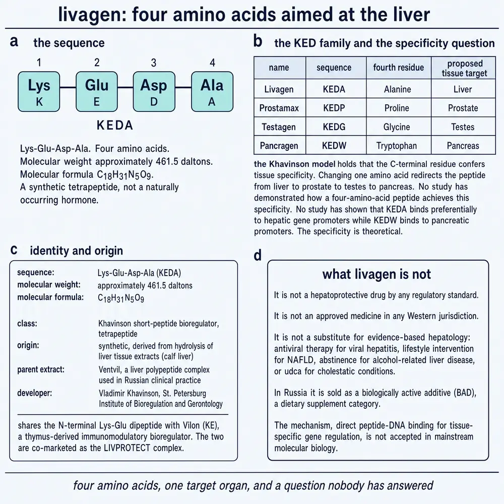
The molecule is a linear tetrapeptide with free ends: lysine at the amino terminus, then glutamate, then aspartate, then alanine, then a free carboxylate. Nothing is cyclised, acetylated, amidated or otherwise protected. Written as one-letter codes it is KEDA, and both forms appear in the literature, sometimes in the same paper.
Three of the four side chains carry a charge at physiological pH, and the arithmetic is worth doing because it is the first thing anyone should notice about a molecule said to bind DNA. Lysine contributes a positive charge; glutamate and aspartate each contribute a negative one; the free carboxyl terminus contributes another. The net is roughly two negative charges. DNA’s backbone is a dense line of negative charges, and the peptides that bind it in biology—histones, transcription factors, protamines—are almost invariably basic. This one is not. The chemical record makes the same point in a different way: the computed partition coefficient is around minus seven point seven and the topological polar surface area is 251 Ų (PubChem, 2026), which describes a molecule that is exceptionally reluctant to enter a lipid membrane. Neither observation refutes anything. Both are constraints that the mechanistic account in Section 08 has to satisfy, and neither has been addressed by any study of this compound.
The identity record itself needed repair. The registry number circulated for this compound across commercial listings and reference profiles is 402856‑42‑2, and it belongs to something else entirely: PubChem resolves it to a dichlorophenyl tropane of formula C₁₇H₂₃Cl₂NO and relative molecular mass 328.3, which is not a peptide (PubChem CID 11522724, 2026). The number carried for the structure above is 433257‑50‑2 (PubChem, 2026). This is a small error with a large reach, because a registry number is the identifier that downstream systems trust when a name is ambiguous, and this compound’s name is ambiguous in three separate directions at once.
Those directions are worth naming, since every one of them has produced a real confusion somewhere in the material surrounding this compound. The trade name has a one-letter neighbour, Livogen, which is a marketed iron-and-folate preparation with no relationship to any of this. The four-letter code is also a place: Keda is a municipality in Adjara, in Georgia—which is not an idle collision, because the chromatin work discussed throughout this monograph was done in Tbilisi and Georgian affiliations run through the entire corpus. And the sequence itself is a strict extension of a sibling’s. Lys–Glu–Asp is Vesugen, a vascular tripeptide with its own literature and its own claims; Lys–Glu is Vilon; Lys–Glu–Asp–Trp is Pancragen and Lys–Glu–Asp–Gly is Testagen. Any text search that matches on the shorter code returns the sibling. Section 12 returns to what that has cost.
Section 04The extract it came from
Every compound in this class has a predecessor that is not a defined molecule at all. The programme’s first generation was a set of tissue extracts—peptide-containing preparations made from the organs of calves and pigs, one extract per organ, administered on the theory that an organ’s own regulatory peptides would restore that organ’s function. The second generation replaced each extract with a short synthetic peptide meant to carry the same organ specificity in a defined form. For the liver, the extract is a polypeptide complex prepared from calf liver and marketed as Ventvil; the peptide is the tetrapeptide described above (Kuznik et al., 2020).
The relationship between the two is the question this section exists to answer, and it is asked far less often than it should be. There are two quite different things that “derived from” can mean. A peptide can be isolated: fractionated out of the extract, sequenced, and then made synthetically, in which case it really is a component of the natural material and the extract’s history is evidence about the peptide. Or a peptide can be designed: composed by someone looking at the extract’s bulk amino-acid composition and choosing a short sequence consistent with it, in which case it is a new molecule that has never been shown to exist in the tissue at all, and the extract’s history is evidence about nothing but the extract.
For this compound the record answers the question directly, in a place that is easy to miss. The 2001 study of protein synthesis in cultured rat hepatocytes describes the peptide, in its own abstract, as having been obtained by directed chemical synthesis on the basis of amino-acid analysis of the liver polypeptide preparations (Brodskiĭ et al., 2001). That is the second meaning. This tetrapeptide was composed from an analysis of the extract; it was not sequenced out of it. The same paper says the same thing about the pineal tetrapeptide, describing Epitalon as constructed on the basis of analysis of the epiphysis peptides.
The distinction matters in a specific and practical way. It means that no evidence about the liver extract transfers automatically to the peptide, because the peptide has never been demonstrated to be a constituent of the extract. Wherever commercial and secondary material describes this compound as the active fragment of the liver complex, it is asserting an isolation that the primary literature does not report and, in the one place the primary literature addresses the point, contradicts. It also means the concordance the reviews emphasise—that the extract and the peptide produce similar effects—is an experimental finding rather than a structural inevitability, and it is a more interesting finding for being so.
That concordance is what the 2020 review reports. Across animal models of acute hepatitis, chronic hepatitis and hepatic fibrosis, and in cell culture, both the liver complex and the tetrapeptide are described as normalising immune and antioxidant status and restoring liver function, with the largest effects in ageing animals (Kuznik et al., 2020). Two cautions attach to that sentence and neither can be resolved from the material available. It is a review: it summarises primary studies rather than reporting one, and the primary studies it summarises for the peptide are not separately retrievable here, so the underlying designs, controls, group sizes and effect magnitudes cannot be inspected. And a review by the compound’s own developers, summarising their own group’s work, is the weakest position from which a favourable summary can be offered. The claim is reported in this monograph as what it is: a secondary account, from inside the programme, of animal experiments this document has not been able to read.
There is a regulatory asymmetry underneath all of this that is easy to state and easy to misread. The liver polypeptide complex has a history of clinical use in Russia. The synthetic tetrapeptide holds no separate pharmaceutical registration anywhere. Those are facts about two different objects, and the temptation—visible in commercial material—is to let the older, more established object lend standing to the newer one. The direction of inference has to run the other way. Whatever the extract has accumulated in the way of clinical exposure belongs to the extract, which is a complex mixture of unknown composition; the defined molecule that was composed from an analysis of it starts from nothing and has, so far, accumulated nothing.
Section 05The programme and the people
The programme this compound belongs to is older than most of the compounds in it. Two review titles from inside the group agree on the starting date from opposite ends of the interval: a 1994 paper describes twenty years of work on the pineal extract, and a 2009 paper describes thirty-five years of research on peptide regulation of ageing (Anisimov et al., 1994; Khavinson & Anisimov, 2009). Both point to 1973 or 1974, and the arithmetic being self-consistent across a fifteen-year gap is a modest but real form of corroboration.
The work began in the Soviet military medical system and moved, after 1992, into the St Petersburg Institute of Bioregulation and Gerontology, which Vladimir Khavinson founded and led. The founding idea was the one described in Section 04: extract the peptide fraction of an animal organ, give it back, and see whether the organ’s function is restored. The extracts came first and the defined short peptides followed, on the theory that the activity of the mixture could be reduced to a sequence.
Commercial material about these compounds tends to describe that history as secret military research, and it is worth being precise about what is and is not true in that. The programme did originate in a military medical academy, and Soviet biomedical research of the period was not published the way Western work was. But the documented record is a gerontology programme with hundreds of indexed publications, most of them in Russian-language journals and a substantial minority in international ones. The interesting fact about this literature is not that it was concealed; it is that it was published continuously for fifty years and was read, outside its own network, by almost nobody.
The compound itself enters the indexed record in 2001, in a study of the rhythm of protein synthesis in cultured rat hepatocytes (Brodskiĭ et al., 2001). Two things about that entry are unusual. The first is that the paper is not about chromatin at all—the mechanism this compound is now known for arrives a year later—and the second is that the transliteration is unstable even within the single abstract, which spells the name one way in its title and another in its body. That instability is characteristic of the whole corpus and is one reason the identity work in Section 03 was necessary.
The chromatin work, which is the substance of the compound’s reputation, was done in Tbilisi. Teimuraz Lezhava’s cytogenetics group at Tbilisi State University—with Tinatin Jokhadze, Jamlet Monaselidze and colleagues—had been studying the chromosomes of very old people for years before these peptides arrived, and had the two things the experiment needed: access to donors in their eighties and nineties, and the classical cytogenetic and calorimetric methods for reading chromatin condensation. The peptides came from St Petersburg; the assays and the donors were Georgian. Every primary chromatin result in this monograph comes from that collaboration.
The collaboration is also the boundary of the evidence. Reading down the reference list, the authorship is essentially two overlapping groups for twenty-two years. That is not unusual for a small field, and it does not by itself indicate anything wrong. What it does mean is that the ordinary error-correcting mechanism—a second group with different hands, different reagents, different donors and no stake in the result attempting the same measurement—has never operated on any of this. Where a finding in this document is described as consistent across studies, the reader should understand that as consistent across studies by the same people, which is a weaker statement than it sounds.
Section 06Unpacking aged chromatin
Two metres of DNA fit into a nucleus a few micrometres across by being wound, folded and stacked. How tightly a given stretch is packed determines whether it can be read: transcription machinery cannot reach DNA that is wound into the dense form called heterochromatin. Some of that packing is permanent architecture—the blocks flanking each centromere, the repetitive regions at chromosome ends—and some of it is regulatory, silencing genes that a particular cell does not need. The second kind is called facultative, and one of the reliable observations about ageing cells is that they accumulate it. Genes that worked in a young cell get wound shut in an old one.
This literature measures that with four classical techniques, and it is worth knowing what each one sees, because the claim is only as good as the reading. Silver staining marks the nucleolus organiser regions—the satellite stalks of the acrocentric chromosomes, where the ribosomal RNA genes live—and the number that stain is taken as a measure of how many are transcriptionally active. C-banding reveals the pericentromeric blocks and allows their size and condensation to be scored. Differential scanning calorimetry measures the temperature at which chromatin melts, which rises with the tightness of packing and so reports on the whole nucleus at once. Sister-chromatid exchange counts recombination events and, when scored by position, says where on the chromosome they occurred.
The founding result for this compound used the first three. In lymphocytes from old donors, the peptide induced activation of ribosomal genes, decondensation of pericentromeric structural heterochromatin, and the release of genes repressed by age-related condensation of euchromatic regions—a study of human cells in culture, with no animal or human administration involved (Khavinson et al., 2002). A study four years later, using donors aged seventy-five to eighty-eight, reported the same three effects and specified the chromosomes: pericentromeric decondensation on chromosomes 1 and 9 (Lezhava et al., 2006).
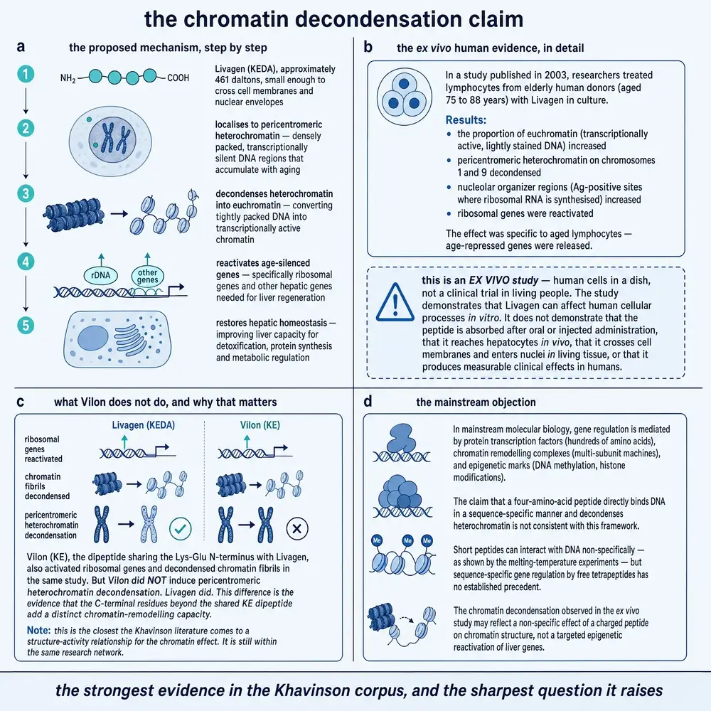
What is notable about the pericentromeric result is that it is the one that carries information. Everything in this literature loosens the ribosomal genes and the facultative heterochromatin—as Section 07 shows, every peptide the group has tested does both. The structural blocks beside the centromeres are different. They are constitutive, made largely of satellite repeats, and they are not supposed to open. A compound that opens them is doing something that most of the class does not do, and it is also doing something whose consequences nobody in this literature has examined, a point Section 13 returns to.
Three caveats attach to the whole domain and are best stated once. All of these are cultured cells: peripheral lymphocytes in medium, with the peptide added to the medium, and nothing about the experiment speaks to what would happen in a living body. None of the reports available here specifies a concentration–response relationship; the peptide is present or it is not. And chromatin decondensation is a change in state, not a benefit. Genes being available to transcription is not the same as the right genes being transcribed, and this literature does not report which genes, in any study, actually changed their expression.
Section 07Five peptides, one system
An organ-specificity claim—the claim that a liver peptide does something a thymus peptide does not—cannot be supported by studying one peptide, however many times. It requires a comparison, run in one laboratory, in one assay, on comparable material, so that the difference between peptides is not confounded with the difference between experiments. This literature ran one, and it is the most valuable single study in the corpus.
Five short peptides—Vilon, Epithalon, Livagen, Prostamax and Cortagen—were tested on leukocytes from subjects aged seventy-five to eighty-eight, scoring ribosomal gene activity, total heterochromatin melting, the polymorphism of the C segments of chromosomes 1, 9 and 16, and the variability of facultative heterochromatin. All five activated ribosomal genes, decondensed densely packed chromatin fibrils, and released genes repressed by age-related condensation of euchromatin. On the pericentromeric blocks the five separated: Epithalon, Livagen and Prostamax decondensed the chromosome 1 block, and of those, Epithalon and Livagen also acted on chromosome 9 (Khavinson et al., 2004). All of this is human cells in culture.
Two further studies fill the grid in. The three-peptide comparison reported that Epitalon and Livagen decondensed pericentromeric heterochromatin of chromosomes 1 and 9 while Vilon did not (Lezhava et al., 2006), and a separate study of Vilon alone, in the same system, states the negative in its own conclusions: the dipeptide unwinds total heterochromatin, reactivates ribosomal genes and releases facultative genes, and does not induce decondensation of pericentromeric structural heterochromatin (Lezhava et al., 2004). A null result reported twice, once inside a comparison and once on its own, is the most solid thing in this corpus. It is the only place where an experiment in this literature could have come out the other way and did not.
What the grid establishes is narrower than the word “specificity” usually implies. It shows that these peptides are not interchangeable and that the differences track their sequences: the two tetrapeptides with an acidic core and a small C-terminal residue reach the pericentromeric blocks and the dipeptide does not. It does not show that the liver peptide is specific to the liver. Nothing in the comparison was a liver cell. What separates the peptides here is a chromatin compartment, not an organ, and the reader should notice that the organ-specificity claim—which is the whole rationale of the second-generation programme—is not what this experiment tested.
There is one result in the corpus that does address organ specificity directly, and it is easy to overlook because it appears in a paper about something else. In cultured rat hepatocytes, the liver tetrapeptide increased the level of protein synthesis at every age tested and had its largest effect in cells from old animals, while the pineal tetrapeptide, tested in the same system, did not change protein synthesis at all (Brodskiĭ et al., 2001). That is a liver cell, two peptides, one assay, and a clean divergence. It is a single study in a rodent primary culture, and it has not been repeated. It is also, on the evidence available, the best support the organ-specificity hypothesis has for this compound.
Section 08How a four-residue peptide could reach DNA at all
The mechanism proposed for the whole class is a chain with four links. The peptide must cross the plasma membrane. It must cross the nuclear envelope. It must bind DNA or histones with enough sequence discrimination to affect particular genes rather than all of them. And that binding must change chromatin state in the direction observed. Each link has been investigated. What matters for this monograph is which links have been investigated with this peptide, and the answer is none of them.
Nuclear entry has been shown by imaging. Fluorescein-labelled short peptides incubated with HeLa cells produced marked fluorescence in cytoplasm, nucleus and nucleolus, establishing that peptides of this size can reach the nuclear interior of an animal cell. The peptides imaged were Epitalon, pinealon and testagen (Fedoreyeva et al., 2011). Histone binding has been shown by fluorescence quenching: six short peptides bound labelled wheat histones H1, H2B, H3 and H4, with binding that depended on the histone, on the peptide’s primary structure and on the methylation status of any associated oligonucleotide. The six were Epitalon, pinealon, bronchogen, testagen, cardiogen and pancragen (Fedoreyeva et al., 2013). Functional consequence has been shown enzymatically: short peptides of two to four residues inhibited or stimulated the hydrolysis of phage DNA by eukaryotic endonucleases depending on methylation status, and did so differently according to their primary structure (Khavinson et al., 2011).
Sequence selectivity has been modelled computationally. Nineteen short peptides were docked to DNA and assigned binding sites, with some peptides sharing sites: Lys–Glu and Glu–Asp–Pro were assigned the sequence agat, Lys–Glu–Asp–Trp and Ala–Glu–Asp the sequence acct, and Ala–Glu–Asp–Leu and Glu–Asp–Leu the sequence ctcc (Khavinson et al., 2016). A larger and more careful study docked all four hundred dipeptides against all possible tetranucleotides in B-form DNA and found that the vast majority of dipeptides cannot bind double-stranded DNA at all, identifying fifty-seven that can, and noting that selectivity rises sharply with peptide length (Kolchina et al., 2019). Cell entry has been modelled too: twenty-six ultrashort peptides were docked to the amino-acid transporters LAT1 and LAT2 and the peptide transporter PEPT1, and the peptides that bound most effectively were those with an acidic residue at the amino terminus (Khavinson et al., 2023).
Read that paragraph again with the subject in mind. Lys–Glu–Asp–Ala is not among the peptides imaged entering a nucleus. It is not among the peptides shown to bind histones. It is not among the nineteen peptides assigned a DNA binding site, and the docking study that covers every possible dipeptide does not cover tetrapeptides. It is not among the transporter ligands named as effective—and the pattern that study reports, a negatively charged N-terminal residue, is the opposite of this peptide’s, which begins with lysine. The mechanistic account for this compound is entirely an argument from family resemblance: relatives of this molecule have been shown to do these things, therefore this molecule presumably does them too.
The class-level model that ties this together is explicit and, to its credit, is presented as a model. Endogenous short peptides are proposed to arise from proteasomal degradation of nuclear proteins, to carry a high local density of opposing charges, and to bind complementary short nucleotide sequences in a way that weakens the hydrogen bonding of the double helix and eases strand separation for transcription. Because genomes lose methylation with age, and demethylated repeats bind these peptides more readily, the model predicts that peptide binding increases in old cells—which is offered as the reason these compounds should act preferentially on aged chromatin (Khavinson et al., 2012). It is an internally coherent story that would explain the observations in Section 06. No part of it has been tested with this compound.
Family resemblance is a reasonable prior and a poor substitute for measurement, and it is weaker here than it looks, because the class’s own results are the reason to doubt it. The docking work reports that selectivity depends sharply on sequence and length; the head-to-head comparison in Section 07 reports that a one-residue difference is enough to abolish an effect. A literature whose central finding is that these peptides differ cannot then treat them as interchangeable.
Section 09An enzyme, a liver, a gut and a fly
Away from chromatin, the compound has been looked at in a scattering of unrelated systems. The scattering is worth reporting in full, because breadth without depth is itself a finding: half a dozen separate lines of investigation were opened, each by a single study, and not one of them was followed up.
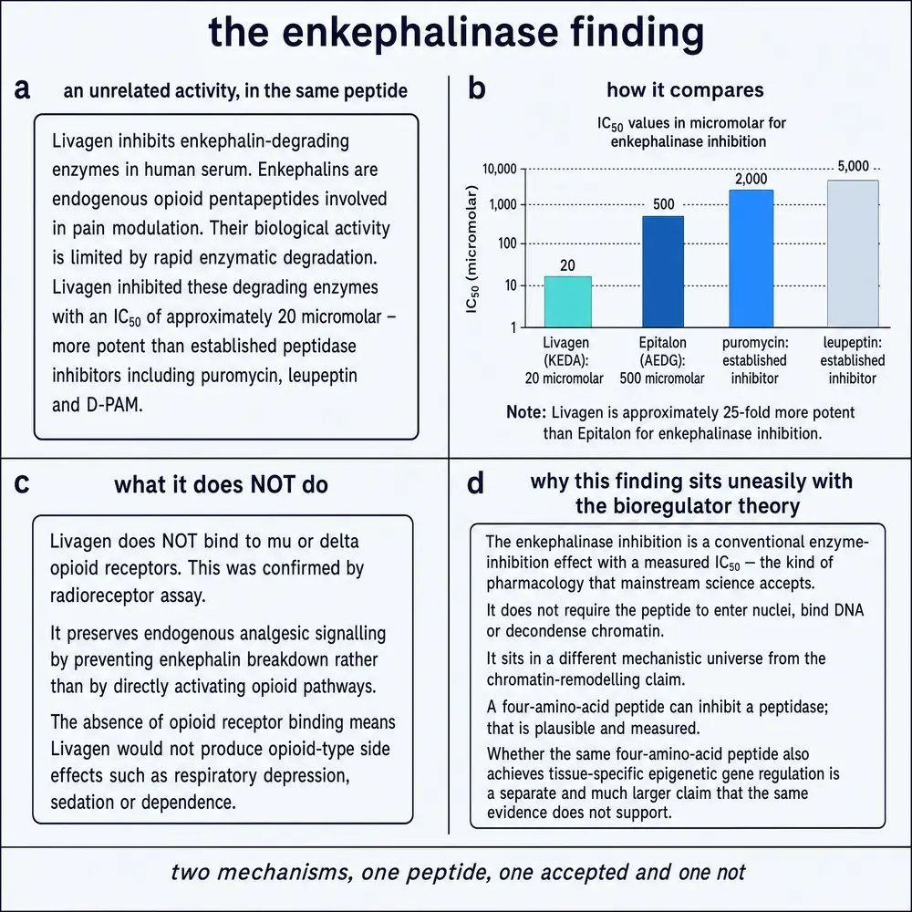
The most quantitative result in the entire corpus concerns an enzyme. In a cell-free assay measuring the rate of tritiated leucine-enkephalin hydrolysis by human serum, the compound inhibited enkephalin-degrading enzymes, and it did so with a published dose–inhibition curve: a half-maximal inhibitory concentration of 20 µM, against 500 µM for Epitalon tested alongside it. It was reported as more effective than the established peptidase inhibitors puromycin, leupeptin and D‑PAM, though no value is published for any of the three. A radioreceptor assay on rat brain membrane found no interaction with μ- or δ-opioid receptors, so whatever effect this has on the endogenous opioid system is an effect on the enzymes that destroy enkephalin rather than on the receptors that respond to it (Kost et al., 2003).
Two points about that number, since it is the only one of its kind here. It is an experimental parameter in a test tube of human serum, not a dose, a route or a schedule for anyone, and nothing in this document should be read as suggesting otherwise. And 20 µM is not a low concentration for a peptide said to work in the nanomolar range; the class’s claimed potency and its one measured potency are three orders of magnitude apart, in different assays, and no study has reconciled them. This result was published in 2003 and has no follow-up of any kind: no cell experiment, no animal experiment, nothing.
The liver work is thin but real. Beyond the hepatocyte protein-synthesis study already described, an organotypic liver culture exposed to the tetrapeptide showed changes in functional morphology by immunocytochemical and morphometric analysis, which the authors interpreted as stabilisation of morphological integrity and reinforcement of cellular and intracellular regeneration (Riadnova et al., 2002). That is a tissue-culture study, its endpoints are morphological, and the interpretive language in the conclusion is considerably firmer than a morphometric measurement can support.
The gut study is the only one in the corpus that administered the compound to a living animal by a defined route, and it produced the closest thing to a pharmacokinetic observation anywhere in this literature. In rats, the peptide is reported as weakly hydrolysed: peptide hydrolases of the small intestine did not hydrolyse it even to a small extent. In vitro it reduced glycyl‑L‑leucine dipeptidase activity in the small intestine by half. After two weeks of oral administration, digestive enzyme activity decreased in young animals and increased in old ones, and in old animals the activities in most cases approached those of young controls (Timofeeva et al., 2005). The bidirectional, age-dependent pattern is the interesting part; it is also exactly the shape of result that is hardest to interpret without a dose–response relationship or a mechanism, and neither is available. The finding of resistance to intestinal hydrolysis is reported here as a study observation in rats and is not evidence about absorption, distribution or fate in any species, including that one.
The remaining two lines are single reports. In cells from individuals aged seventy-two to eighty-six, the radiation-induced adaptive response—the phenomenon in which a small dose of radiation makes cells more resistant to a later large one—was studied with a subsequent copper chloride challenge, and corrective activity of the peptide was observed; the abstract reports the observation without quantifying it (Dzhokhadze et al., 2007). And in a Drosophila model of neurodegeneration involving heat shock in kynurenine-pathway mutants, the liver peptide and the cortex peptide were both reported active. That report is a congress abstract rather than a paper: there are no methods, no numbers and no results beyond the summary, and it cannot be assessed (Savvateeva-Popova et al., 2007).
Section 10Cobalt, telomeres, and a rabbit
The most speculative thread in the corpus is also the most quantitatively reported, which is an unusual combination and repays attention. Cultured lymphocytes from donors aged eighty to ninety-one, with a control group aged eighteen to thirty, were exposed to cobalt chloride alone and to cobalt chloride with the peptide. Cobalt is a genotoxic metal; the experiment asks not how much damage it causes but where on the chromosome the resulting recombination events land.
Cobalt alone drove sister-chromatid exchange into the pericentromeric heterochromatin, where 15.4 ± 1.8 per cent of exchanges were registered against 4.5 ± 0.6 per cent in controls. Cobalt with the peptide moved them instead into telomeric heterochromatin, 12.0 ± 1.2 per cent against 2.8 ± 0.5 per cent. In the same experiment, chromosomal aberrations under cobalt were 3.4 ± 0.6 per cent with the peptide present against 4.2 ± 0.7 per cent in the control group (Lezhava & Jokhadze, 2007). All of it is human cells in culture.
The authors read the shift as decondensation making the telomeric regions accessible to repair. That reading is consistent with the data and it is not the only one available. An agent that relocates recombination from one chromosomal compartment to another has changed where exchange happens; it has not thereby shown that the new location is preferable. Sister-chromatid exchange is used as a marker of repair and as a marker of damage, and the same number supports either description. The reduction in aberrations is the more directly protective observation, and it is small.
The cobalt pairing recurs through the group’s later work on cells from patients, where the peptide and the metal are applied separately and together as modifying agents. In lymphocytes from patients with atherosclerosis aged eighty and over, genomic instability was elevated on every parameter measured and was reported as normalised by the peptide alone and in combination with cobalt (Dzhokhadze et al., 2014). In lymphocytes from patients with ductal breast cancer, DNA single-strand break density, chromosomal abnormality frequency and chromatin condensation were all elevated and all reported as improved (Jokhadze et al., 2017). These are studies of cells from patients, not studies of patients, and Section 13 returns to what the first of them concluded from that.
Beyond the peer-reviewed record there is one further claim, and it is reported here precisely because it is the kind of claim that ends up detached from its qualifications. A 2012 book chapter from the Tbilisi group describes, citing an unpublished project, that the peptide combined with cobalt normalised telomere length in cells from old individuals and reduced blood cholesterol by around forty-one per cent in a rabbit model of atherosclerosis (Lezhava et al., 2012). No paper reporting either result has been located. There is no way to know the design, the group sizes, the controls or the duration. A telomere-length normalisation and a forty-one per cent cholesterol reduction would be substantial findings if they survived publication; as they stand they are unpublished preliminary animal data mentioned in a review chapter, and they should not be cited as anything else.
Section 11The most recent work, and what recency is worth
The two most recent primary studies are from 2020 and 2023, and they are near-replicas of each other and of the work from two decades earlier. Both tested four bioregulators—Epitalon, this compound, Cortagen and Vilon—on cultured lymphocytes from donors aged seventy-five to eighty-eight against younger controls, using differential scanning calorimetry, silver-stained nucleolus organiser regions, C-heterochromatin scoring and sister-chromatid exchange. Both report the same three-part conclusion: the peptides unwind total, constitutive and facultative heterochromatin; sister-chromatid exchange rises in telomeric regions and falls in the medial regions of the chromosome arms; and each peptide selectively deheterochromatinises a specific region rather than acting uniformly (Lezhava et al., 2020; Lezhava et al., 2023).
Recency is given weight in this document for one reason, and it is worth stating the principle rather than leaving it implicit. A recent finding matters when it is consistent with the older record, because consistency across twenty-one years suggests the measurement is real and not an artefact of one cohort, one batch of reagent or one period’s technique. It does not matter merely because it is newest; a newer result that contradicted the older ones would raise the question of which to believe rather than settling it. Here the 2023 study reports what the 2002 study reported, using the same methods on donors of the same ages, which is genuine corroboration of the observation.
It is corroboration of a bounded kind. The same laboratory ran both. The 2023 study introduces no new method, no new endpoint, no dose–response relationship, no gene-expression measurement and no living organism. Twenty-one years produced a fourth report of the same experiment. What the corpus lacks is not repetition—it has that—but the second and third steps that normally follow a robust in-vitro observation: someone else confirming it, and someone asking what it does in an animal.
Section 12Siblings, and the code that is not this one
The family in Section 04 has a naming problem that is not cosmetic. Its members are designated by their one-letter sequences, and because the sequences are short and share stems, several designations are strict prefixes or extensions of others. Lys–Glu is Vilon. Add aspartate and it is Vesugen, KED. Add alanine to that and it is this compound, KEDA; add tryptophan instead and it is Pancragen, KEDW; add glycine and it is Testagen, KEDG.
The consequence is asymmetric and it runs in one direction only. A paper about Vesugen never says KEDA, so a search on the longer code is safe. A search, a summary, or a reader working from the shorter stem is not, because KED returns Vesugen — a vascular tripeptide with a substantial literature of its own, including claims this compound has never had. One study describes KED as having vasoprotective effects and being an effective substance in the treatment of atherosclerosis and other cardiovascular disorders in elderly people, on the basis of endothelin‑1, connexin and sirtuin 1 expression measured in cultured normal, atherosclerotic and restenotic endothelium (Kozlov et al., 2016). Another attaches KED to neurogenesis in Alzheimer’s disease (Khavinson et al., 2021b). Neither has anything to do with the compound described in this monograph, and the class-context review this document cites for the general model uses “KED” in the Vesugen sense throughout.
The identity checks written for this project were built around that hazard, and around two others peculiar to this compound: the marketed haematinic whose name differs by one vowel, and the Georgian municipality that shares the four-letter code in exactly the region where the cytogenetics was done. A text is admitted here as being about this compound only on the trade name, the full spelled-out sequence with guards against longer runs, or the bare code accompanied by subject matter—liver, chromatin, or a named investigator from the programme. A size word such as “tetrapeptide” does not count as corroboration, because it corroborates every four-residue sequence ever published, which is precisely the set the bare code collides with.
Within the family, the honest comparison is unflattering in one direction and informative in the other. Epithalon has by far the larger evidence base and, where the two have been tested side by side, it has sometimes come out ahead: in lymphocyte cultures from patients with hypertrophic cardiomyopathy and their first-degree relatives, three bioregulators were compared and Epithalon was reported as the most effective at normalising the functional parameters of the genome and lowering chromosomal instability (Dzhokhadze et al., 2013). That study is cited here because the group published it, and because a monograph that reports the head-to-head where its subject won and omits the one where it did not has not reported a comparison at all.
What is genuinely this compound’s own, on the available record, amounts to three things. It is the only member of the class with a published dose–inhibition curve against enkephalin-degrading enzymes. It is the member with the liver work: hepatocyte protein synthesis, organotypic culture, digestive enzymes, and the extract pairing. And in the one direct organ-level comparison in the corpus, it acted on hepatocyte protein synthesis where the pineal peptide did not. That is a short list and it is not nothing.
Section 13What is not known, at full volume
The gaps in this record are not the ordinary gaps of an active field, where some questions are open because others were answered first. They are structural, and they sit underneath everything in Part Four.
There is no pharmacokinetics. Not a limited dataset, not a study in the wrong species — none, in any species, of any kind. No half-life, no bioavailability by any route, no distribution, no metabolism, no elimination. The closest thing in the literature is the observation that intestinal peptide hydrolases in rats did not measurably cleave the molecule, and that is one incidental measurement inside a study about digestive enzymes. In its absence, everything anyone believes about what happens to this molecule in a body rests on inference from its structure: an unmodified linear tetrapeptide with free termini is exposed at both ends, to aminopeptidases at the lysine and carboxypeptidases at the alanine, and would be expected to be cleaved quickly in plasma. That is a reasonable expectation. It is not a measurement, and this document does not present it as one.
There is no formal toxicology. No dose-escalation study, no maximum tolerated dose, no reproductive or developmental study, no interaction study, no long-term study, to international standards or to any standard. What exists is incidental: the in-vitro lymphocyte studies report no cytotoxicity at the concentrations they used, and the animal work summarised in the 2020 review reports no treatment-related adverse effects. Absence of reported harm in a handful of small experiments that were not designed to detect harm is close to no information at all. It is worth noting that the class has produced one real safety dataset, for a different compound: long-term subcutaneous administration of the thymic dipeptide to mice from six months of age caused no unfavourable effects on development and the authors concluded that chronic administration was safe (Khavinson et al., 2000). That is a study of Vilon in mice and it says nothing about this molecule.
There is a theoretical safety question that nobody in this literature has raised, and it arises directly from the central finding rather than from anything external. Pericentromeric heterochromatin is not decorative. It is where the kinetochore assembles, and its condensed state contributes to correct sister-chromatid cohesion and to accurate chromosome segregation during division. The signature effect of this compound—the one that distinguishes it from Vilon and defines its claim—is the decondensation of exactly that structure. Loosening it in a lymphocyte for the duration of a culture experiment may be entirely benign. Sustained loosening in a dividing tissue is a different proposition, and no published study has looked. This is a theoretical concern rather than an observed harm, and it should be read that way; but it is a concern generated by the mechanism itself, and a literature that reports the decondensation as unambiguously beneficial has not engaged with it.
There is no registered clinical trial. A search of ClinicalTrials.gov for the compound name, for the sequence, and for the programme returns nothing; the same search for “peptide bioregulator” as a class term also returns nothing (ClinicalTrials.gov, 2026). There is no marketing authorisation from any major regulator. The compound circulates as a research chemical and as a component of supplement preparations, and its commercial presentation carries a conventional unit size that appears nowhere in the scientific literature and corresponds to no studied quantity. This document specifies no amount, route or schedule for any person, and nothing in it should be read as recommending human use.
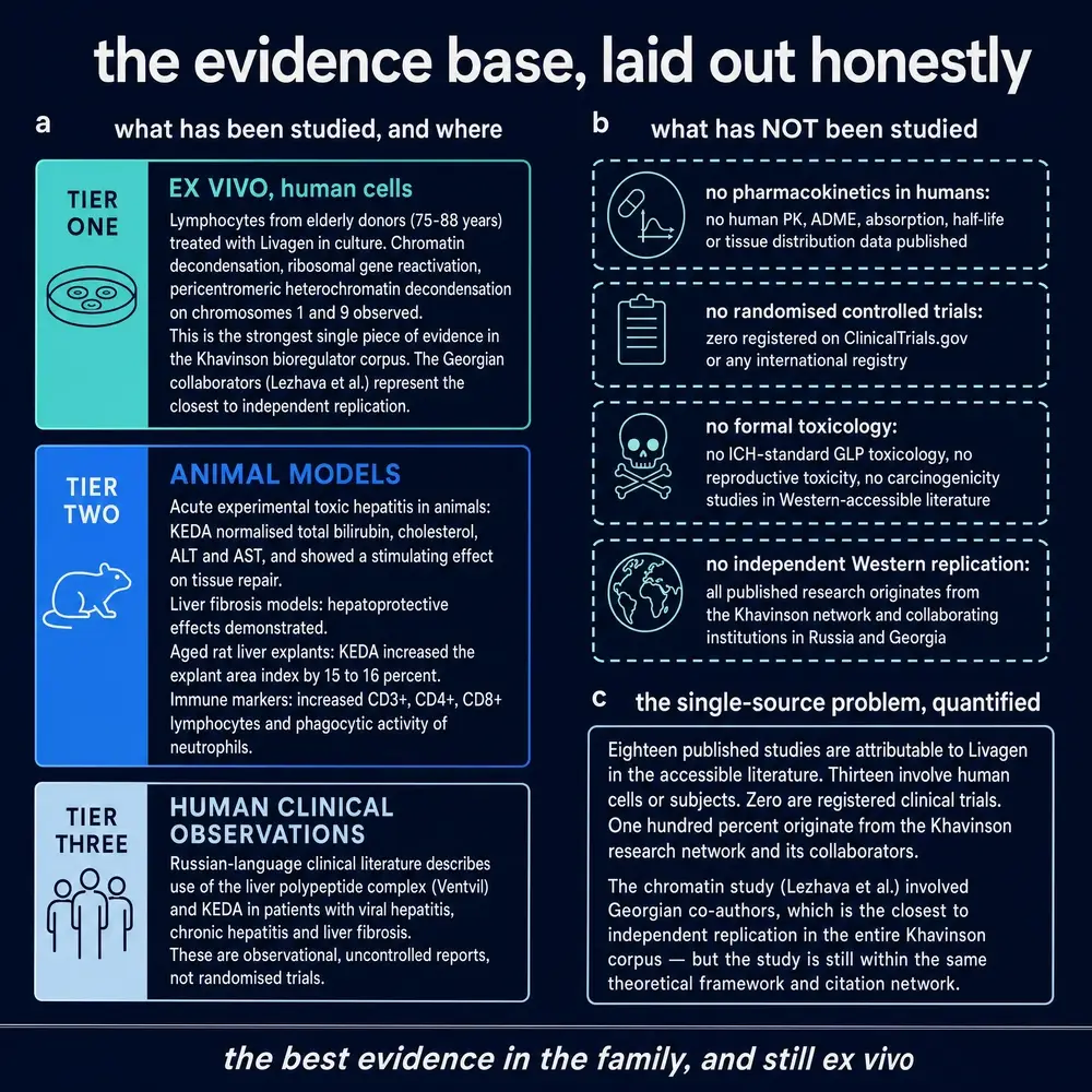
And there is no independent replication. This is the first-order limitation, and it is worth separating from a claim it can be confused with. The programme’s own reviews describe collaboration with laboratories in several countries (Khavinson & Anisimov, 2009), and there is no reason to doubt that the programme as a whole has collaborators. What could not be found is a publication on this compound from any group outside the St Petersburg and Tbilisi network. Every experimental result in Part Four originates inside it. The consistency of the record across two decades is therefore consistency of a technique in the hands that developed it, which is worth something, and it is not what the word usually means.
One more thing belongs here, because it is a property of the corpus rather than of the compound. Several papers in this literature state conclusions considerably stronger than their designs can bear, and the pattern is consistent enough to affect how the whole should be read. The clearest instance concerns cells from patients with atherosclerosis: having measured chromosomal aberrations, aneuploidy and polyploidy in cultured lymphocytes and observed improvement, the study concludes that the identified protective action proves the compound’s efficacy in the prevention of atherosclerosis (Dzhokhadze et al., 2014). Chromosomal parameters in a lymphocyte culture cannot establish the prevention of a vascular disease in a person, and no part of that study went near a person. The measurement is a measurement; the conclusion is not entailed by it. Where this monograph reports a result from that literature, it reports the measurement.
Section 14Believing a closed literature exactly as far as it has earned
Return to the culture dish. Lymphocytes from a person in their eighties, a four-residue peptide in the medium, and chromatin that measurably comes apart at a lower temperature than it did before. That observation has been made four times over twenty-one years, by people using classical methods they know well, and it has been made alongside a negative control of the best kind—a closely related peptide that produces most of the same effects and, reproducibly, not this one. There is nothing obviously wrong with it.
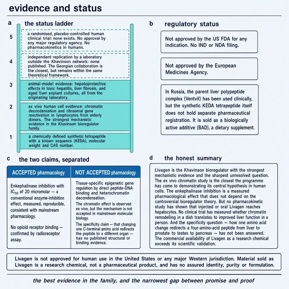
What there is, is nothing around it. The observation has no dose–response curve, no identified gene whose expression changed, no measurement in any tissue other than a cultured lymphocyte, no demonstration that the molecule reaches a nucleus, no measurement of what happens to it in a body, no animal in which the chromatin effect was sought, no second laboratory, and no trial. Each of those absences is individually unremarkable in a small field. Together they describe a finding that has been repeated but never developed: twenty-two years of confirming the same measurement without asking the next question.
The temptation with a literature like this is to resolve it, in one direction or the other. It can be dismissed—single school, obscure journals, translated abstracts, a category that no pharmacology recognises—and dismissal costs nothing, because nothing here has to be acted on. Or it can be accepted, because the internal record really is consistent, the negative controls really do behave as they should, and the mechanism proposed for the class really is coherent. Both moves are cheaper than the accurate one, which is to hold the finding at exactly the weight it has: a real and reproducible observation in cultured human cells, of unknown relevance to anything outside the dish, made by people with an interest in the answer and never checked by anyone without one.
The last thing worth saying concerns what this compound is used for now. It is sold, and it is discussed, in language borrowed from what its relatives have shown and from what its parent extract has been used for—an inheritance of standing that the molecule has not earned on its own record. It is described as the active fragment of a liver extract, which the primary literature says it is not; as an established member of a recognised class, which is a category its own programme created; and as a chromatin-modifying agent, which it may well be, in a dish, at concentrations nobody has specified, with consequences nobody has measured. The gap between what is on the label and what is in the record is not a gap in the science. It is the whole of what this monograph found.
This monograph is a research document. It describes published experiments and it does not recommend that any person use this compound. No dose, route, schedule, formulation or duration is specified for any person anywhere in it. Concentrations, administration routes and durations appear only as parameters of the studies that reported them, always with the study type and the species or cell system named in the same sentence. Nothing here is medical advice.
Section 15References
- Anisimov VN, Khavinson VKh, Morozov VG. Twenty years of study on effects of pineal peptide preparation: epithalamin in experimental gerontology and oncology Ann N Y Acad Sci 1994;719:483-93. PMID 8010617 · doi
- Brodskiĭ VIa, Khavinson VKh, Zolotarev IuA, Nechaeva NV, Malinin VV, Novikova TE et al.. [Rhythm of protein synthesis in cultures of hepatocytes from rats of different ages. Norm and effect of the peptide livagen] Izv Akad Nauk Ser Biol 2001;(5):517-21. PMID 15926314
- Dzhokhadze TA, Buadze TZh, Dvalishvili NA, Lezhava TA. [Variability of radiation-induced adaptive response in old age individuals and their correction by Peptide bioregulator -Livagen] Georgian Med News 2007;(148-149):50-4. PMID 17921545
- Dzhokhadze TA, Buadze TZh, Gaĭozishvili MN, Rogava MA, Lazhava TA. [Functional regulation of genome with peptide bioregulators by hypertrophic cardiomyopathy (by patients and relatives)] Georgian Med News 2013;(225):94-7. PMID 24423684
- Dzhokhadze TA, Buadze TZh, Gaiozishvili MN, Kakauridze NG, Lezhava TA. [Genomic instability in atherosclerosis] Georgian Med News 2014;(236):82-6. PMID 25541832
- Fedoreyeva LI, Kireev II, Khavinson VKh, Vanyushin BF. Penetration of short fluorescence-labeled peptides into the nucleus in HeLa cells and in vitro specific interaction of the peptides with deoxyribooligonucleotides and DNA Biochemistry (Mosc) 2011;76(11):1210-9. PMID 22117547 · doi
- Fedoreyeva LI, Smirnova TA, Kolomijtseva GY, Khavinson VKh, Vanyushin BF. Interaction of short peptides with FITC-labeled wheat histones and their complexes with deoxyribooligonucleotides Biochemistry (Mosc) 2013;78(2):166-75. PMID 23581987 · doi
- Jokhadze T, Gaiozishvili M, Buadze T, Sigua T, Namchelvadze E, Lezhava T. [EVALUATION OF GENOMIC PARAMETERS IN DUCTAL BREAST CANCER PATIENTS AND THE ABILITY OF IT'S CORRECTION] Georgian Med News 2017;(265):120-125. PMID 28574395
- Khavinson V, Linkova N, Diatlova A, Trofimova S. Peptide Regulation of Cell Differentiation Stem Cell Rev Rep 2020;16(1):118-125. PMID 31808038 · doi
- Khavinson VK, Lin'kova NS, Tarnovskaya SI. Short Peptides Regulate Gene Expression Bull Exp Biol Med 2016;162(2):288-292. PMID 27909961 · doi
- Khavinson VK, Linkova NS, Rudskoy AI, Petukhov MG. Feasibility of Transport of 26 Biologically Active Ultrashort Peptides via LAT and PEPT Family Transporters Biomolecules 2023;13(3). PMID 36979488 · doi · PMC10046148
- Khavinson VK, Popovich IG, Linkova NS, Mironova ES, Ilina AR. Peptide Regulation of Gene Expression: A Systematic Review Molecules 2021;26(22). PMID 34834147 · doi · PMC8619776
- Khavinson VK, Anisimov VN, Zavarzina NY, Zabezhinskii MA, Zimina OA, Popovich IG et al.. Effect of vilon on biological age and lifespan in mice Bull Exp Biol Med 2000;130(7):687-90. PMID 11140587 · doi
- Khavinson VK, Lin'kova NS, Umnov RS. Peptide KED: Molecular-Genetic Aspects of Neurogenesis Regulation in Alzheimer's Disease Bull Exp Biol Med 2021;171(2):190-193. PMID 34173097 · doi
- Khavinson VKh, Lezhava TA, Monaselidze JG, Dzhokhadze TA, Dvalishvili NA, Bablishvili NK et al.. Effects of Livagen peptide on chromatin activation in lymphocytes from old people Bull Exp Biol Med 2002;134(4):389-92. PMID 12533768 · doi
- Khavinson VKh, Lezhava TA, Malinin VV. Effects of short peptides on lymphocyte chromatin in senile subjects Bull Exp Biol Med 2004;137(1):78-81. PMID 15085253 · doi
- Khavinson VKh, Fedoreyeva LI, Vanyushin BF. Site-specific binding of short peptides with DNA modulated eukaryotic endonuclease activity Bull Exp Biol Med 2011;151(1):66-70. PMID 22442805 · doi
- Khavinson VKh, Solov'ev AIu, Zhilinskiĭ DV, Shataeva LK, Vaniushin BF. [Epigenetic aspects of peptide regulation of aging] Adv Gerontol 2012;25(1):11-22. PMID 22708439
- Khavinson VKh, Anisimov VN. [35-year experience in research of peptide regulation of aging] Adv Gerontol 2009;22(1):11-23. PMID 19827673
- Kolchina N, Khavinson V, Linkova N, Yakimov A, Baitin D, Afanasyeva A et al.. Systematic search for structural motifs of peptide binding to double-stranded DNA Nucleic Acids Res 2019;47(20):10553-10563. PMID 31598715 · doi · PMC6847403
- Kost NV, Sokolov OIu, Gabaeva MV, Zolotarev IuA, Malinin VV, Khavinson VKh. [Effect of new peptide bioregulators livagen and epitalon on enkephalin-degrading enzymes in human serum] Izv Akad Nauk Ser Biol 2003;(4):427-9. PMID 12942748
- Kozlov KL, Bolotov II, Linkova NS, Drobintseva AO, Khavinson VK, Dyakonov MM et al.. [Molecular aspects of vasoprotective peptide KED activity during atherosclerosis and restenosis] Adv Gerontol 2016;29(4):646-650. PMID 28539025
- Kuznik BI, Khasanova NB, Ryzhak GA, Mezsheriakova IE, Khavinson VK. [The influence of polypeptide liver complex and tetrapeptide KEDA on organism physiological function in norm and age-related pathology.] Adv Gerontol 2020;33(1):159-164. PMID 32362099
- Lezhava T, Monaselidze J, Kadotani T, Dvalishvili N, Buadze T. Anti-aging peptide bioregulators induce reactivation of chromatin Georgian Med News 2006;(133):111-5. PMID 16705247
- Lezhava T, Khavison V, Monaselidze J, Jokhadze T, Dvalishvili N, Bablishvili N et al.. Bioregulator Vilon-induced reactivation of chromatin in cultured lymphocytes from old people Biogerontology 2004;5(2):73-9. PMID 15105581 · doi
- Lezhava T, Jokhadze T. Activation of pericentromeric and telomeric heterochromatin in cultured lymphocytes from old individuals Ann N Y Acad Sci 2007;1100:387-99. PMID 17460203 · doi
- Lezhava T, Jokhadze T, Monaselidze J, Buadze T, Gaiozishvili M, Sigua T. EPIGENETIC MODIFICATION UNDER THE INFLUENCE OF PEPTIDE BIOREGULATORS ON "AGED" HETEROCHROMATIN Georgian Med News 2020;(309):120-124. PMID 33526740
- Lezhava T, Jokhadze T, Monaselidze J, Buadze T, Gaiozishvili M, Sigua T et al.. EPIGENETIC MODIFICATION UNDER THE INFLUENCE OF PEPTIDE BIOREGULATORS ON THE "OLD" CHROMATIN Georgian Med News 2023;(335):79-83. PMID 37042594
- Riadnova IIu, Filippov SV, Iuzhakov VV. [Functional morphology of an organotypic liver culture exposed to the peptide livagen] Adv Gerontol 2002;10:88-94. PMID 12577697
- Timofeeva NM, Khavinson VKh, Malinin VV, Nikitina AA, Egorova VV. [Effect of peptide Livagen on activity of digestive enzymes in gastrointestinal tract and non-digestive organs in rats of different ages] Adv Gerontol 2005;16:92-6. PMID 16075683
- Lezhava T, Jokhadze T, Monaselidze J, Gaiozishvili M, Sigua T, Buadze T. The functioning of “aged” heterochromatin. In: Nagata T, ed. Senescence. Rijeka: InTech; 2012. Open-access book chapter. doi:10.5772/35651. Carries the Co2+ work later published in full, and an unpublished STCU-4307 project claim which this document reports as unpublished. Resolved 3 August 2026. link
- Savvateeva-Popova E, Nikitina E, Tokmacheva E, Medvedeva A, Kamyshev N, Kamysheva E. Therapeutic activity of short synthetic peptides Cortagen and Livagen in a Drosophila model for neurodegenerative disorders facilitated by heat shock in mutants of the kynurenine pathway. Parkinsonism Relat Disord. 2007;13:S136. Congress abstract, not a full paper; no methods or results beyond the abstract are available. doi:10.1016/s1353-8020(08)70752-1. Resolved 3 August 2026. link
- National Center for Biotechnology Information. PubChem Compound Summary CID 87919683, Livagen (Lys-Glu-Asp-Ala). PubChem, Bethesda MD. Molecular formula C18H31N5O9, relative molecular mass 461.5, monoisotopic mass 461.2122, InChIKey IKVDKWACACMDLR‑BJDJZHNGSA‑N, XLogP −7.7, topological polar surface area 251 Å2, registry number 433257-50-2. Queried 3 August 2026. link
- National Center for Biotechnology Information. PubChem Compound Summary CID 11522724, (2R,3S)-3-(3,4-dichlorophenyl)-2-(ethoxymethyl)-8-methyl-8-azabicyclo[3.2.1]octane. PubChem, Bethesda MD. The compound to which registry number 402856-42-2 resolves. Molecular formula C17H23Cl2NO, relative molecular mass 328.3. Queried 3 August 2026. link
- United States National Library of Medicine. ClinicalTrials.gov, search for Livagen, Lys-Glu-Asp-Ala, peptide bioregulator and Khavinson. Zero studies returned for “Livagen”, zero for “peptide bioregulator” and zero for “Khavinson”. Queried 3 August 2026. link
Section 16How this document was assembled
The finding that shaped the method came before any of the prose. The research library this monograph belongs to holds no scientific full text naming this compound. Its own dossier for it is a twenty-page document in which every one of the seventeen substantive sections is flagged as having insufficient evidence and the bibliography is empty. A library that holds nothing about a compound is telling the reader something about the compound, and in this case it was telling the truth: the published record is small.
The corpus was therefore built from the indexed literature directly. Thirty PubMed records were resolved and verified against live NCBI records, and every author, title, journal, volume, issue, page range, year and identifier printed in the reference list above was fetched from the record rather than recalled. That check earned its place. Five attributions carried in this project’s own working notes were wrong and were corrected: two chromatin papers attributed to authors who are not their first authors, a serum-enzyme study attributed to three investigators none of whom leads it, a page range that belongs to a different article in the same issue, and a study cited without any identifier at all. One of those errors is instructive about the shape of the rest. The founding 2002 chromatin paper had been attributed to Ryadnova, who is a real author on this compound—of the organotypic liver culture study, where NCBI spells her Riadnova—which is exactly the kind of misattribution that survives inspection, because nothing about it looks wrong.
No full text was available for any of the thirty records, so every one was read as an indexed abstract. That constrains what can responsibly be said, and the constraint has been observed throughout: no methodological judgement is offered that an abstract cannot support, group sizes and statistical treatments are reported only where an abstract states them, and where a paper’s design cannot be inspected the text says so. Five further sources have no PubMed record—a book chapter, a congress abstract, two database entries and a trial-registry search—and each is listed above with what it is and when it was resolved.
Identity was enforced mechanically rather than by eye, because this compound’s designations collide in three separate ways and one of the collisions is with a sibling in the same family. A shared identity module, imported by every stage of the build, admits a text as being about this compound on the trade name, on the full spelled-out sequence with guards against longer residue runs on either side, or on the bare four-letter code accompanied by subject matter. It rejects the sibling designations of the family, the one-vowel neighbour that is a marketed haematinic, the Georgian place name, and the bare code corroborated only by a word describing a peptide’s size. The module carries nineteen test cases drawn from real records, including the sibling papers whose findings are most likely to be misattributed, and the build refuses to proceed if any of them fails.
Two searches were run for absences rather than presences, because in a corpus this small an absence is a finding. ClinicalTrials.gov was queried for the compound name, for the sequence, for the class term and for the programme’s founder; all four returned nothing. And every retrievable record was checked for an author affiliation outside the St Petersburg and Tbilisi network; none was found.
All nine figures are original works, generated by this project’s own figure toolkit from the numerical results printed in their captions. No published figure has been reproduced or adapted. Each figure is drawn on a canvas whose height is computed from its own registered ink rather than asserted, so a figure cannot be authored in a way that clips itself, and every colour resolves through a theme token rather than a fixed value, so the light and dark editions render from one source. Both editions were rendered and inspected.
Section 17How the evidence was weighed
Four rules governed the reading, and they are stated here so that a reader who disagrees with one can discount the parts of the document that depend on it.
Study type is named in the sentence that reports each result, without exception. This is the single most useful discipline available for this literature, because the distance between a lymphocyte in a dish and a claim about a person is routinely crossed inside one abstract, and naming the system in the reporting sentence makes the crossing visible whenever it happens. Where a source’s own conclusion travels further than its design, the measurement is reported and the conclusion is named as the source’s.
Evidence for the class is not evidence for the compound. A great deal of what circulates about this molecule is really about its relatives—nuclear entry, histone binding, DNA sequence selectivity, transporter-mediated uptake—and in each of those cases the peptides actually tested are named in this document and this one is not among them. Where an argument rests on family resemblance, the text says so.
Negative and divergent results are reported at the same volume as positive ones. The most solid result in the corpus is a null: a sibling peptide that reproducibly fails to decondense the structure this compound is said to decondense. The comparison in which this compound was outperformed by a sibling is reported alongside the comparisons in which it was not. A head-to-head design is valuable precisely because it can fail, and reporting only the arm that succeeded would discard the reason for running it.
Recency is weighed for consistency, not for date. The 2023 study is given weight because it reports what the 2002 study reported, using the same methods on donors of the same age, and consistency across twenty-one years suggests a real measurement. It is not given weight for being newest, and it is not treated as advancing the evidence base, because it introduces no method, endpoint or system that the earlier work did not have.
Two things were deliberately not done. No attempt was made to reconcile the class’s claimed nanomolar working range with the single measured micromolar potency in the corpus, because no study reconciles them and a reconciliation invented here would be invention. And no attempt was made to translate any result into a statement about what this compound would do in a person, because nothing in the record supports such a statement and the absence of that translation is, in the end, the most important thing this document has to report.
Continue exploring