
MOTS-c The message in the machine — a peptide the mitochondrion writes, and what a decade of evidence does and does not establish about it
Sixteen amino acids are encoded inside a mitochondrial gene for a ribosome, in a stretch of DNA the organelle that owns it cannot read. The cell exports the message, translates it outside, and sends the product to the nucleus to change which genes are switched on. In the eleven years since that was discovered, MOTS-c has become one of the best-characterised and least clinically established molecules in metabolic biology — banned in sport, sold online, and still, as of August 2026, waiting on its first completed trial.
Quantities appear only as reported study parameters — what a named group of researchers gave to a named population for a named duration. Nothing here is a dose recommendation, and no protocol for any person appears anywhere in this document.
01A peptide that should not exist
Start with a reading problem rather than a definition.
Inside every one of your cells sit several hundred mitochondria, and inside each of those sits a small circular chromosome of its own — 16,569 base pairs in humans, a bacterial inheritance from an ancient merger, carrying thirteen protein-coding genes that build parts of the machinery for making energy. The mitochondrion transcribes and translates this chromosome itself, using its own ribosomes and, crucially, its own slightly divergent genetic code. Human mitochondria read the triplets AGA and AGG as instructions to stop; everywhere else in the cell those same triplets mean arginine.
Somewhere in the middle of the gene MT-RNR1 — the gene for the small subunit of the mitochondrial ribosome, the 12S ribosomal RNA, whose only recognised job was to help build the machine that does the translating — there is a stretch of fifty-one base pairs. Read with the mitochondrion's own code, that stretch is a dead letter: a start codon followed immediately by a stop. The organelle cannot make anything of it. Read with the standard code used everywhere outside the mitochondrion, the same fifty-one bases spell a sixteen-amino-acid peptide.
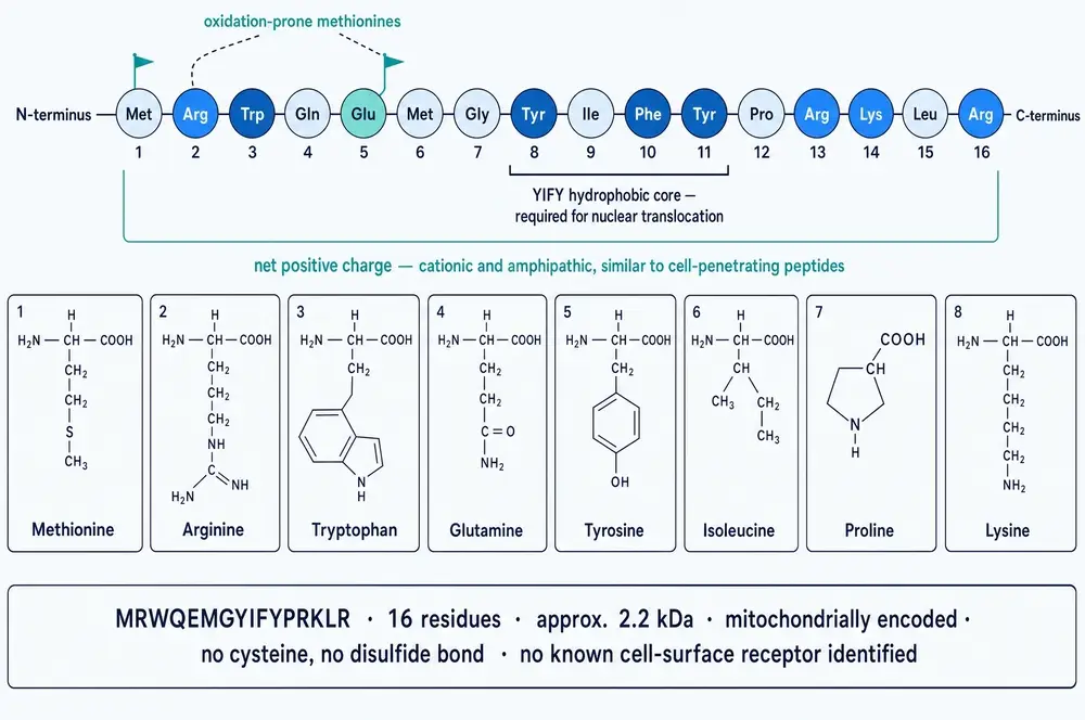
That peptide is MOTS-c. Its name is an acronym of its own address: mitochondrial open reading frame of the twelve S rRNA, type c. Its sequence is MRWQEMGYIFYPRKLR, and its calculated mass is about 2.2 kilodaltons — small enough that on a protein gel it sits below almost everything a biochemist normally looks at (Lee et al., 2015; Kumagai et al., 2024).
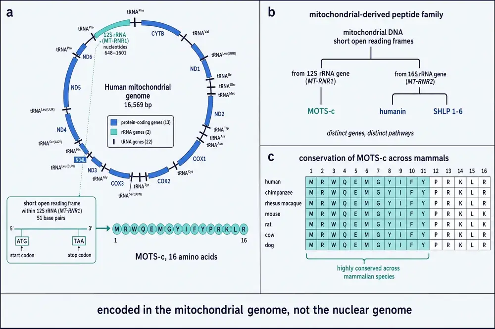
The consequence is the thing worth holding onto. Because the mitochondrion cannot translate it, the transcript has to leave the organelle and be read in the cytoplasm. A gene written in mitochondrial DNA is therefore, of necessity, expressed outside the mitochondrion. Whatever else MOTS-c is, it is a message that only means anything once it has been carried out of the room where it was written.
02What came before: humanin, and the idea of a mitochondrial hormone
None of this would have been looked for without a precedent, and the precedent has an unusually human origin.
In 2001, researchers working on Alzheimer's disease did something counterintuitive. Rather than study the devastated regions of a patient's brain, they built a complementary DNA library from the occipital lobe — a region that had survived comparatively intact — and screened it for anything that would keep neurons alive in the presence of amyloid-β. What came out was a twenty-four amino-acid peptide encoded not in the nucleus but inside the mitochondrial 16S ribosomal RNA gene, MT-RNR2. Its discoverer named it humanin, in the hope that it might restore something of the humanity that the disease takes away (reviewed in Lee et al., 2016; Miller et al., 2020).
Humanin was found three times over, by three groups using three different biochemical handles — as a survival factor against amyloid toxicity, as a binding partner of insulin-like growth factor binding protein 3, and as a protein that blocks BAX from translocating to mitochondria and triggering cell death. Independent discovery by three routes is a strong signal that a molecule is doing something real rather than something artefactual.
For more than a decade humanin stood alone: one peptide, encoded in mitochondrial DNA, behaving like a hormone. Its existence licensed an obvious question. If the mitochondrial genome had one hidden reading frame, why only one?
The answer, so far, is that it has many. MOTS-c was the second to be characterised. Six further peptides, the small humanin-like peptides SHLP1 through SHLP6, were identified by searching the same 16S locus; two more, SHMOOSE and Gau, followed. Re-annotating the whole 16,569-base-pair mitochondrial genome for open reading frames between nine and forty amino acids yields close to four hundred candidates across both strands (Miller et al., 2020; and the 2023 Genes review). Most of those will be noise. The point is that the mitochondrial genome, long treated as a closed catalogue of thirteen proteins, was never systematically searched for anything smaller.
MOTS-c differs from the rest of its family in ways that matter for everything that follows. It is the only one encoded in the 12S gene rather than the 16S. It is the only one shown to enter the nucleus and act on nuclear gene expression directly. Its described actions are metabolic rather than anti-apoptotic. And unlike humanin, which signals through an identified cell-surface receptor complex, no receptor for MOTS-c has ever been found — a gap that shapes how confidently anything about its mechanism can be stated.
03The search, and the people who ran it
The laboratory that went looking was Pinchas Cohen's, at the University of Southern California's Leonard Davis School of Gerontology. Cohen had been central to the humanin work and to the argument that mitochondria-derived peptides constitute a genuine class rather than a curiosity. The first author on the paper that reported MOTS-c was Changhan Lee. It appeared in Cell Metabolism in March 2015, with a author list running through Zeng, Drew, Sallam, Martin-Montalvo, Wan, Kim, Mehta, Hevener and de Cabo to Cohen (Lee et al., 2015).
The search itself was computational, and it had a loose end to pull on. In the early 1980s, groups working on interferon had cloned complementary DNAs from a human myeloblast line and found that the majority of the strongly interferon-responsive clones mapped to the mitochondrial ribosomal RNA locus. No genes were assigned. The observation sat unexplained for three decades. The MOTS-c team went back to it (Lee et al., 2016; Benayoun et al., 2019).
What made the search tractable in 2015 rather than 1985 was partly computational and partly conceptual. Short open reading frames had begun to be taken seriously in nuclear genomes — in Drosophila first, then in mammals — as sequencing and mass spectrometry improved enough to detect very small translated products. And the mitochondrial transcriptome had turned out to be far more complex than the tidy thirteen-gene picture implied.
There is also a structural reason the field had missed it, and it is worth stating because it generalises. Standard bioinformatic pipelines routinely discard exactly the sequences where these peptides live: mitochondrial reads are often filtered out of chromatin-immunoprecipitation and accessibility datasets as background contamination, and ribosomal RNA — which is around 95 per cent of the RNA in a cell — is deliberately depleted before most RNA sequencing. The genes encoding every known mitochondria-derived peptide sit inside ribosomal RNA genes on the mitochondrial chromosome. They were being thrown away twice over (Benayoun et al., 2019).
The field's reaction was immediate and unusually direct. The same March 2015 issue of Cell Metabolism carried a commentary titled "A mitochondrially encoded hormone ameliorates obesity and insulin resistance" (Zarse & Ristow, 2015). Nine months later, a separate group asked in Aging Cell whether a common variant in the new peptide might help explain Japanese longevity (Fuku et al., 2015) — a question this document returns to in section 17, because the answer turned out to be no. When the nuclear-translocation work appeared three years later, the accompanying commentary was titled "A Mitochondrial-Derived Peptide Exercises the Nuclear Option" (Mangalhara & Shadel, 2018).
04Establishing that it is real
A predicted peptide is a hypothesis. Most of the 2015 paper is the work of converting it into an observation, and the chain is worth walking because it is the part of a discovery readers almost never see.
Is the gene actually mitochondrial? The obvious objection is that the mitochondrial genome has been leaking copies of itself into the nuclear genome for millions of years. These nuclear mitochondrial DNA transfers — NUMTs — mean that finding a mitochondrial-looking sequence does not prove a mitochondrial origin. The authors addressed this three ways. A BLAST search found no NUMT sharing complete homology with the MOTS-c reading frame, and all 101 hits in the human expressed-sequence-tag database matched the mitochondrial 12S locus. Rats carry no MOTS-c NUMT at all, so in that species mitochondrial DNA is the only possible source. And HeLa cells stripped of their mitochondrial DNA by chronic low-dose ethidium bromide — so-called ρ⁰ cells — lost both the 12S transcript and MOTS-c, while nuclear-encoded GAPDH was untouched. Degrading mitochondrial RNA with actinonin did the same thing in a time-dependent way (Lee et al., 2015).
Is the peptide actually there? It was detected in multiple mouse and rat tissues and in human and rodent plasma using a MOTS-c-specific immunoassay. Fasting mice for forty-eight hours lowered it in skeletal muscle, testes and plasma while leaving brain and heart alone — a tissue-selective, physiologically responsive pattern rather than a flat background signal.
It does not run where it should. The calculated mass is 2.2 kDa, but endogenous MOTS-c has been reported at roughly 6 kDa in mouse myotubes, around 3 kDa in mouse skeletal muscle after injection but 6 kDa in fat from the same animals, and at 10–20 kDa in mouse and 20–25 kDa in human muscle by a different group. Oligomerisation or post-translational modification is proposed; neither is established (Kumagai et al., 2024; and the 2024 Theranostics membrane-repair paper).
Mass spectrometry cannot find what the antibodies report. Liquid chromatography–mass spectrometry detects synthetic MOTS-c used as a positive control, but has failed to detect circulating cell-free MOTS-c in human plasma from samples in which immunoassay reported it successfully (reported in the 2021 Frontiers in Genetics mitogenome study). The analyte that human studies are measuring has not been confirmed to be the peptide.
There is a smaller methodological trap in the same family, and it is a neat illustration of why these peptides were hard to see. Because the reading frames sit inside ribosomal RNA genes, reverse transcription primed with random hexamers cannot distinguish messenger-RNA-derived complementary DNA from ribosomal-RNA-derived complementary DNA. Priming with oligo(dT) alone — which selects for polyadenylated transcripts — sharply reduces the apparent signal from the 12S locus while barely moving control genes. Measure it the ordinary way and you are largely measuring the ribosome (the 2021 Journal of Lifestyle Medicine expression study).
Taken together, the origin of MOTS-c is about as well established as a molecular claim gets. Its measurement in human blood is not. Both statements need to be carried forward simultaneously, and the rest of this document does so.
05The folate detour
The 2015 paper did not start from a guess about mechanism. It started by measuring everything.
Human embryonic kidney cells were made to overexpress MOTS-c, or treated with the synthetic peptide, and then profiled by microarray and by unbiased metabolomics. Of 356 named metabolites, 194 shifted in the overexpressing cells. The pattern that emerged pointed at one place: the folate cycle — the pathway that shuttles single carbon atoms around the cell for use in building things.
What follows is a chain of consequences rather than a receptor and a signal. MOTS-c inhibits the folate cycle. The activated folate pool, 5-methyl-tetrahydrofolate, falls; methionine falls with it and homocysteine rises. Because the manufacture of purines from scratch is tethered directly to that folate pool, blocking it stalls the purine assembly line. And when a production line stalls, the part immediately upstream of the blockage piles up. Here that part is AICAR — aminoimidazole carboxamide ribonucleotide — which accumulated to more than twenty times control levels (Lee et al., 2015).
AICAR is not an obscure metabolite. It is a well-known activator of AMP-activated protein kinase, AMPK, the enzyme that acts as the cell's low-fuel sensor and, when switched on, tells the cell to take up glucose and burn fat. It is sold as a research compound for precisely that purpose. The peptide, in other words, does not activate AMPK by binding it. It manufactures AMPK's activator in place.
Downstream, the expected things happened: phosphorylation of AMPKα at threonine 172 and of Akt at serine 473, in a time- and dose-dependent way; then, by seventy-two hours, phosphorylated acetyl-CoA carboxylase and raised carnitine palmitoyltransferase 1 — the switch that lets fatty acids into the mitochondrion to be oxidised.
Two experiments turn this from a plausible story into a causal one, and they are the reason this mechanism deserves the confidence it gets.
The rescue. Adding folic acid to the culture medium at 100 nanomolar — an ordinary vitamin, at a low concentration — fully reversed both the accelerated glycolysis and the suppressed oxygen consumption. If the effect runs through folate depletion, refilling the folate pool should abolish it. It did.
The mutants. Substituting alanine for either of two conserved residues, the glutamate at position 5 or the glycine at position 7, abolished the effect entirely. So did a scrambled version of the sequence with the same amino-acid composition and no homology to anything known. The activity belongs to that specific sequence, not to the general act of adding peptide to cells.
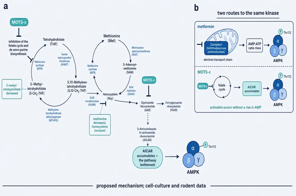
The parallel the authors drew themselves is the most useful way to hold this. MOTS-c shares five properties with methotrexate, the antifolate used against cancer and, at lower amounts, rheumatoid arthritis: both target the folate cycle, both deplete 5-methyl-tetrahydrofolate, both raise AICAR, both activate AMPK in the presence of plentiful ATP, and both reduce mitochondrial respiration. It also runs parallel to metformin, the first-line diabetes drug, which is likewise thought to act partly through the folate cycle. The difference the authors emphasise is anatomical: metformin acts principally on the liver, humanin on the hypothalamus and pancreatic islets, and MOTS-c on skeletal muscle.
06Why AMPK is not the whole answer
It would be convenient if MOTS-c were simply an AMPK activator. The arithmetic says otherwise, and the arithmetic is in the original paper.
Knocking down AMPKα2 with small interfering RNA reduced the glucose-stimulated glycolytic response by 16 per cent. Knocking down both AMPK catalytic subunits reduced it by 30 per cent. The chemical AMPK inhibitor compound C reduced it by 40 per cent. Removing SIRT1, a different metabolic regulator, reduced it by 40 per cent, and inhibiting SIRT1 pharmacologically by 45 per cent. No single blockade removed more than 45 per cent of the effect (Lee et al., 2015).
There is a second, subtler signal in the same experiments. AMPK is normally activated when the cell is short of energy — when AMP rises relative to ATP. Under MOTS-c, AMPK was activated while AMP fell and ADP and ATP rose. That is the same paradoxical pattern produced by salicylate, leptin and metformin, and it means the peptide is not simply signalling energy scarcity.
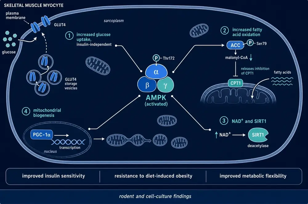
The strongest evidence that AMPK is not the whole story arrived in 2024, from a different direction entirely. In the work on plasma-membrane repair described in section 09, neither compound C nor AMPKα knockdown abolished the protective effect of MOTS-c on damaged muscle-cell membranes — the first clean demonstration that a major MOTS-c action does not require AMPK at all (the 2024 Theranostics study).
There is also a plain logical constraint worth stating. AMPK is a serine/threonine kinase. The MOTS-c sequence contains neither serine nor threonine. Whatever AMPK is doing in this pathway, it is not phosphorylating the peptide (Benayoun et al., 2019).
07The nuclear option
In 2018 the picture changed from metabolism to information.
Kim and colleagues showed that MOTS-c is present at low levels in the nucleus of resting cells and moves there in quantity under stress. Three different challenges — restricting glucose, withdrawing serum, or applying the oxidant tert-butyl hydroperoxide — drove nuclear translocation detectable within thirty minutes, peaking around three hours and largely reversing by twenty-four. Mitochondrial MOTS-c fell as nuclear MOTS-c rose. A second oxidant, paraquat, did the same thing dose-dependently, and pre-treating with the antioxidant N-acetylcysteine blocked it — even though N-acetylcysteine on its own raised total MOTS-c, which rules out the trivial explanation that translocation simply follows abundance (Kim et al., 2018).
How it gets in is more interesting than that it does. MOTS-c carries no classical nuclear localisation signal. It does carry a cluster of basic residues at positions 13–16, RKLR, which looks like one — and replacing that cluster with alanines did not block entry. What did block entry was replacing the hydrophobic core at positions 8–11, YIFY. Nuclear import therefore depends on a hydrophobic interaction with something else, not on a charge-based address label.
Once inside, the peptide binds DNA. In gel-shift assays, synthetic MOTS-c bound, concentration-dependently, to promoter fragments containing antioxidant response elements from seven genes in the NRF2 stress-response programme, including haem oxygenase 1 and NQO1. Binding required both the hydrophobic core and the basic tail — so the two domains have separable jobs, one for getting in and both for holding on. Chromatin immunoprecipitation confirmed the binding happening in living cells and rising after stress. A reporter driven by four copies of an antioxidant response element confirmed the transcription that follows.
MOTS-c also physically associates with NFE2L2 — NRF2 itself — but only after stress, and the two proteins enter the nucleus independently of one another. Sequencing the transcriptome of stressed cells overexpressing MOTS-c identified 802 genes significantly regulated, 390 up and 412 down, with the upregulated set overlapping known NRF2 targets and enriched for ATF1 and ATF7 binding motifs. MOTS-c co-precipitated with ATF1 from the nucleus. The downregulated set was dominated by immunity and interferon genes — which loops back neatly to those unassigned interferon-responsive clones from the 1980s.
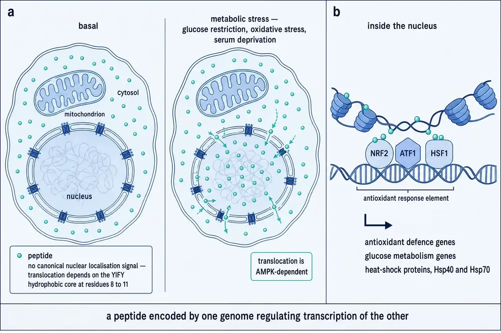
The conceptual claim being made here is large, and it is worth stating precisely. More than a thousand nuclear-encoded proteins are imported into mitochondria; over 98 per cent of the mitochondrial proteome is written in the nucleus. Traffic in the other direction was assumed not to carry regulatory information — all thirteen mitochondrially encoded proteins are structural components of the respiratory chain. MOTS-c was the first factor encoded in mitochondrial DNA shown to regulate nuclear gene expression. On that reading the two genomes are not a hierarchy with the nucleus in charge, but a single bipartite system with cross-regulation running both ways.
A pleasing symmetry supports the general idea. In yeast, the nuclear ribosomal RNA gene encodes a peptide, Tar1p, that acts inside the mitochondria — the exact mirror image. No human counterpart has been found (Benayoun et al., 2019).
08A direct binding partner, at last
For nine years MOTS-c had pathways but no protein target. In 2024 it acquired one.
Kumagai and colleagues showed that MOTS-c binds the catalytic α subunit of casein kinase 2 — but not its regulatory β subunit — with a dissociation constant by surface plasmon resonance of 1 nanomolar. That is a tight, specific interaction, in the range one expects of a real biological partnership rather than a sticky peptide. In a cell-free assay MOTS-c increased CK2's phosphorylation of a substrate in a dose-dependent way (Kumagai et al., 2024).
The striking result is what happens in an animal. A single injection in mice raised CK2 activity in gastrocnemius muscle and lowered it in epididymal fat, with no change in liver. The same inversion was reproduced in cultured muscle cells and fat cells. Immunoprecipitation found MOTS-c bound to CK2α in muscle and fat but not liver. Proteomics showed the peptide rewiring which proteins CK2 associates with, tissue by tissue: in muscle it displaced a phosphatase that acts on Akt and recruited a chaperone, which offers a mechanism for the rise in Akt phosphorylation seen since 2015.
Functionally, CK2 activity is required. A CK2 inhibitor prevented the MOTS-c-driven increase in glucose uptake into mouse muscle, and knocking down CK2α in primary human skeletal muscle cells abolished it there too.
The reason this matters beyond mechanism is that it explains a human genetic finding that had been unexplained for a decade. The naturally occurring K14Q variant of MOTS-c binds CK2α with a dissociation constant of 16.2 nanomolar — more than an order of magnitude weaker — and fails to do any of the things wild-type MOTS-c does. Part Four returns to what that means in people.
09Glucose transport, mitochondrial shape, and torn membranes
Three further strands do not fit the transcription story and should not be forced into it.
Glucose transport requires mitochondrial fusion. In cultured cells, MOTS-c raised markers of mitochondrial biogenesis substantially while the number of mitochondria counted by fluorescent dye fell by around 40 per cent. The paradox resolves under the electron microscope: the mitochondria were fewer because they were longer, having fused, with raised MFN2 and OPA1 and visible tethering to the endoplasmic reticulum. In brown adipocytes, MOTS-c moved the glucose transporter GLUT4 to the plasma membrane and increased glucose uptake — and both effects were completely abolished by blocking fusion, either with TNFα or by knocking down MFN2 (the 2021 Scientific Reports study).
It helps repair torn muscle membranes, and this part does not need AMPK. Hard eccentric exercise damages the sarcolemma. In mice, exhaustive downhill running damaged the membrane, lowered plasma MOTS-c and reduced mitochondrial DNA content, while moderate running did the opposite. MOTS-c binds the C-terminal domain of TRIM72, the protein that patches membrane wounds, and accelerates its trafficking to the injury site; in laser-wounding assays it reduced dye entry. In TRIM72-knockout mice the protective effect on membrane integrity was lost — but MOTS-c still promoted vesicle fusion at the membrane, and it turns out to bind a membrane lipid, phosphatidylinositol 4,5-bisphosphate, directly. In humans undergoing orthopaedic surgery, those meeting activity guidelines had higher muscle MOTS-c, higher mitochondrial DNA and higher TRIM72, with MOTS-c and TRIM72 correlated (the 2024 Theranostics study).
It rides a motor into the nucleus. Work on lung injury published in 2025 filled in the missing transport step from section 07. Under oxidative stress, CK2A phosphorylates myosin-9 at serine 1943; MOTS-c grips the resulting myosin–actin complex using the same YIFY hydrophobic core that section 07 showed was required for entry, and is carried in along the cytoskeleton. Knock down myosin-9 and the peptide stays in the cytoplasm and its antioxidant target genes stay silent. That is an unusually complete account of how a molecule with no address label reaches the nucleus.
10Insulin sensitivity, and the experiment that located the organ
The animal work in the 2015 paper is the reason anyone paid attention, and one experiment in it does more than the rest combined.
Mice given MOTS-c intraperitoneally at 5 mg/kg/day for seven days cleared glucose faster on a tolerance test. That alone would be suggestive. What made it interpretable was the hyperinsulinaemic–euglycaemic clamp: infuse insulin at a fixed rate, then infuse however much glucose is needed to hold blood sugar steady. The more glucose you have to supply, the more sensitive to insulin the animal is. In high-fat-fed mice, MOTS-c raised the required glucose infusion rate by about 30 per cent.
Then the decisive refinement. By infusing labelled glucose during the clamp, the authors could separate two things that a whole-body number confuses: how much glucose the tissues take up, and how much the liver puts out. Insulin-stimulated glucose disposal rose. Hepatic glucose production did not move at all. Because skeletal muscle accounts for 70 to 85 per cent of insulin-stimulated glucose disposal, that pattern locates the action in muscle and excludes the liver (Lee et al., 2015).
The chronic experiment is the one people remember. Male CD-1 mice on a diet deriving 60 per cent of calories from fat, given MOTS-c at 0.5 mg/kg/day intraperitoneally for eight weeks, were protected from becoming obese — while eating an identical number of calories as controls. Food intake was measured, not assumed. They were also protected from the hyperinsulinaemia that normally accompanies that diet, accumulated less fat in the liver, and showed higher AMPK phosphorylation and more GLUT4 in muscle. Over three weeks the same amount raised the respiratory exchange ratio — meaning proportionally more glucose and less fat being burned — and raised heat production, with no change in total physical activity.
The energy has to come from somewhere, and here it came out of the exhaust rather than the fuel tank.
Finally, and most relevant to everything in Part Four: MOTS-c declines with age. Comparing four-month with thirty-two-month mice, it fell in both skeletal muscle and serum. Soleus muscle from twelve-month mice was measurably more insulin resistant than from three-month mice — and seven days of MOTS-c restored the older muscle's insulin-stimulated glucose uptake to the level of the young animals.
Every result in this section is from mice. None of it has been shown in a person. The clamp study is a well-controlled demonstration of a real effect in a mouse, and that is exactly what it should be taken for.
11The pancreas gets a vote
If MOTS-c acts on muscle, the islet work is a genuine surprise, because it suggests the peptide also sits inside the loop that decides how much insulin leaves the pancreas in the first place.
In vitro work on rat and pig islets found a reciprocal arrangement: insulin stimulates MOTS-c release from beta cells, and MOTS-c in turn suppresses insulin secretion while raising expression of the insulin receptor. That is a feedback loop in which the mitochondrion gets a say in the cell's principal secretory decision. It also cuts against a naive reading of MOTS-c as straightforwardly insulin-like — it is not an insulin mimetic, and in this tissue it opposes insulin output.
Two rodent disease models produced larger effects. In non-obese diabetic mice — an autoimmune model in which essentially all animals become diabetic — MOTS-c delayed onset from around eight weeks to twenty-three, with two-thirds still disease-free at thirty weeks; mechanistically the peptide was reported to bind Raptor and inhibit mTORC1 (Kong et al., 2021). In a model driven by the insulin-receptor antagonist S961, diabetes incidence fell from 70 per cent to 30 per cent with MOTS-c treatment. In the same 2025 work, beta cells from 90-week-old mice carried roughly fourteen times less MOTS-c than beta cells from 12-week-old mice, and the cells still holding MOTS-c were systematically not the ones producing the inflammatory cytokine IL-1β.
A methodological caution the authors themselves raise: rat and porcine MOTS-c behaved differently, running at markedly different apparent molecular weights and responding to glucose in opposite directions, and the porcine sequence has still not been identified. Species is not a detail here.
12Exercise, muscle, and the arithmetic of ageing
This is the part of the MOTS-c literature that has travelled furthest into public conversation, and it is also where the human and animal evidence diverge most sharply. It repays reading carefully.
What one hard effort does
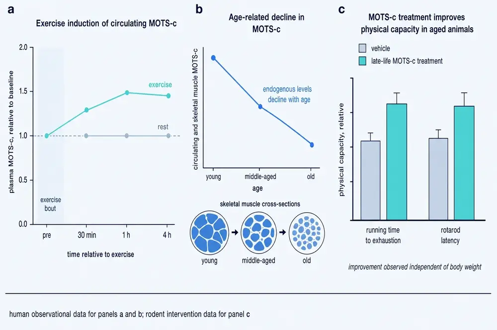
In healthy young men performing ten sixty-second maximal cycling intervals, MOTS-c in the thigh muscle rose 11.9-fold and was still elevated four hours later. In the same men, plasma MOTS-c rose only about 1.5-fold and was back to baseline by four hours (Reynolds et al., 2021).
That gap between compartments is the single most important measurement problem in this field, and it recurs everywhere. The muscle holds the signal; the blood only leaks a little of it. Any study that measures MOTS-c in serum and reasons about muscle is making an inference that this experiment does not support.
The same split appears with age. Between young adulthood and the eighth decade, circulating MOTS-c falls by about 21 per cent — while MOTS-c inside the thigh muscle rises around 1.5-fold. In young men blood and muscle levels track each other; by middle age the correlation is gone (D'Souza et al., 2020). One proposed explanation is that ageing muscle loses the ability to release the peptide. Another, from the same literature, is simpler: MOTS-c is concentrated in slow-twitch fibres — it is barely detectable in predominantly fast mouse gastrocnemius and abundant in soleus — so the age-related shift toward slow fibres may be showing up as chemistry rather than signalling.
And then a result that refuses to fit the story at all. Seventy-five professional athletes tested by an anti-doping laboratory had roughly 40 per cent lower resting serum MOTS-c than sedentary controls, with endurance specialists lowest of all. If exercise raised circulating MOTS-c in any simple chronic sense, that is the opposite of what one would expect. It remains unexplained.
What treatment does to old mice
The 2021 Nature Communications study is the source of nearly every popular claim about MOTS-c and ageing, so its actual findings matter.
Twenty-two-month-old mice — roughly equivalent to a person in their seventies — given MOTS-c for two weeks ran about twice as long and 2.16 times as far as untreated old mice, outperforming untreated middle-aged animals. Started later still, at 23.5 months, treated mice had better grip strength, longer strides, and could complete a walking test after running was no longer possible for controls. The authors described the result as physical reprogramming rather than simple rejuvenation.
Two details are routinely dropped when this study is retold, and both belong in any honest account.
It bought healthspan, not lifespan. Median survival rose 6.4 per cent and maximum 7.0 per cent, with a hazard ratio of 0.654 — and the result did not reach statistical significance (P = 0.23). Better years; not demonstrably more of them.
The schedule mattered and is not settled. That study required ten days of pre-treatment before the effect appeared; seven days was insufficient. A different group reported a 12 to 15 per cent performance improvement from a single administration ten minutes before exercise. These are not obviously compatible, and no one has reconciled them.
Muscle wasting
Across atrophy models the direction is consistent: MOTS-c attenuates loss. It reduced immobilisation-induced atrophy in mice, partially protected against muscle deterioration in a cancer-cachexia model, and — in the only human-tissue administration experiment in this entire corpus — reduced dexamethasone-induced atrophy in primary human skeletal muscle cells. That last study also found that human myotubes upregulated PGC-1α under dexamethasone, the opposite of the rodent response, which is a small but pointed reminder that mouse muscle and human muscle are not interchangeable.
One rodent finding is worth recording for its oddity. Rats that ran voluntarily for eight weeks doubled MOTS-c in the plantaris muscle, and still had it six weeks later after sitting in ordinary cages — while a conventional mitochondrial protein raised 3.5-fold by the same training fell straight back to sedentary levels. Whatever the muscle is doing with this peptide, it holds onto it.
13Everywhere else
Preclinical MOTS-c work has spread across organ systems fast, and the breadth is easy to misread. What follows is a map of where researchers have looked, not a list of things the peptide does for people.
Lung. The most sophisticated pharmacology in the whole corpus is here. An engineered analogue, R13A-MOTS-c, replaces a single arginine with alanine, raising hydrophobicity enough to let the peptide enter cells through LAT1 — the amino-acid transporter that also carries L-DOPA, gabapentin and pregabalin across membranes — bypassing the lysosome. It was effective in irradiated mouse lung at half the amount the natural peptide required. The control that makes this convincing: swapping the arginine one position over, at 16, produced an identical gain in hydrophobicity and no permeability at all. The transporter is recognising a position, not a property. Separately, in a 150-patient cardiac-bypass cohort, the 24-hour change in MOTS-c improved prediction of acute respiratory distress syndrome (raising the area under the curve from 0.824 to 0.885) while the static level predicted nothing.
Liver. MOTS-c attenuated diabetic liver fibrosis in rodents through Keap1–Nrf2–Smad2/3, and mitochondrial dysfunction featured in a multigenerational model of maternal obesity and fatty liver disease.
Kidney. A clean negative worth stating. In 82 children with type 1 diabetes, MOTS-c was significantly lower than in controls — and correlated with nothing: not albuminuria, not hyperfiltration, not estimated filtration rate, creatinine, cystatin C, HbA1c or body mass index. The authors read this as evidence that mitochondrial disturbance in type 1 diabetes begins early and independently of kidney disease. It is equally a reminder that a low biomarker which predicts nothing is not a useful biomarker.
Bone, disc, retina, placenta. Effects on bone metabolism and osteogenic differentiation; a MOTS-c-modified self-assembling peptide hydrogel for intervertebral disc stem cells; protection of retinal pigment epithelium against oxidative injury, with relevance argued for macular degeneration; and, in a mouse model of intrauterine growth restriction, protection of the placenta via Nrf2 — with the nice dissection that removing Nrf2 abolished every vascular effect while leaving the nutrient-transporter effects intact.
Brain. In a traumatic brain injury model, peripherally injected MOTS-c reached the cortex — but, as the authors note, only because the injury had already broken the blood–brain barrier, so the experiment cannot say whether it would reach an intact brain. In a valproate-induced rat model of autism, a study designed around the hypothesis that MOTS-c acts through tetrahydrobiopterin and BDNF found that neither marker moved, while the behavioural and histological measures improved anyway. The stated mechanism failed and the effect did not.
Two patterns run through this breadth. The first is that Nrf2 and AMPK recur across organs with nothing anatomically in common, which is consistent with MOTS-c acting as a general stress-response amplifier rather than as an organ specific agent. The second is a caution: the peptide does not always fall in disease. It rose in spermatocytes exposed to particulate air pollution as their mitochondria failed, in early hepatic steatosis, and in serum after cardiac bypass. A signal that goes up as an organelle fails is a distress flare, and it breaks any simple reading in which more MOTS-c means better health.
14Measuring MOTS-c in humans, and why the numbers disagree
Almost everything known about MOTS-c in people rests on measuring it in blood. That measurement is in worse shape than the literature built on it generally admits, and the honest order of business is to establish this before reporting any of the findings that depend on it.
Here is the problem in one number. Across studies in this corpus, the reported normal concentration of serum MOTS-c in healthy control subjects spans roughly 1,500-fold — from about 0.5 ng/mL to about 764 ng/mL. These are not diseased populations being compared with healthy ones. These are the healthy ones.
It is worse than a between-kit problem. Two studies using the identical commercial kit, from the same manufacturer with the same catalogue number, report control values about tenfold apart. And a review of the wider literature puts the range at 154 pg/mL to 584 ng/mL — a spread of nearly four thousandfold — concluding plainly that "the immunoreactive species of circulating MOTS-c detected using different kits are not identical" (the 2022 Aging breast-cancer study).
Several converging problems produce this:
- Nobody has confirmed what is being measured. No study in this corpus used mass spectrometry to verify that the immunoreactive material is MOTS-c. As section 04 recorded, when mass spectrometry has been tried on human plasma it has failed to find the peptide in samples where immunoassay reports it.
- The same manufacturer describes its own kit inconsistently, with two irreconcilable dynamic ranges quoted in different papers.
- A common genetic variant is invisible to the assay. The K14Q form discussed in section 16 is partially inactive, and standard immunoassays cannot distinguish it from the working peptide. A "normal" reading may be a normal quantity of inert material — and, as section 16 shows, carriers circulate roughly twenty times more of it.
- Pre-analytical handling varies. Some groups deliberately sampled in the early morning to avoid circadian variation, and a circadian rhythm has since been documented directly in sheep. Some used protease inhibitors and some explicitly did not. Exercise-induced changes return to baseline within about four hours, so time since last activity is a real confounder that few studies control.
- Transcript and protein do not agree. One 2025 study found no correlation whatsoever between MOTS-c transcript levels and plasma MOTS-c protein in the same people.
15The biomarker record
With that caveat carried, the human observational literature is large, consistent in method and strikingly inconsistent in result.
Lower than controls in Hashimoto's thyroiditis (90 cases, and a receiver-operating characteristic area under the curve of 1.00, which is itself a reason for suspicion rather than confidence); relapsing–remitting multiple sclerosis; stable chronic obstructive pulmonary disease and, further still, in exacerbations of it; adult polycystic ovary syndrome, in serum and in muscle; obstructive sleep apnoea, graded by severity; ovarian cancer, in serum and tissue, where low tissue levels tracked shorter survival; major depressive disorder, where neuron-derived extracellular vesicles carried 83–84 per cent less MOTS-c and the deficit normalised only in patients who responded to treatment; and acute COVID-19 in those who went on to develop persistent symptoms.
Higher than controls in acute coronary syndrome, rising stepwise from control through unstable angina to myocardial infarction across 356 patients; in adrenocortical adenoma and phaeochromocytoma; in prostate precancerous lesions; and in kidney transplant recipients.
No significant difference in adolescent polycystic ovary syndrome, in Alzheimer's disease plasma protein, in breast cancer patients treated with metformin, in prostate cancer compared with benign hyperplasia, and in adrenocortical carcinoma.
Two of those entries are the same disease. Two studies of polycystic ovary syndrome, both published in 2026, using different commercial kits on different age groups, reached opposite conclusions — one finding serum MOTS-c significantly lower in cases, the other finding a non-significant elevation. That is not a defect of this summary. It is the state of the evidence.
The cardiovascular literature splits the same way and more sharply, because there the direction reverses within the same organ system: MOTS-c is reported higher in acute coronary syndrome but lower in ST-elevation myocardial infarction, lower in atrial fibrillation plasma and atrial tissue, and lower in aortic valve disease. In chronic obstructive pulmonary disease, low MOTS-c helps identify an exacerbation while high MOTS-c predicts readmission.
Only one intervention has been shown to move it in a controlled human setting, and it is not exercise. Over twelve months in 163 patients with type 2 diabetes, the SGLT2 inhibitor empagliflozin raised circulating MOTS-c by 36 per cent and a combination arm by 29 per cent, while the GLP-1 receptor agonist liraglutide moved it by −3 per cent and insulin by +0.6 per cent.
16The natural experiment: one letter at position 1,382
The strongest human evidence about MOTS-c does not come from giving it to anyone. It comes from a population in which the peptide is already broken.
At position 1,382 of the mitochondrial genome, some people carry a C where most carry an A. The substitution falls in codon 14 of the MOTS-c reading frame and changes lysine to glutamine: K14Q. It is a chemically radical substitution rather than a conservative one, removing a positive charge, and it is essentially confined to East Asia, where it defines the mitochondrial sub-haplogroup D4b2 and runs at frequencies of roughly 5 to 8 per cent — on the authors' own arithmetic, several tens of millions of carriers.
What makes this the best human evidence in the corpus is that the causal chain was reconstructed at every level.
The molecule. K14Q binds CK2α at 16.2 nanomolar against 1 nanomolar for the wild type (Kumagai et al., 2024).
The cell. In muscle and fat cell lines, K14Q failed to raise insulin-stimulated Akt phosphorylation, failed to promote glucose uptake, and in transfected human cells was indistinguishable from an empty vector.
The animal. In male mice on a high-fat diet, wild-type MOTS-c reduced weight gain and improved glucose tolerance; K14Q was statistically indistinguishable from injecting water. In female mice neither peptide did anything — a sex difference the authors connect to evidence that MOTS-c works in ovariectomised but not intact females (Zempo, Kim et al., 2021).
The compensation. Carriers circulate nearly twenty times more MOTS-c than matched controls. Osmotic-pump infusion in mice showed the variant is cleared 2.6 times more slowly, but the calculated production rate is 7.4 times higher — so the elevation is mostly the body making more. The authors place this in the well-described family of bioinactive-hormone syndromes, alongside bioinactive growth hormone, insulin and leptin, in which over-secretion of a partly inactive hormone eventually produces something resembling deficiency.
The population. Meta-analysis across three Japanese cohorts totalling 27,527 people found the C allele significantly raised type 2 diabetes prevalence in men (pooled Z = 2.86, P < 0.01, with zero heterogeneity between cohorts) and not in women. Carriers also had more visceral fat at matched age and body mass index.
And then the finding that makes this more than an association.
Among men overall, the difference was not significant: 13.1 per cent of carriers had diabetes against 10.8 per cent of non-carriers. But when the cohort was split by accelerometer-measured activity, the risk turned out to live entirely in the least active third — men averaging under seven minutes of moderate-to-vigorous activity a day — where carriers had 18.5 per cent diabetes against 11.2 per cent, a 65 per cent relative excess. In more active men the difference disappeared. The authors named the phenomenon a kinesio-genomic interaction. In women there was no such pattern at any activity level.
Two control analyses make the attribution unusually clean. Four other polymorphisms defining the same haplogroup lineages carried no diabetes risk, and haplogroup D4b as a whole carried none — so the signal belongs to this base, not to the ancestry it travels with. And mice given either peptide showed no change in voluntary wheel running, ruling out the alternative that the variant works by making people less inclined to move.
The same allele in athletes, and in the old
The variant does not simply impair. In 211 Japanese adults with muscle biopsies, carriers had a significantly higher proportion of fast-twitch MHC-IIx myosin (26.3 against 21.1 per cent). In 86 physically active young men, carriers produced about 20 per cent more knee-extension torque — 245 against 204 newton metres — and were stronger at every speed tested. Across 721 Japanese athletes and 873 matched controls, allele frequency rose monotonically from endurance athletes through controls to sprint and power athletes.
And the direction inverts with age. In 683 community-dwelling Koreans over 65, male carriers had significantly more appendicular muscle, more lean mass and stronger grip in both hands than non-carriers — differences that survived adjustment for every measured covariate. In women, nothing.
The trade-off the authors propose is coherent: fast-twitch fibre proportion is inversely related to whole-body glucose uptake, so a variant that shifts fibre type toward fast buys speed and power and costs glucose disposal. Their framing is that the allele may have suited a life of high activity and limited food, and has become a liability under the opposite conditions. That hypothesis is explicitly speculative and is not tested anywhere in this corpus.
One further observation is worth recording as a question rather than a finding. A comparative-genomics study notes that MOTS-c residues R13 and K14 together resemble the kind of dibasic site used to process secreted hormones. The K14Q substitution replaces exactly that lysine. No paper connects the two, and this document does not either — but the coincidence is the sort of thing that deserves an experiment.
17The longevity claim, and its retraction by arithmetic
In 2015, within months of the discovery, a group proposed in Aging Cell that this same variant was associated with exceptional longevity in Japan, and that MOTS-c might therefore be part of the explanation for Japanese lifespans (Fuku et al., 2015). The idea was attractive, widely repeated, and still circulates.
It rested on 96 centenarians.
When the same question was put to an expanded cohort of 736 Japanese centenarians, the C-allele frequency was 7.7 per cent among the very old and 7.5 per cent among controls. The authors' conclusion was that the variant "is unlikely to be involved with exceptional longevity" (Zempo, Kim et al., 2021), and a 2026 review restates the reversal.
This is a small section because the finding is simple, but it is included deliberately, for three reasons. It is a clean worked example of recency weighting done properly: the newer result supersedes the older one not because it is newer but because it is better powered on the identical question with the identical measurement. It removes an apparent paradox — the same allele appearing to cause diabetes and longevity at once — that would otherwise need explaining. And it is a caution about this compound's popular literature, in which the longevity claim continues to appear unqualified more than five years after the data stopped supporting it.
18Giving it to people
Two records exist. They are frequently conflated and should not be.
The analogue that has already been tested
Between 2018 and 2021, CohBar Inc. ran a three-part Phase 1a/1b study of CB4211, described as an analogue of MOTS-c with improved properties, in 88 participants — single ascending doses in healthy volunteers, then seven days of daily subcutaneous dosing, then twenty-eight days in obese subjects with fatty liver disease (NCT03998514). The trial completed in April 2021.
By the sponsor's own topline announcement, the Phase 1b stage compared 25 mg once daily by subcutaneous injection for four weeks against placebo in 20 subjects — 11 on drug, 9 on placebo. It met its primary safety endpoint: no serious adverse events, with transient, mild-to-moderate injection-site reactions the only adverse event in more than 10 per cent of subjects. Among the exploratory measures, alanine aminotransferase fell 21 per cent against a 4 per cent rise on placebo, aspartate aminotransferase fell 28 against 11 per cent, and glucose fell 6 per cent against zero, each reported as significant at P < 0.05. Body weight showed a trend with no figures given.
The endpoint the study was built around did not separate. Liver fat measured by MRI proton density fat fraction fell by 5.03 percentage points on CB4211 and by 4.88 points on placebo; 36 per cent of treated subjects achieved a greater than 30 per cent relative reduction against 33 per cent on placebo.
The peptide itself
The first registered trial in which MOTS-c itself is administered to people began on 2 February 2026.
Four other registered studies name MOTS-c and none of them administers it: an interventional study in renal transplantation where the interventions are two anaesthetic techniques and MOTS-c is an outcome measure; two observational studies from the University of Athens measuring it among other markers; and a newborn deafness-gene screening cohort that touches the 12S locus rather than the peptide.
So the human record, stated exactly. One completed Phase 1 study of an analogue in 88 people, with an equivocal efficacy readout reported only by press release. One ongoing Phase 2a study of the peptide itself in 120 people, which has reported nothing and is not due to report primary results until February 2027. Roughly twenty observational studies measuring the peptide in blood with assays that do not agree with one another. And one large natural genetic experiment, in section 16, which is the only place in this literature where MOTS-c biology has been tested in humans at scale — and which was possible precisely because nobody had to administer anything.
19Prohibited, sold, and untested
Three facts about MOTS-c's position in the world sit oddly together, and the oddity is instructive.
It is banned in sport. The 2026 World Anti-Doping Agency Prohibited List names MOTS-c under section S4.4.1, Metabolic Modulators, in the sub-class "activators of the AMP-activated protein kinase (AMPK)". It is listed twice, by acronym and by full expansion, and it is prohibited at all times — in and out of competition. Its listed companions are worth noticing: BAM15, and AICAR — the very metabolite that the founding paper showed MOTS-c causes to accumulate. The regulator has, in effect, classified the peptide alongside the drug it manufactures inside the cell.
It is sold. The peptide is available from research-chemical suppliers and is discussed in consumer literature as something close to a substitute for training. The corpus assembled for this monograph is itself evidence of the imbalance: of 364 de-duplicated documents in the source library that name MOTS-c, 174 were vendor catalogue and product pages and 69 were consumer web content, against 107 peer-reviewed scientific full texts.
It has not been shown to do anything in a person. Section 18 sets out the whole of the interventional human record: one Phase 1 study of an analogue whose efficacy endpoint did not separate from placebo, and one ongoing Phase 2a trial that has reported nothing.
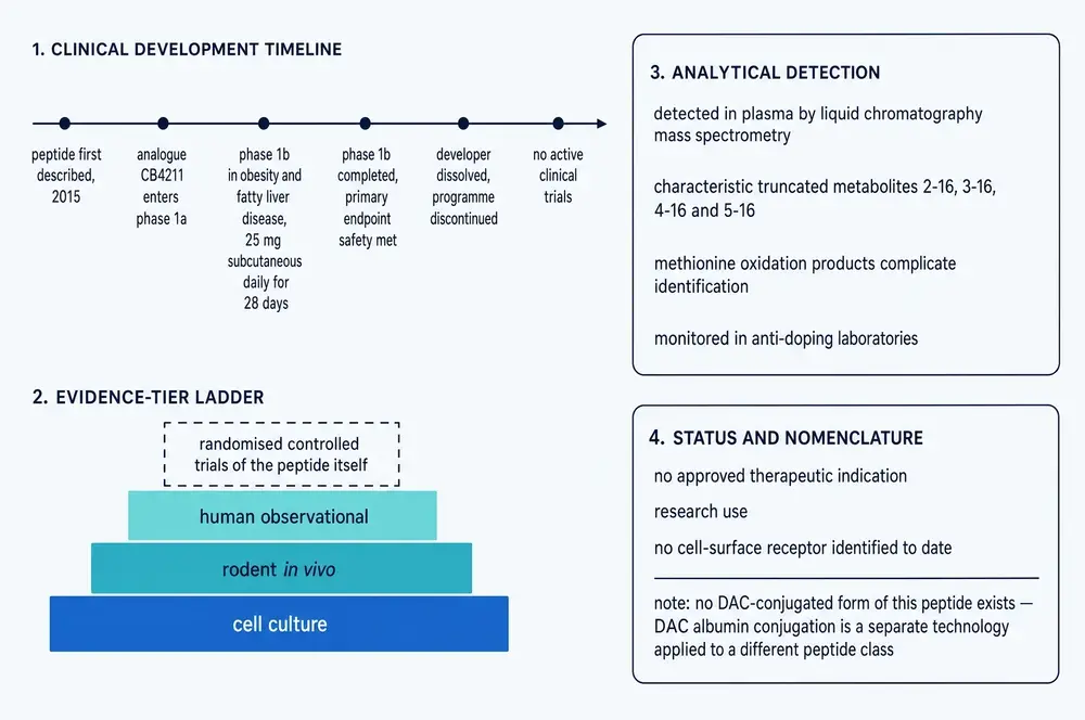
The order in which these three things happened is the point. Regulation and commerce arrived first; evidence is still in progress. Nothing about the WADA listing constitutes a finding that MOTS-c enhances human performance — prohibited lists are precautionary instruments that act on plausible mechanism and availability, not on demonstrated efficacy, and treating a ban as evidence of effect inverts what it is for.
One further caution comes from inside the scientific literature rather than from outside it. The comparative-genomics analysis discussed in section 16 found no statistically significant purifying selection on the full sixteen-amino-acid peptide across 348 vertebrate species — only on an internal five-residue core, MGYIF. Its authors drew the commercial implication themselves, noting that these peptides "are already being marketed as anti-aging therapeutics and performance enhancers" and that their results suggest the peptide "as it is currently being studied (and marketed) might not be in its most biologically relevant form." Whether the sixteen-mer everyone works with is the physiological molecule is an open question, not a settled one.
20What would have to be true
The useful way to close is not with a summary but with a list of things that could be found out, and what each would settle.
The receptor problem. There is no known cell-surface receptor for MOTS-c. Until there is — or until it is positively established that there is none and the peptide enters cells by transport, as the LAT1 work with the R13A analogue suggests — no account of how an injected peptide produces an effect in a distant tissue is complete. This is the largest single gap in the mechanism, and it is the reason claims about systemic action should be held loosely.
The assay problem is solvable and nobody has solved it. A mass-spectrometric method that detects endogenous circulating MOTS-c, distinguishes the K14Q variant from the wild type, and is cross-validated against the commercial immunoassays would retrospectively determine which of the twenty-odd human biomarker studies mean anything. Until that exists, section 15's disagreements cannot be adjudicated, and no reference range should be quoted by anyone. This is unglamorous work that would be worth more than another organ system.
The compartment problem. Muscle MOTS-c rose twelvefold after one exercise bout while plasma rose 1.5-fold; muscle rises with age while blood falls; elite athletes have less in their blood than sedentary controls. Any study that treats serum MOTS-c as a readout of muscle MOTS-c is making an inference the data contradict. Paired muscle-and-blood sampling would resolve it.
What the ongoing trial will and will not settle. NCT07505745 measures the Matsuda index after twelve weeks in 120 adults with prediabetes. If it is positive, it will establish that MOTS-c improves an oral-glucose-tolerance-derived measure of insulin sensitivity in that population over that period — which is precisely the human translation of the 2015 mouse clamp study, and would be a real result. It will not establish an effect on any clinical outcome, on body composition beyond weight and waist, on ageing, on physical performance, or on anything outside metabolism. It is a single site, so it will need replication. And because the registry does not disclose the administered amount, the result will not be interpretable in dose terms from public information alone.
Which animal findings are most and least likely to travel. On the evidence in this corpus, the insulin-sensitivity work is the best positioned: it has a mechanism, a located target organ, a human genetic variant that behaves as the mechanism predicts, and a trial designed around it. The muscle and ageing work is the most oversold relative to its evidence — the headline mouse result improved function without significantly extending life, and its human counterpart does not exist. The organ-protection literature in section 13 is the broadest and the thinnest per organ, and its recurring reliance on Nrf2 suggests a general stress-response effect that may not be specific to any of the diseases it has been tested against.
The K14Q result deserves following up in the other direction. If a partially inactive MOTS-c produces more fast-twitch fibre, more strength, more visceral fat and more diabetes in sedentary men, then the peptide's normal role in humans is being demonstrated by its absence. That is a stronger form of evidence than most pharmacology produces, and it has not been pursued outside East Asian cohorts because the allele barely exists elsewhere. Engineered loss-of-function work in a second population, or a prospective study of carriers randomised to activity, would test the causal claim directly.
Readers should note in particular that the single largest body of evidence about this peptide in humans concerns a genetic variant that reduces its activity, and that the interventional human record consists of one equivocal Phase 1 study of an analogue and one trial that has not reported.
21References
Generated from verified NCBI records rather than from recall. Author lists, journal names, volumes, pages and identifiers are taken from the PubMed record for each citation, and every identifier was derived by mapping the PubMed Central identifier of the full text that was actually read onto the harvest manifest. Two earlier monographs in this series shipped reference lists assembled from memory; between them, sixteen identifiers pointed at real but unrelated papers.
- Alser M, Ramanjaneya M, Rizwana Anwardeen N, Donati F, Botrè F, Jerobin J, et al.. The Effect of Chronic Endurance Exercise on Serum Levels of MOTS-c and Humanin in Professional Athletes. Reviews in cardiovascular medicine. 2022;23(5):181.
PMID 39077591 · doi:10.31083/j.rcm2305181 · PMC11273660 - Amado CA, Martín-Audera P, Agüero J, Lavín BA, Guerra AR, Boucle D, et al.. Circulating levels of mitochondrial oxidative stress-related peptides MOTS-c and Romo1 in stable COPD: A cross-sectional study. Frontiers in medicine. 2023;10:1100211.
PMID 36844198 · doi:10.3389/fmed.2023.1100211 · PMC9944395 - Amado CA, Martín-Audera P, Agüero J, Ferrer-Pargada D, Josa Laorden B, Boucle D, et al.. Alterations in circulating mitochondrial signals at hospital admission for COPD exacerbation. Chronic respiratory disease. 2023;20:14799731231220058.
PMID 38112134 · doi:10.1177/14799731231220058 · PMC10734331 - Benayoun BA, Lee C. MOTS-c: A Mitochondrial-Encoded Regulator of the Nucleus. BioEssays : news and reviews in molecular, cellular and developmental biology. 2019;41(9):e1900046.
PMID 31378979 · doi:10.1002/bies.201900046 · PMC8224472 - Bhullar KS, Shang N, Kerek E, Wu K, Wu J. Mitofusion is required for MOTS-c induced GLUT4 translocation. Scientific reports. 2021;11(1):14291.
PMID 34253808 · doi:10.1038/s41598-021-93735-2 · PMC8275580 - Bień J, Pruszynska-Oszmalek E, Kolodziejski P, Leciejewska N, Szczepankiewicz D, Grzęda E, et al.. MOTS-c modulates pancreatic islet function in rats and pigs in vitro. Histochemistry and cell biology. 2025;163(1):64.
PMID 40478460 · doi:10.1007/s00418-025-02391-4 · PMC12144051 - Cao P, Wang B, Zhang N, Yang J, Tong Q, Gong Z. Circulating Mitochondrial Open Reading Frame of the 12S Ribosomal RNA Type-c Is Higher in Acute Coronary Syndrome and Is a Prognostic Biomarker for Major Cardiac Events in Patients With Acute Myocardial Infarction: A Case-Control Study. Journal of the American Heart Association. 2025;14(24):e041905.
PMID 41368821 · doi:10.1161/JAHA.125.041905 · PMC12826893 - Chen D, Zhao HM, Sun XL, Xing ZX, Li SP, Li SC, et al.. MOTS‑c protects against placental injury via Nrf2 activation in hypoxia‑induced intrauterine growth restriction mice. International journal of molecular medicine. 2026;57(1).
PMID 41268602 · doi:10.3892/ijmm.2025.5697 · PMC12651125 - Chen F, Li Z, Wang T, Fu Y, Lyu L, Xing C, et al.. MOTS-c mimics exercise to combat diabetic liver fibrosis by targeting Keap1-Nrf2-Smad2/3. Scientific reports. 2025;15(1):18460.
PMID 40425777 · doi:10.1038/s41598-025-03526-2 · PMC12116857 - Coradduzza D, Cruciani S, Sibono L, Tedde A, Zinellu A, Maioli M, et al.. Diagnostic relevance of Humanin, GAS5 and miR-21/miR-103 in prostate disease risk stratification. Clinical and experimental medicine. 2025;25(1):279.
PMID 40768089 · doi:10.1007/s10238-025-01810-z · PMC12328530 - Cuyàs E, Verdura S, Martin-Castillo B, Menendez JA, METTEN study group. Circulating levels of MOTS-c in patients with breast cancer treated with metformin. Aging. 2022;15(4):892-897.
PMID 36490309 · doi:10.18632/aging.204423 · PMC10008497 - D'Souza RF, Woodhead JST, Hedges CP, Zeng N, Wan J, Kumagai H, et al.. Increased expression of the mitochondrial derived peptide, MOTS-c, in skeletal muscle of healthy aging men is associated with myofiber composition. Aging. 2020;12(6):5244-5258.
PMID 32182209 · doi:10.18632/aging.102944 · PMC7138593 - Elhusseiny R, Ihsan M, Bellefroid T, Farooq A, Racinais S, Deldicque L. Mitochondrial-derived peptides MOTS-c and humanin attenuate dexamethasone-induced atrophy in human skeletal muscle cells. Physiological reports. 2026;14(4):e70791.
PMID 41732124 · doi:10.14814/phy2.70791 · PMC12930096 - Emser SV, Schaschl H, Millesi E, Steinborn R. Extension of Mitogenome Enrichment Based on Single Long-Range PCR: mtDNAs and Putative Mitochondrial-Derived Peptides of Five Rodent Hibernators. Frontiers in genetics. 2021;12:685806.
PMID 35027919 · doi:10.3389/fgene.2021.685806 · PMC8749263 - Filibeli BE, Dedemoglu F, Garipçin P, Bulut S, Başok Bİ, Kizildağ S, et al.. Are serum MOTS-c levels and MOTS-c m.1382A>C polymorphism related to polycystic ovary syndrome?. Archives of endocrinology and metabolism. 2026;70(3):e260031.
PMID 41945630 · doi:10.20945/2359-4292-2026-0031 · PMC13055642 - Fuku N, Pareja-Galeano H, Zempo H, Alis R, Arai Y, Lucia A, et al.. The mitochondrial-derived peptide MOTS-c: a player in exceptional longevity?. Aging cell. 2015;14(6):921-3.
PMID 26289118 · doi:10.1111/acel.12389 · PMC4693465 - Girişgen İ, Altıncık SA, Avcı E, Öcal M, Becerir T, Malaş Öztekin G, et al.. Could MOTS-c Levels in Children with Type 1 Diabetes Mellitus Be an Indicator for Early Diabetic Kidney Disease?. Journal of clinical research in pediatric endocrinology. 2025;17(2):168-175.
PMID 39711006 · doi:10.4274/jcrpe.galenos.2024.2024-5-6 · PMC12118320 - Goetzl EJ, Wolkowitz OM, Srihari VH, Reus VI, Goetzl L, Kapogiannis D, et al.. Abnormal levels of mitochondrial proteins in plasma neuronal extracellular vesicles in major depressive disorder. Molecular psychiatry. 2021;26(12):7355-7362.
PMID 34471251 · doi:10.1038/s41380-021-01268-x · PMC8872999 - Goetzl EJ, Yao PJ, Kapogiannis D. Prediction of Post-Acute-Sequelae of COVID-19 by Cargo Protein Biomarkers of Blood Total Extracellular Vesicles in Acute COVID-19. The American journal of medicine. 2023;136(8):824-829.
PMID 37072092 · doi:10.1016/j.amjmed.2023.03.026 · PMC10106499 - Gruschus JM, Morris DL, Tjandra N. Evidence of natural selection in the mitochondrial-derived peptides humanin and SHLP6. Scientific reports. 2023;13(1):14110.
PMID 37644144 · doi:10.1038/s41598-023-41053-0 · PMC10465549 - Güvenir Seven S, Sahin H, Erkanlı Şentürk G, Uysal N, Uzun H, Ekici O, et al.. Therapeutic Effects of MOTS-c in the Valproic Acid-Induced Autism Model in Rats: Role of Tetrahydrobiopterin and Brain-Derived Neurotrophic Factor. Molecular neurobiology. 2026;63(1):453.
PMID 41706383 · doi:10.1007/s12035-026-05741-y · PMC12916525 - Harding E, Bazzani V, Vascotto C. Small but mighty: mitochondrial DNA at the centre of retrograde signalling. Cell communication and signaling : CCS. 2026;24(1).
PMID 41937181 · doi:10.1186/s12964-026-02858-4 · PMC13173781 - Hyatt JK. MOTS-c increases in skeletal muscle following long-term physical activity and improves acute exercise performance after a single dose. Physiological reports. 2022;10(13):e15377.
PMID 35808870 · doi:10.14814/phy2.15377 · PMC9270643 - Ikonomidis I, Pavlidis G, Pliouta L, Katogiannis K, Maratou E, Thymis J, et al.. Effects of Glucagon-Like Peptide-1 Receptor Agonists, Sodium-Glucose Cotransporter-2 Inhibitors, and Their Combination on Neurohumoral and Mitochondrial Activation in Patients With Diabetes. Journal of the American Heart Association. 2025;14(5):e039129.
PMID 40008510 · doi:10.1161/JAHA.124.039129 · PMC12132653 - Jamnick NA, Livingston PD, Gammon CJ, Weinzierl NM, Novinger LJ, Bonetto A. MOTS-c partially protects against skeletal muscle deterioration in C26 cachexia. Frontiers in medicine. 2026;13:1838178.
PMID 42266945 · doi:10.3389/fmed.2026.1838178 · PMC13243040 - Jia H, Zhou LC, Chen YF, Zhang W, Qi W, Wang P, et al.. Mitochondria-encoded peptide MOTS-c participates in plasma membrane repair by facilitating the translocation of TRIM72 to membrane. Theranostics. 2024;14(13):5001-5021.
PMID 39267782 · doi:10.7150/thno.100321 · PMC11388074 - Kamiński K, Blatkiewicz M, Szyszka M, Olechnowicz A, Komarowska H, Klimont A, et al.. Expression Patterns of MOTS-c in Adrenal Tumors: Results from a Preliminary Study. International journal of molecular sciences. 2024;25(16).
PMID 39201408 · doi:10.3390/ijms25168721 · PMC11354279 - Kim J, Choi JW, Namkung J. Expression Profile of Mouse Gm20594, Nuclear-Encoded Humanin-Like Gene. Journal of lifestyle medicine. 2021;11(1):13-22.
PMID 33763338 · doi:10.15280/jlm.2021.11.1.13 · PMC7957044 - Kim KH, Son JM, Benayoun BA, Lee C. The Mitochondrial-Encoded Peptide MOTS-c Translocates to the Nucleus to Regulate Nuclear Gene Expression in Response to Metabolic Stress. Cell metabolism. 2018;28(3):516-524.e7.
PMID 29983246 · doi:10.1016/j.cmet.2018.06.008 · PMC6185997 - Kim S. The Relationship Between MOTS-c K14Q Polymorphism and Sarcopenia, Blood Lipids, and Mental Health in Older Korean Adults. Biomedicines. 2024;12(10).
PMID 39457696 · doi:10.3390/biomedicines12102384 · PMC11504729 - Kim SJ, Miller B, Mehta HH, Xiao J, Wan J, Arpawong TE, et al.. The mitochondrial-derived peptide MOTS-c is a regulator of plasma metabolites and enhances insulin sensitivity. Physiological reports. 2019;7(13):e14171.
PMID 31293078 · doi:10.14814/phy2.14171 · PMC6640593 - Kong BS, Min SH, Lee C, Cho YM. Mitochondrial-encoded MOTS-c prevents pancreatic islet destruction in autoimmune diabetes. Cell reports. 2021;36(4):109447.
PMID 34320351 · doi:10.1016/j.celrep.2021.109447 · PMC10083145 - Kong BS, Lee H, L'Yi S, Hong S, Cho YM. Mitochondrial-encoded peptide MOTS-c prevents pancreatic islet cell senescence to delay diabetes. Experimental & molecular medicine. 2025;57(8):1861-1877.
PMID 40855115 · doi:10.1038/s12276-025-01521-1 · PMC12411631 - Kotan GC, Bayraktar B. Effects of graviola oil extract on fattening performance and circadian rhythms of adipokine, cardiac and mitochondrial function markers in lambs. BMC veterinary research. 2026;22(1).
PMID 41803858 · doi:10.1186/s12917-026-05400-3 · PMC13085293 - Kumagai H, Kim SJ, Miller B, Zempo H, Tanisawa K, Natsume T, et al.. MOTS-c modulates skeletal muscle function by directly binding and activating CK2. iScience. 2024;27(11):111212.
PMID 39559755 · doi:10.1016/j.isci.2024.111212 · PMC11570452 - Kumagai H, Natsume T, Kim SJ, Tobina T, Miyamoto-Mikami E, Shiose K, et al.. The MOTS-c K14Q polymorphism in the mtDNA is associated with muscle fiber composition and muscular performance. Biochimica et biophysica acta. General subjects. 2022;1866(2):130048.
PMID 34728329 · doi:10.1016/j.bbagen.2021.130048 · PMC8741734 - Kumagai H, Miller B, Kim SJ, Leelaprachakul N, Kikuchi N, Yen K, et al.. Novel Insights into Mitochondrial DNA: Mitochondrial Microproteins and mtDNA Variants Modulate Athletic Performance and Age-Related Diseases. Genes. 2023;14(2).
PMID 36833212 · doi:10.3390/genes14020286 · PMC9956216 - Kutuk IS, Akin S, Demirel H, Mumusoglu S, Ciftci T, Yildiz BO. Reduced serum and skeletal muscle MOTS c levels in women with polycystic ovary syndrome are associated with mitochondrial dysfunction. Scientific reports. 2026;16(1).
PMID 41680431 · doi:10.1038/s41598-026-39687-x · PMC12976357 - Lee C, Zeng J, Drew BG, Sallam T, Martin-Montalvo A, Wan J, et al.. The mitochondrial-derived peptide MOTS-c promotes metabolic homeostasis and reduces obesity and insulin resistance. Cell metabolism. 2015;21(3):443-54.
PMID 25738459 · doi:10.1016/j.cmet.2015.02.009 · PMC4350682 - Lee C, Kim KH, Cohen P. MOTS-c: A novel mitochondrial-derived peptide regulating muscle and fat metabolism. Free radical biology & medicine. 2016;100:182-187.
PMID 27216708 · doi:10.1016/j.freeradbiomed.2016.05.015 · PMC5116416 - Li F, Jia Y, Fang J, Gong L, Zhang Y, Wei S, et al.. Neuroprotective Mechanism of MOTS-c in TBI Mice: Insights from Integrated Transcriptomic and Metabolomic Analyses. Drug design, development and therapy. 2024;18:2971-2987.
PMID 39050800 · doi:10.2147/DDDT.S460265 · PMC11268520 - Li X, Zhan F, Qiu G, Lu P, Shen Z, Qi Y, et al.. MOTS-c attenuates lung ischemia-reperfusion injury via MYH9-Dependent nuclear translocation and transcriptional activation of antioxidant genes. Redox biology. 2025;84:103681.
PMID 40403491 · doi:10.1016/j.redox.2025.103681 · PMC12150175 - Liao Y, Xu J, Jiao Y, Sun X, Gao M, Ding Y, et al.. Humanin and MOTS-c Attenuate Atrial Fibrillation by Suppressing Fibrosis and Mitochondrial Dysfunction. Biomedicines. 2026;14(5).
PMID 42193373 · doi:10.3390/biomedicines14051048 · PMC13204372 - Lin Y, Yang RY, Li J, Shao SZ, Shi XQ, Huang ZW, et al.. MOTS-c-modified functional self-assembly peptide hydrogels enhance the activity of nucleus pulposus-derived mesenchymal stem cells of intervertebral disc degeneration. Materials today. Bio. 2025;32:101872.
PMID 40510834 · doi:10.1016/j.mtbio.2025.101872 · PMC12159495 - Luo Z, Ji R, Ye R, Shi Y, Pang Q, Yin M. Reduced serum levels of mitochondria-derived peptide MOTS-c in patients with obstructive sleep apnea. Sleep and biological rhythms. 2025;23(3):305-311.
PMID 40538389 · doi:10.1007/s41105-025-00578-9 · PMC12174021 - Mangalhara KC, Shadel GS. A Mitochondrial-Derived Peptide Exercises the Nuclear Option. Cell metabolism. 2018;28(3):330-331.
PMID 30184481 · doi:10.1016/j.cmet.2018.08.017 - Miller B, Kim SJ, Kumagai H, Mehta HH, Xiang W, Liu J, et al.. Peptides derived from small mitochondrial open reading frames: Genomic, biological, and therapeutic implications. Experimental cell research. 2020;393(2):112056.
PMID 32387288 · doi:10.1016/j.yexcr.2020.112056 · PMC7778388 - Mohtashami Z, Singh MK, Neto FT, Salimiaghdam N, Hasanpour H, Kenney MC. Mitochondrial Open Reading Frame of the 12S rRNA Type-c: Potential Therapeutic Candidate in Retinal Diseases. Antioxidants (Basel, Switzerland). 2023;12(2).
PMID 36830076 · doi:10.3390/antiox12020518 · PMC9952431 - Pham T, Taberner A, Hickey A, Han JC. Mitochondria-derived peptide MOTS-c restores mitochondrial respiration in type 2 diabetic heart. Frontiers in physiology. 2025;16:1602271.
PMID 40661667 · doi:10.3389/fphys.2025.1602271 · PMC12257629 - Raijmakers RPH, Jansen AFM, Keijmel SP, Ter Horst R, Roerink ME, Novakovic B, et al.. A possible role for mitochondrial-derived peptides humanin and MOTS-c in patients with Q fever fatigue syndrome and chronic fatigue syndrome. Journal of translational medicine. 2019;17(1):157.
PMID 31088495 · doi:10.1186/s12967-019-1906-3 · PMC6518812 - Ramirez-Torres A, Reagan AL, Howard LE, Wiggins E, Vidal AC, Wan J, et al.. Racial differences in circulating mitochondria-derived peptides may contribute to prostate cancer health disparities. The Prostate. 2022;82(13):1248-1257.
PMID 35789022 · doi:10.1002/pros.24398 · PMC9388542 - Reynolds JC, Lai RW, Woodhead JST, Joly JH, Mitchell CJ, Cameron-Smith D, et al.. MOTS-c is an exercise-induced mitochondrial-encoded regulator of age-dependent physical decline and muscle homeostasis. Nature communications. 2021;12(1):470.
PMID 33473109 · doi:10.1038/s41467-020-20790-0 · PMC7817689 - Rodríguez-Esparragón F, Cazorla-Rivero SE, Torrealba E, Cánovas-Molina Á, González-Hernández AN, Martín-Alfaro R, et al.. Insights into the Biomarker Potential of Humanin and Mots-c Expression and Telomere Length in Alzheimer's Disease. International journal of molecular sciences. 2025;26(22).
PMID 41303353 · doi:10.3390/ijms262210866 · PMC12652385 - Shi F, Zhang Z, Wang J, Wang Y, Deng J, Zeng Y, et al.. Analysis by Metabolomics and Transcriptomics for the Energy Metabolism Disorder and the Aryl Hydrocarbon Receptor Activation in Male Reproduction of Mice and GC-2spd Cells Exposed to PM2.5. Frontiers in endocrinology. 2021;12:807374.
PMID 35046903 · doi:10.3389/fendo.2021.807374 · PMC8761788 - Shu X, Liu J, Xu B, Wang H, Liu L, Zheng X, et al.. Characterization of the Avian Mitochondrial-Derived Peptide MOTS-c and Its Potential Role as a Metabolic Regulator. Animals : an open access journal from MDPI. 2025;15(15).
PMID 40805020 · doi:10.3390/ani15152230 · PMC12345487 - Sonay HO, Duran EN, Algemi M, Sahtiyanci B, Utku IK, Çokiçli E, et al.. Reduced Circulating MOTS-c Levels in Hashimoto's Thyroiditis Reflect Integrated Autoimmune and Metabolic Dysregulation: A Cross-Sectional Study. Journal of clinical medicine. 2026;15(11).
PMID 42278864 · doi:10.3390/jcm15114002 · PMC13257931 - Sánchez-Quintero MJ, Iboleón A, Martín Chaves L, Pozo Vilumbrales B, Carmona-Segovia ADM, Martínez López P, et al.. Circulating PGC-1α and MOTS-c Peptide as Potential Mitochondrial Biomarkers in Patients Undergoing Aortic Valve Replacement. Biologics : targets & therapy. 2025;19:87-96.
PMID 40104672 · doi:10.2147/BTT.S504289 · PMC11914779 - Tekin S, Bir LS, Avci E, Şenol H, Tekin I, Çınkır U. Comparison of Serum Mitochondrial Open Reading Frame of the 12S rRNA-c (MOTS-c) Levels in Patients With Multiple Sclerosis and Healthy Controls. Cureus. 2022;14(7):e26981.
PMID 35989823 · doi:10.7759/cureus.26981 · PMC9385168 - Thoudam T, Zeng G, Gao H, Jiang Y, Huda N, Yang Z, et al.. Mitochondria-derived peptides in liver disease: Emerging regulators of hepatic metabolism and therapeutic targets. Hepatology communications. 2026;10(2).
PMID 41543486 · doi:10.1097/HC9.0000000000000885 · PMC12788896 - Yi X, Hu G, Yang Y, Li J, Jin J, Chang B. Role of MOTS-c in the regulation of bone metabolism. Frontiers in physiology. 2023;14:1149120.
PMID 37200834 · doi:10.3389/fphys.2023.1149120 · PMC10185875 - Yin Y, Li Y, Ma B, Ren C, Zhao S, Li J, et al.. Mitochondrial-Derived Peptide MOTS-c Suppresses Ovarian Cancer Progression by Attenuating USP7-Mediated LARS1 Deubiquitination. Advanced science (Weinheim, Baden-Wurttemberg, Germany). 2024;11(43):e2405620.
PMID 39321430 · doi:10.1002/advs.202405620 · PMC11578304 - Yong CQY, Tang BL. A Mitochondrial Encoded Messenger at the Nucleus. Cells. 2018;7(8).
PMID 30104535 · doi:10.3390/cells7080105 · PMC6115982 - Zarse K, Ristow M. A mitochondrially encoded hormone ameliorates obesity and insulin resistance. Cell metabolism. 2015;21(3):355-6.
PMID 25738453 · doi:10.1016/j.cmet.2015.02.013 - Zempo H, Kim SJ, Fuku N, Nishida Y, Higaki Y, Wan J, et al.. A pro-diabetogenic mtDNA polymorphism in the mitochondrial-derived peptide, MOTS-c. Aging. 2021;13(2):1692-1717.
PMID 33468709 · doi:10.18632/aging.202529 · PMC7880332 - Zhang Y, Huang J, Zhang Y, Jiang F, Li S, He S, et al.. The Mitochondrial-Derived Peptide MOTS-c Alleviates Radiation Pneumonitis via an Nrf2-Dependent Mechanism. Antioxidants (Basel, Switzerland). 2024;13(5).
PMID 38790718 · doi:10.3390/antiox13050613 · PMC11117534 - Zhang YL, Huang G, Li SP, Zhang WL, Chen D, Jin LG, et al.. LAT1-mediated delivery of engineered R13A-MOTS-c attenuates radiation-induced lung injury via Nrf2 activation and mitochondrial protection. Redox biology. 2026;94:104204.
PMID 42142418 · doi:10.1016/j.redox.2026.104204 · PMC13199819 - Zhong P, Peng J, Hu Y, Zhang J, Shen C. Mitochondrial derived peptide MOTS-c prevents the development of heart failure under pressure overload conditions in mice. Journal of cellular and molecular medicine. 2022;26(21):5369-5378.
PMID 36156853 · doi:10.1111/jcmm.17551 · PMC9639045 - Çakmak T, Yaşar E, Çakmak E, Tekin S, Karakuş Y, Türkoğlu C, et al.. Evaluation of Coronary Flow Level with Mots-C in Patients with STEMI Undergoing Primary PCI. Arquivos brasileiros de cardiologia. 2023;120(1):e20220358.
PMID 36629605 · doi:10.36660/abc.20220358 · PMC9833276
Two sources cited in the text are not journal articles and so do not appear above. The 2026 World Anti-Doping Agency Prohibited List was read directly, in the copy held in the source project's regulatory store; MOTS-c appears on page 11 under section S4.4.1 and in the index on page 23. Trial records NCT07505745 and NCT03998514 were retrieved from ClinicalTrials.gov on 1 August 2026, and the CB4211 topline figures in section 18 come from the sponsor's own press release of 10 August 2021, which is not peer-reviewed and is labelled as such where it is used.
22How this document was assembled
The corpus was built by an eight-stage pipeline against project 05, the Therapeutic Peptide Research Library. Every file with a document extension in the project's document stores was opened and its extracted text searched for explicit mentions of MOTS-c or its expanded name; neighbourhood matches on humanin, SHLP1–6, mitochondrial-derived peptide, MT-RNR1 or the K14Q variant were recorded but never counted on their own, because most such documents concern the peptide family or the ribosomal locus rather than this compound. That sweep opened 45,807 files and returned 446 raw matches, which collapsed to 364 after de-duplication.
Classifying those by kind of source is the step that matters. Only 107 were peer-reviewed scientific full texts. The remainder were vendor catalogue material, consumer web content captured for style training, bulk acquisition files and internal working documents — none of which is evidence about the compound. The pipeline then queried PubMed directly, retrieving 252 indexed records spanning 2014 to 2026 and fetching 136 open-access full texts from PubMed Central. Merging the local and fetched sets and removing the 64 documents present in both gives the reading corpus this monograph is written from: 178 unique scientific full texts, roughly 2,537 printed-page equivalents.
Two recurring traps this pipeline exists to avoid. First, a PubMed article record contains reference and comment lists that are themselves full of identifier nodes belonging to other papers; parsing those without scoping each lookup to the article's own subtree silently assigns a bibliography entry's identifiers to the article being read. Every lookup in stage 02 is scoped for this reason. Second, a raw match count is not a corpus size — for this compound it overstated the scientific evidence base more than threefold, because a peptide name search of a commercial research library returns mostly shop windows.
| Stage | What it does | Result |
|---|---|---|
| 01b | Targeted scan of the project's document stores | 45,807 files opened |
| 02 | PubMed E-utilities harvest, complete publication record | 252 records |
| 03 | PubMed Central open-access full-text retrieval | 136 full texts |
| 04 | De-duplication, classification, inventory report | 178 unique |
| 05 | Reference list generation from verified records | 68 citations |
| 09 / 10 | Authored data figures and schematics | {FIGCOUNT} figures |
| 06 | Assembly of this document | 1 deliverable |
| 07 | Print wrapper and PDF render | 2 deliverables |
23Evidence handling
Findings in this document are labelled by the kind of study that produced them, in the sentence that reports them. Human trials, human observational measurements, animal experiments, cell and tissue work, computational analyses and trial-registry entries are different kinds of claim, and the differences are stated rather than left to the reader. Animal and in-vitro findings are never phrased so as to imply a human outcome. Registry records and regulatory listings are treated as facts about administration and regulation, never converted into evidence of efficacy or safety.
Where evidence conflicts, both sides are given. The two 2026 studies of polycystic ovary syndrome that reach opposite conclusions are presented as a conflict rather than resolved by preference. The 2015 longevity hypothesis and its 2021 refutation are weighed against each other, with the reason the newer result supersedes the older one stated explicitly — it is better powered on an identical question, not merely more recent. Where a mechanism established in cells failed to replicate in animals, the authors' own negative finding is reported next to the positive one. Where the single human efficacy endpoint ever tested did not separate from placebo, that is said in the same section as the endpoints that did.
Recency was weighted but not treated as decisive. Eighty-two of the 252 indexed records are dated 2025 or 2026, and this document draws on them heavily. Where a recent finding contradicts a preponderance of earlier evidence without being better powered or better controlled, it is reported as a contradiction rather than as a correction.
Continue exploring