
Humanin The message from the ruins — a peptide found in the one part of an Alzheimer’s brain that had not died
Almost all Alzheimer’s research looks at the damage. In 1998 a laboratory in Tokyo looked at what was left. It took the occipital lobe of an autopsied patient — the region the disease characteristically spares — built a library of every gene switched on there, and asked a blunt question: is anything in this tissue actively keeping cells alive? Four rounds of selection later it had one answer, and the answer was strange. The rescuing sequence was twenty-four amino acids long, and it was written inside a gene that is not supposed to encode a protein at all — the mitochondrion’s gene for a ribosomal RNA. The peptide was named humanin, after humanity. In the twenty-five years since, it has been shown to protect neurons, heart muscle, kidney, retina, lung, testis, ovary, bone and blood vessels; it founded the idea that mitochondria send signals as well as receive them; and it has never once been given to a human being in a registered clinical trial.
Findings are labelled by the kind of study that produced them, in the sentence that reports them. A result in a mouse is called a result in a mouse. A result in a dish is called a result in a dish. Where a number comes from a measurement in people, the population and its size are given, because for this compound the sizes are often very small and that is part of the finding.
Six molecules, kept apart. Humanin, S14G-humanin (also written HNG and [Gly14]-humanin — three names for one molecule), HNGF6A, AGA-(C8R)HNG17, colivelin and rattin are different compounds with different potencies, spanning roughly nine orders of magnitude. Their evidence is never pooled here, and none of it is pooled with MOTS-c, a sibling peptide that has its own monograph in this series.
No dose is recommended anywhere in this document. Amounts appear only as parameters of studies that were done, always with the species, the route and the duration attached.
01The part of the brain that did not die
Alzheimer’s disease does not consume a brain evenly. It begins in the entorhinal cortex and the hippocampus, spreads through the temporal and parietal association areas, and characteristically leaves the primary sensory regions comparatively intact. The occipital lobe — the visual cortex at the back of the head — is among the last places to go, and in many patients it never really does. A pathologist examining such a brain sees devastation at the front and something close to normality at the back.
That asymmetry is usually treated as a fact about where the disease goes. In the late 1990s, a group at Keio University School of Medicine in Tokyo treated it instead as a question about what the surviving tissue might be doing.
The laboratory belonged to Ikuo Nishimoto, and it had a specific reason to be interested in how neurons die. Six years earlier Nishimoto had published a paper in Nature arguing that the amyloid precursor protein — the molecule at the centre of Alzheimer’s genetics — behaves like a receptor, coupling through the G protein Go (Nishimoto et al., 1993). If a familial Alzheimer’s mutation turned that receptor into a death signal, then the interesting question was not only what pulls the trigger but what, in some cells, blocks it.
The method they used was called a death trap, and its logic is attractively brutal. Take a library of complementary DNA — a snapshot of every gene being transcribed in a tissue — and put it into cells that are about to be killed. Kill them. Whatever survives is likely to have received a cDNA that protects. Recover the plasmids from the survivors and do it again.
The killing was done by a familial Alzheimer’s mutation. The cells were F11 hybrids of rat primary neurons and a mouse neuroblastoma line, engineered to switch on the V642I “London” mutant of the amyloid precursor protein when given the insect hormone ecdysone. Seventy-two hours of that is lethal. The library came from the occipital cortex of a patient who had died with Alzheimer’s disease.
Four rounds of selection reduced the library to 250 clones. Those clones fell into thirty-six groups that cross-hybridised with one another; the largest group held twenty-eight of them, all carrying versions of a single complementary DNA 1,567 bases long. Inside it was an open reading frame of seventy-five bases — enough for twenty-four amino acids and a stop (Hashimoto et al., 2001).
Synthesised and added back to dying cells, that twenty-four-residue peptide abolished the death. Not reduced it — abolished it, across a wide range of familial Alzheimer’s mutations in the amyloid precursor protein and in presenilin 1, and against amyloid-β itself. It did not work against glutamate excitotoxicity, and it did not work against a prion fragment, which mattered: the effect had a shape, and was not simply a peptide making cells generically hardier.
They called it humanin. The etymology given in the literature is “humanity” — the hope that a molecule which could keep neurons alive might give back what dementia takes. Reviews most often attribute the name to Nishimoto, the senior author; at least one credits Hashimoto, the first author. The 2001 paper itself offers no explanation at all.
02One gene, two readings
Here the story acquires the feature that has defined it ever since, and it is worth being precise about the order in which things were learned, because the literature routinely gets it wrong.
The 2001 paper did not report a mitochondrial peptide. It reported that the rescuing cDNA was 99 per cent identical — 1,545 bases out of 1,553 — to a human sequence already deposited in GenBank as a nuclear complementary DNA. Nothing in it says the word mitochondrion. The mitochondrial identity was published the following year, when the same group raised an antibody and noted that their 1,567-base cDNA matched the mitochondrial 16S ribosomal RNA (Tajima et al., 2002).
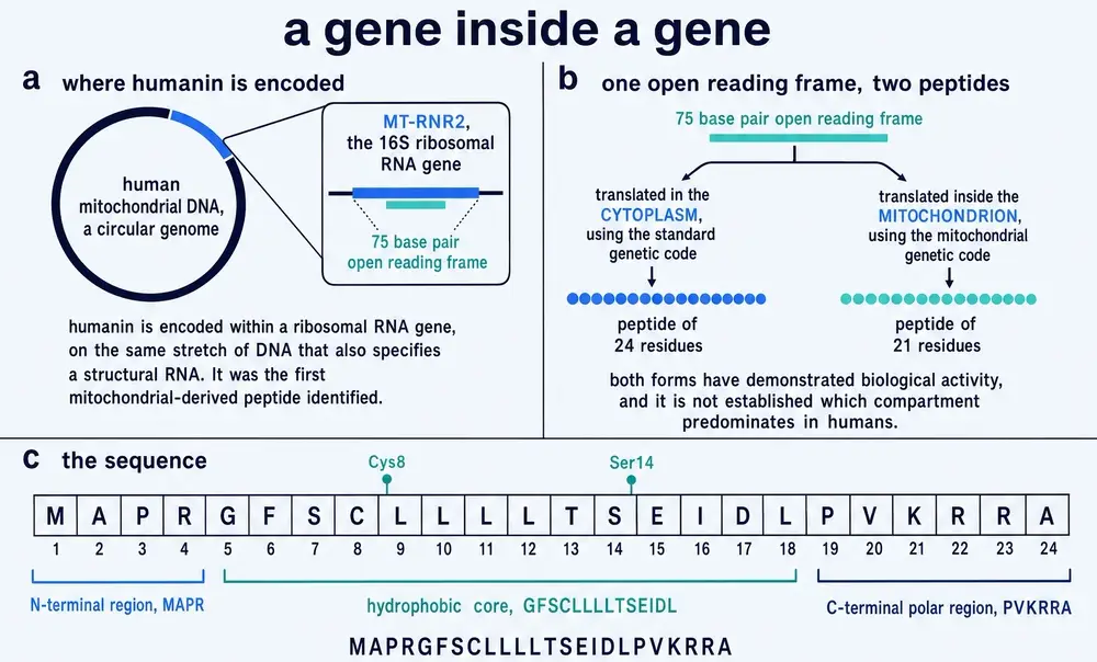
The gene is MT-RNR2. It occupies positions 1,671 to 3,229 of the human mitochondrial genome and it encodes the large subunit ribosomal RNA — a structural RNA, part of the machine that builds proteins. Both of the databases that curate human genes classify it as non-coding. It is not supposed to be a gene for a protein. The humanin reading frame sits inside it, beginning at position 2,633.
Read that frame out and something genuinely elegant happens, and it explains a discrepancy that runs through the whole literature. The same seventy-five bases give two different peptides depending on which machine reads them.
A ribosome in the cytoplasm uses the standard genetic code and produces twenty-four residues, MAPRGFSCLLLLTSEIDLPVKRRA, terminating at a TAA. A ribosome inside the mitochondrion uses the vertebrate mitochondrial code, in which the codons AGA and AGG are stop signals rather than arginine. The twenty-second codon of the humanin frame is AGG. An organelle translating its own gene therefore stops three residues early and produces twenty-one.
Both forms are biologically active, which is why the field has never been able to settle the question by function alone, and why the peptide is reported in the literature as both twenty-four and twenty-one residues long without either being an error.
The consequence is that humanin is a genuinely awkward object. It is a sequence with strong evidence of biological activity, nested inside a gene annotated as non-coding, in an organelle whose own translation machinery reads it differently from the rest of the cell. Every subsequent controversy about the molecule — where it is made, whether the thing measured in blood is really it, whether the nuclear copies matter — descends from this one structural fact.
03Found three times
A molecule discovered once by a functional screen is a candidate. A molecule that three laboratories arrive at independently, chasing three unrelated questions, is harder to dismiss. Humanin is usually described as the second kind, and that description needs one correction.
In May 2003 John Reed’s laboratory at the Burnham Institute in La Jolla published in Nature that humanin binds Bax (Guo et al., 2003). They had not been looking for a neuroprotective peptide. They had been looking for proteins that interact with Bax, the pro-apoptotic switch that punches holes in mitochondrial membranes, and humanin came out of a yeast two-hybrid screen with Bax as the bait. No author on that paper had any connection to Tokyo. This is a genuinely independent discovery.
The Nature paper also contains a detail that has shaped the field’s confusions ever since. Guo and colleagues cloned humanin from a nuclear library and treated the nuclear copy as the real one, noting only in passing that “the mitochondrial genome contains an identical open reading frame” and that the mitochondrial version also works. Their proposal was that humanin arose in mitochondria and transferred to the nucleus. Twenty-two years later, which copy the body actually uses is still not settled.
Five months later a second group published in the Proceedings of the National Academy of Sciences that humanin binds insulin-like growth factor binding protein 3 (Ikonen et al., 2003). Pinchas Cohen’s laboratory at UCLA had run a yeast two-hybrid screen with IGFBP-3 as bait against a HeLa library, and humanin fell out of it unprompted. The screen was genuinely unbiased and genuinely unrelated to Alzheimer’s disease.
The paper, however, was not independent of the original group. Hashimoto, Niikura and Nishimoto are all co-authors. Several widely read reviews state that two laboratories found humanin “independently of each other and of Nishimoto’s group”, or that there were “three independent laboratories”. That is true of Reed’s paper and false of Cohen’s, whose author list includes Nishimoto himself. What was independent was the route — an unrelated bait, an unrelated question — not the personnel. The distinction matters because convergent discovery is being used as an argument that the molecule is real, and an argument is only as strong as its weakest instance.
A fourth recovery is worth naming because it came from a different species and a different continent. A group in Rome, screening for neuroprotective factors, found a rat gene encoding a humanin-like peptide they called rattin (Caricasole et al., 2002). It is thirty-eight residues rather than twenty-four, shares 73 per cent identity across the conserved region, and — unlike humanin — protects against NMDA excitotoxicity.
Nishimoto did not see most of what followed. He died on 17 October 2003, aged forty-seven, five months after the Nature paper and eleven days before the PNAS paper appeared. His last review of the molecule he had named was published posthumously in Trends in Molecular Medicine in March 2004, running from page 102 to page 105 of that issue. His obituary, written by his Keio colleague Masaaki Matsuoka, is on page 105.
04Is it actually made?
This is the section most reviews of humanin skip, and it is the one a careful reader most needs, because everything downstream depends on it. A peptide that rescues cells when you add it to them is a pharmacological fact. Whether the body makes it, and from which genome, is a separate question with a genuinely unsettled answer.
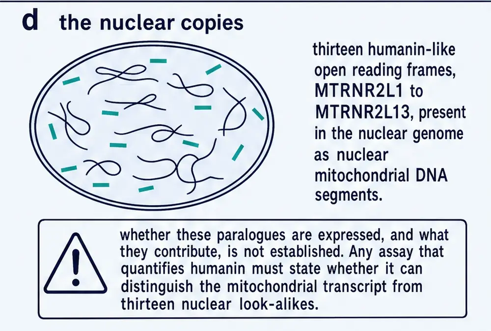
The difficulty is specific and it is not going away. When mitochondrial DNA fragments insert themselves into the nuclear genome — a common event, and the resulting sequences are called NUMTs — they carry their reading frames with them. The human nuclear genome contains thirteen such humanin-like loci, MTRNR2L1 through MTRNR2L13, scattered across eleven chromosomes. At least ten of them are transcribed in human tissue (Bodzioch et al., 2009). Both of the authorities that curate human gene names classify all thirteen as pseudogenes.
And two of them — MTRNR2L8 on chromosome 11 and MTRNR2L12 on chromosome 3 — encode peptides that are identical to each other and differ from mitochondrial humanin by a single amino acid.
Almost every measurement of humanin in a tissue or a blood sample has been made with an antibody. An antibody raised against a twenty-four-residue peptide cannot reliably be assumed to distinguish it from another twenty-four-residue peptide differing at one position. The curators at UniProt have made this doubt permanent: every one of the thirteen nuclear entries carries a standing caution that “the possibility exist[s] that the physiologically active humanin peptide is encoded by one of the related genes present in the nuclear genome”. Section 13 returns to what this does to the human data.
Against that background, the evidence that humanin is genuinely a mitochondrial product comes from two directions, and both are worth stating precisely.
The clean experiment was done in a rat. A group at UCLA reasoned that the rat humanin sequence is predicted to lack the confounding nuclear insertions, and used that to ask the question directly (Paharkova et al., 2015). Production of the rat peptide fell in cells depleted of mitochondrial DNA, which places its origin in the mitochondrial genome; and it was unaffected by cycloheximide, which blocks cytoplasmic ribosomes but not mitochondrial ones, which places its translation inside the organelle. The same paper found that adding a humanin analogue to isolated mitochondria from a rat β-cell line reduced hydrogen peroxide production by 55 per cent. That paper’s opening sentence concedes what it was written to fix: evidence for the mitochondrial origin of humanin “has been lacking”. The equivalent experiment has never been done in human cells, where the thirteen nuclear copies exist and ten are transcribed.
The evolutionary argument is the strongest one available, and it is clever. Because the humanin frame is embedded in a gene for a structural RNA, any selection acting on the RNA would constrain the DNA sequence — but it would not care which amino acids the frame happens to spell. Synonymous codon bias — a preference among codons that all specify the same residue — can therefore only arise if something is selecting on the peptide. Analysing vertebrate genomes, Gruschus and colleagues found exactly that for humanin, with robust evidence of purifying selection (Gruschus et al., 2023).
That result cuts two ways, and the monograph reports both edges. The same analysis found no significant bias for SHLP1, SHLP2, SHLP3 or SHLP5 — four of the peptides that the field describes as humanin’s family. Whatever the test establishes about humanin, it declines to establish it about most of its relatives. And humanin’s own conservation is patchier than the headline suggests: the initiating methionine is conserved in only 79 per cent of vertebrates, ranging from 96 per cent in birds to 46 per cent in reptiles, and the stop codon in only 58 per cent.
What is missing is the measurement that would settle it. Despite twenty-five years of work and several hundred papers reporting humanin concentrations in human blood, there is no published mass-spectrometric identification of endogenous human humanin — no measurement that reads the actual peptide sequence out of a real sample and distinguishes the mitochondrial product from its nuclear near-twins. Every mass-spectrometry paper in this corpus characterises synthetic peptide or maps an interaction using synthetic peptide. The nearest thing to a worked example concerns a sibling peptide, where a detected sequence turned out to match equally well a micropeptide translatable from a NUMT on chromosome 17 — an interval that falls inside MTRNR2L1.
One further complication, which anyone reading the animal literature should hold on to: in mice, the mitochondrial humanin gene is itself a pseudogene, and the humanin-like peptide is encoded by a nuclear gene. The most heavily used experimental species for this compound is not a model of the arrangement being studied.
None of this means humanin is not made. The codon-bias result is a real argument that something has been conserving this peptide sequence across vertebrate evolution, and the rat experiment is a real demonstration of mitochondrial translation in at least one mammal. It means that the sentence “humanin is a mitochondrial-derived peptide”, which appears in the opening line of most papers on the subject, is a well-supported inference rather than a settled measurement — and that the field has known this and said so in print for over a decade.
05Two receptors, and then four
If humanin is secreted and acts on other cells, it must bind something. The question of what has been open since 2004, and the honest answer in 2026 is that the field carries several incompatible accounts side by side and has never tested them against each other.
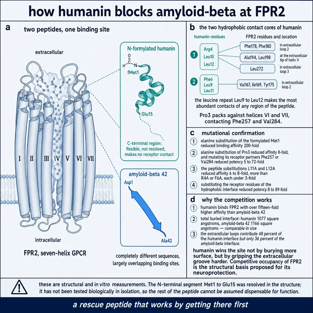
The first answer came from the National Cancer Institute. Humanin, they reported, draws mononuclear phagocytes across a membrane through FPRL1 — formyl peptide receptor-like 1 — and its murine counterpart FPR2 (Ying et al., 2004). The finding had an attractive corollary: amyloid-β42 uses the same receptor, so humanin might work by competing with it for access.
A companion paper the same year supplied the only hard receptor potency numbers the humanin literature has ever produced. In cells transfected with human FPRL1, humanin suppressed cyclic AMP with a half-maximal concentration of 3.5 nM; N-formylated humanin did the same at 0.012 nM, a roughly 290-fold gain from a single chemical modification (Harada et al., 2004).
One year later the Keio group tested that receptor in the system where humanin had been discovered, and it failed. Knocking down FPR2 did not attenuate humanin’s rescue of neurons from Alzheimer-related insults; the authors concluded in print that “a receptor other than FPR2 exists” (Hashimoto et al., 2005). That negative result is the reason the second account exists at all.
The second account is a three-part receptor: ciliary neurotrophic factor receptor α, WSX-1, and gp130. Overexpressing the first two increased humanin binding to neurons, knocking them down reduced it, and a reconstituted assay showed humanin driving the three subunits into a complex (Hashimoto et al., 2009). Downstream it runs through JAK2 and STAT3.
Since then the picture has become more crowded rather than less. In the testis, humanin’s protection of germ cells is blocked by antibodies against gp130, IL-27R and EBI-3, and is absent in mice lacking either of the latter two — but it is not blocked by antibody against CNTFR (Jia et al., 2021). The testicular receptor is not the neuronal one. In glioblastoma, humanin acts through gp130 and the formyl peptide receptors are undetectable. And in 2024 a fourth mechanism arrived: humanin as a ligand for integrin αV, signalling through the TGF-β receptor to drive migration and invasion (Ha et al., 2024).
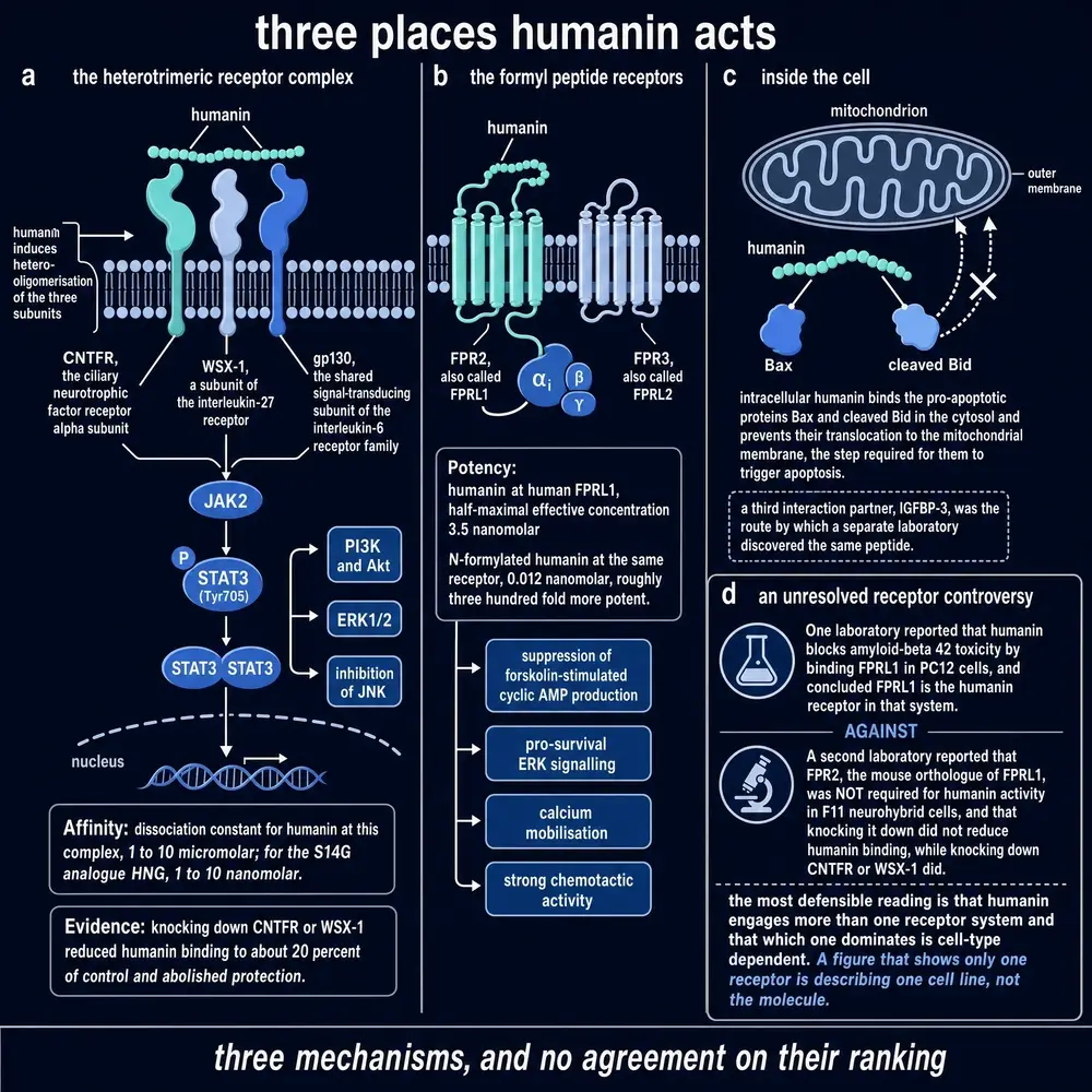
The field’s standard reconciliation is that different cell types use different receptors, phagocytes carrying the formyl peptide receptors and neurons not. That is a reasonable hypothesis and it has never been an experimental result. Two decades of reviews restate the conflict without adjudicating it; a 2026 review still writes the architecture as “humanin–FPR2/gp130”, two receptors joined by a slash.
The sharpest datum is easy to miss. The 2024 glioblastoma paper looked for expression of IGFBP3, BCL2, WSX-1, gp130, CNTFR and FPRL1 across cancer cell types that did and did not respond to humanin, and found no distinguishable pattern. None of the canonical receptors predicted which cells the peptide would act on.
06The other humanin, inside the cell
Alongside the receptor literature runs a second, largely separate account in which humanin never leaves the cell that made it.
Reed’s 2003 Nature paper established the first version: humanin binds Bax and prevents it moving from the cytosol to the mitochondrial membrane, where it would otherwise trigger the release of cytochrome c and commit the cell to apoptosis. Two years later the same group extended it to Bid and its truncated form, and showed that humanin retains protective activity on mitochondria from bax-null cells — so the effect is not solely Bax-dependent (Zhai et al., 2005). A third partner, BimEL, is bound with unusual specificity: humanin binds the extra-long splice form and not the two shorter ones, and protects against death induced by the one it binds (Luciano et al., 2005).
Then there is IGFBP-3, the binding protein that carries most of the circulating insulin-like growth factor. Humanin binds its heparin-binding domain, residues 215 to 232, and blocks the protein’s import into the nucleus. The F6A mutant, which does not bind IGFBP-3, is the tool that separates this arm from the others.
Since 2019 this account has been substantially reinterpreted, and the reinterpretation is uncomfortable. Studies of humanin with Bax and with Bid found that the two do not simply bind — they form fibres together, with Bax converting entirely to β-sheet as it is incorporated (Morris et al., 2019, 2020). The proposed mechanism is sequestration by co-fibrillation rather than classical inhibition. The internal control is reassuring: the anti-apoptotic protein BCL-xL, which humanin has no reason to neutralise, does not form fibres with it.
In 2025 the same group reported that humanin itself forms amyloid-like β-sheet fibrils, and that the mutants which fail to fibrillate are the same ones previously catalogued as secretion-deficient (Morris et al., 2025). They also questioned the founding structural model directly, suggesting that results the original laboratory attributed to humanin dimerisation may reflect aggregation instead.
Humanin is sold as a research chemical and is injected by people outside any clinical protocol. The most recent structural work on it reports that it forms amyloid-like fibrils under physiological conditions, and its authors raise this explicitly in connection with therapeutic administration. No study in this corpus has examined what injected humanin does over time in a living animal in this respect. This document recommends no human use; the point here is that the question has not been asked.
07The potency problem
Three numbers, taken together, describe a molecule whose pharmacology is stranger than its reputation.
The first: wild-type humanin protects cells in the founding assay at 1 to 10 micromolar. The second: substituting a single serine at position 14 with glycine — giving S14G-humanin, the analogue that carries most of the animal literature — drops that to 10 nanomolar or below, a thousandfold gain. The third: fusing a nine-residue fragment of another neurotrophic peptide to a shortened humanin derivative gives colivelin, which suppresses the same death at 100 femtomolar (Chiba et al., 2005).
That is nine orders of magnitude across four closely related molecules, and it is the strongest possible argument for the rule this monograph applies throughout: their evidence must not be pooled. A result obtained with colivelin says nothing about humanin.
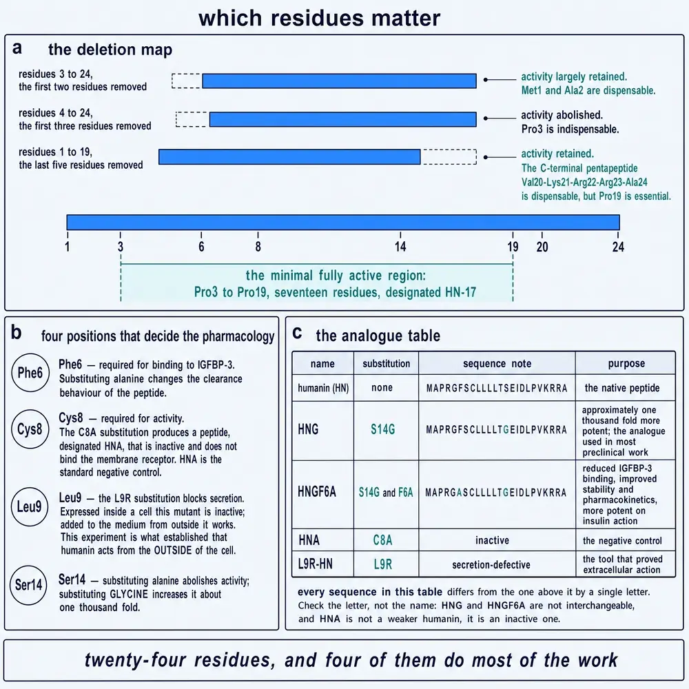
The fourth number is the one the field does not put in its abstracts. In 2013 the laboratory that had identified and named the three-part humanin receptor published a paper stating that “the protective effect of Humanin via the htHNR is weak (EC50 = 1–10 µM)” (Hashimoto et al., 2013). The paper exists because that weakness sent them looking for a better agonist of their own receptor, and they found one — calmodulin-like skin protein, active at 10 to 100 picomolar and circulating in normal human blood at around 5 nanomolar. The discovering laboratory, in other words, concluded that humanin is a micromolar agonist at the receptor it had named for humanin, and that something else is probably the physiological ligand.
The fifth observation is an absence. There is no measured binding constant for humanin at any of its proposed cell-surface receptors. No dissociation constant, no Scatchard analysis, no surface plasmon resonance, no calorimetry — not for the three-part receptor, not for the formyl peptide receptors. The 3.5 nM figure above is a functional half-maximal concentration in a transfected cell line, not an affinity. The only rigorously measured affinity in the entire humanin literature is for a partner nobody predicted: apolipoprotein E4, at 12.7 nM for wild-type humanin (Miller et al., 2024). The only measured affinity for an intracellular partner is 5.05 micromolar, for IGFBP-3.
The structural work is correspondingly thin on the native molecule. There are four experimental structures. Three are solution NMR of synthetic humanin or its analogues in 30 per cent trifluoroethanol, a solvent used to coax helix out of peptides that do not have much of their own — in water humanin is largely unstructured, with transient turns. The fourth is a 2022 cryo-electron microscopy structure of FPR2 with humanin bound, and it is a genuinely important result. It also used N-formylated humanin, not the native peptide, and the physiological abundance of the formylated form has not been established.
Two consistent structure–function findings survive all of this and are worth carrying forward. An alanine scan identified eight essential residues — Pro3, Ser7, Cys8, Leu9, Leu12, Thr13, Ser14 and Pro19 — with the active domain running from Pro3 to Pro19. And four substitutions (P3A, L12A, S14A and P19A) behave as dominant-negative antagonists, blocking wild-type humanin by forming an inactive heterodimer with it; inserting a second mutation that prevents dimerisation abolishes the antagonism. Whatever humanin is doing, it is doing it as a pair.
08The brain
The first demonstration that a humanin analogue does anything in a whole animal came from a laboratory unconnected to the discoverers. At Meijo University in Nagoya, [Gly14]-humanin injected into the cerebral ventricles of mice reversed the memory impairment caused by scopolamine, and did so without changing how much the animals moved — a control that matters, because a drug that makes a mouse more active will improve a maze score without improving memory (Mamiya & Ukai, 2001).
What followed over two decades is one of the broader preclinical neurology literatures attached to any peptide in this series. Humanin analogues have been given into the ventricles, into the hippocampus, into the peritoneum and up the nose, in mice and rats, against amyloid-β infusion, against anticholinergic amnesia, and in the standard transgenic models of Alzheimer’s disease. Reported effects include restored performance in water mazes and object recognition, preserved cholinergic neurons, reduced amyloid burden, reduced tau phosphorylation, restored dendritic spine density, and — repeatedly and across several groups — rescue of long-term potentiation measured in the living animal, which is a more physiological endpoint than a behavioural score.
Two internal controls give this literature more weight than a simple list of positive findings would. In the amyloid-β25-35 model, S14G-humanin prevented the deficit at 50 picomoles, while S14A-humanin — a neuroprotection-defective mutant — did nothing at 5 nanomoles, a hundredfold higher dose (Tajima et al., 2005). And in an alanine scan of the analogue series, truncated derivatives and most tert-leucine substitutions were simply inactive in vivo. The effect is sequence-specific, not an artefact of injecting a hydrophobic peptide into a brain.
Two findings on the other side belong here rather than in a late section.
The first is a clean failure. In the intracerebroventricular streptozotocin model — a rat model of sporadic rather than familial Alzheimer’s disease, built around disrupted brain insulin signalling — S14G-humanin was tested at 0.01, 0.05, 0.1 and 1 nanomole daily for eleven days and failed at every dose. It did not restore spatial memory and it did not restore hippocampal Akt phosphorylation. The paper is titled, without euphemism, “Humanin Does Not Protect Against STZ-Induced Spatial Memory Impairment” (Negintaji et al., 2015). Since sporadic disease is what almost all patients have, this is not a peripheral negative.
The second is a dissociation. In triple-transgenic mice given intranasal S14G-humanin for three months, brain amyloid fell markedly in both sexes — but cognition improved only in males (Niikura et al., 2011). A treatment that lowers the pathology without changing the behaviour in half the animals is a result worth holding on to when reading any summary that reports only the amyloid figure.
Colivelin deserves its own note, because it is the most potent molecule in the family and because its record is not uniformly favourable. It crosses the blood–brain barrier after intraperitoneal injection, reaches the central nervous system when given intranasally by way of the olfactory bulb, and in transgenic mice carrying the G93A superoxide dismutase mutation it dose-dependently improved motor performance and prolonged survival — where the neurotrophic fragment alone improved motor performance but did not extend life. But colivelin is also sold as an off-the-shelf STAT3 activator, and in that role it has produced the opposite result: in rats with chronic cerebral hypoperfusion, activating the pathway with colivelin exacerbated neuronal damage and cognitive decline, while inhibiting the same pathway helped. The contradiction is direct, it is within the same species, and it is not resolved.
09The heart, and the pig that did not follow
The cardiac work produced the cleanest dose–response curve in the entire humanin corpus, and then the clearest illustration of why a clean rodent curve is not a clinical result.
In mice subjected to forty-five minutes of coronary occlusion and twenty-four hours of reperfusion, S14G-humanin reduced infarct size as a fraction of the tissue at risk from 58 ± 4 per cent in vehicle animals to 44 ± 4 at 0.2 mg/kg, 39 ± 2 at 1 mg/kg and 28 ± 4 at 2 mg/kg. Four milligrams per kilogram gave no further benefit — the curve plateaus, which is what a real receptor-mediated effect looks like. At one week, ejection fraction had fallen from 78 to 25 per cent in vehicle animals and from 76 to 56 per cent in treated ones (Muzumdar et al., 2010).
Ten years later the same intervention was taken into a large animal, which is the step at which most cardioprotective agents fail. Female Yucatan minipigs underwent balloon occlusion of the left anterior descending coronary artery followed by forty-eight hours of reperfusion, with S14G-humanin at 2 mg/kg given intravenously ten minutes before reperfusion — a protocol designed to mirror what could actually be done to a patient arriving for angioplasty (Sharp et al., 2020).
With sixty minutes of ischaemia, infarct size fell from 56.8 to 33.7 per cent of the area at risk, a 41 per cent relative reduction (p = 0.017). This is a genuine large-animal result and it should be reported as one.
With seventy-five minutes of ischaemia, at the same dose, there was no significant reduction at all (p = 0.541).
And in both arms, the circulating markers of myocardial injury — troponin I and heart-type fatty acid binding protein — showed no significant reduction. The authors state plainly that by these clinically relevant measures the peptide provided no benefit.
Read together, those three results say something more useful than any of them alone. The effect is real, it is measurable by tissue staining in a species with a human-like heart, and it is fragile with respect to the one variable that is least controllable in a patient — how long the artery has been shut. Human door-to-balloon times are rarely sixty minutes.
The timing sensitivity recurs in the rodent work in a different form. In rats, 84 µg/kg was effective given before ischaemia and failed when given after ischaemia had begun; only 252 µg/kg — three times the dose — worked in the delayed protocol. And when the same group looked at the brain injury that follows cardiac ischaemia, treatment during ischaemia or at reperfusion gave no neuroprotection at all, and even at the tripled dose never reduced blood–brain-barrier breakdown or brain oxidative stress.
The vascular work is more straightforwardly positive. In apolipoprotein E-deficient mice on a high-cholesterol diet for sixteen weeks, the analogue HNGF6A reduced proximal aortic plaque area from 0.09 to 0.01 mm² — six of seven untreated animals had plaques against two of six treated — restored endothelium-dependent relaxation, and cut nitrotyrosine staining from 12.3 to 3.2 per cent of plaque area, without lowering cholesterol and without any direct vasoactive effect (Oh et al., 2011).
One human tissue finding sits awkwardly beside that and is reported here rather than buried. In thirty-four carotid endarterectomy specimens, humanin was higher in the unstable, symptomatic plaques than in the stable ones — 29.4 against 14.1 per cent of plaque area. That is consistent with humanin being induced by stress rather than preventing it, and it is a pattern that will recur throughout the human data.
10An effect on the liver that is not in the liver
The metabolic work contains the single most surprising mechanistic result in the humanin literature, and it is one that ought to change how the compound is thought about.
Under hyperinsulinaemic–euglycaemic clamp in rats, humanin infused into the cerebral ventricles increased the glucose infusion rate required to hold blood sugar steady — the standard readout of improved insulin sensitivity — principally by suppressing the liver’s own glucose output, which fell from 12.1 ± 0.9 to 4.7 ± 0.4 (Muzumdar et al., 2009).
Infused intravenously instead, the analogues reproduce the effect. The obvious inference is that circulating humanin acts on the liver.
It does not. Blocking STAT-3 in the hypothalamus — in the brain, not the liver — completely abolished the hepatic effect of intravenously administered humanin analogue. And humanin has no direct effect on isolated primary hepatocytes. The peptide reaches the brain, the brain instructs the liver, and a related study showed the instruction travelling by way of the vagus nerve: vagotomy blocked the effect of both intravenous and intracerebroventricular humanin on hepatic triglyceride secretion.
In Zucker diabetic fatty rats, a single intravenous bolus of 100 µg of the analogue HNGF6A lowered blood glucose by nearly half over four hours in a group of five animals. A related experiment showed that the analogue acts directly on the β-cell as well, increasing glucose-stimulated insulin secretion from islets of both normal and diabetic mice in a manner independent of the ATP-sensitive potassium channel. And in non-obese diabetic mice — an autoimmune model — humanin normalised glucose tolerance after six weeks, reduced the severity of islet infiltration, and over twenty weeks delayed or prevented the onset of diabetes.
Two negatives complete the picture. The analogue does not require IGFBP-3 binding to improve insulin sensitivity, since the non-binding analogue works — which detaches this arm from one of the founding mechanisms. And in the one detailed diabetic-nephropathy study to report a full cytokine panel, humanin significantly reversed two of seven measured cytokines and left the rest, including every anti-inflammatory one, unchanged.
11Everywhere else
Beyond brain, heart and metabolism, humanin and its analogues have been reported protective in reproductive tissue, kidney, lung, retina, cochlea, bone, skin, intestine and liver. Read as a set this is impressive; read carefully it is a statement about the breadth of preclinical interest, not about the breadth of demonstrated benefit, and this monograph reports it as the former.
The reproductive work is the most mechanistically informative, because it is where the receptor question was tested most rigorously and gave a different answer from the brain — gp130 and IL-27R and EBI-3, but not CNTFR. It also produced a rare functional endpoint: in mice with chemotherapy-induced premature ovarian insufficiency, S14G-humanin restored litter size from 4.6 ± 1.1 to 6.2 ± 0.8 against a control value of 8.0 (p = 0.029). Offspring counted is a harder endpoint than a stain.
The same literature contains a useful demonstration of specificity. Humanin alone had no effect on baseline germ-cell apoptosis — it acts only where death has been provoked. Its protection is stage-selective within the seminiferous epithelium. And the structure–function work in this tissue found the membrane-receptor route dominant and the Bax-binding route minor, with one analogue — HN-L12A — acting as an outright antagonist that blocked wild-type humanin’s protection.
The retinal work illustrates why the organ atlas needs its evidence tier attached. Humanin analogues protect retinal pigment epithelial cells in culture across many studies. When taken into a live animal — the Royal College of Surgeons rat, a genuine model of retinal degeneration — twice-weekly injection for four weeks produced no difference from sham on electroretinography at either dose tested. Optokinetic visual acuity improved at the high dose only. That is a partially negative in-vivo result behind a largely positive in-vitro literature, and both belong in the record.
Elsewhere the pattern repeats with variations: benefit in acute lung injury and in sepsis models; benefit in septic and diabetic kidney injury, including a recent mechanism in which humanin blocks mitochondrial Z-DNA leakage and the necroptosis it triggers; protection of cochlear hair cells against aminoglycoside damage in explant; prevention of glucocorticoid-induced bone growth impairment; prolonged anagen in hair follicles. In a haemorrhagic shock model the effect partitioned: humanin-G improved lung architecture in every group but reduced neutrophil infiltration only in animals with an intact AMPKα1, which is a clean demonstration that the peptide works through more than one pathway and that they can be separated.
12The tumour problem
A peptide whose principal action is to stop cells dying raises an obvious question, and the humanin literature contains two answers that point in opposite directions.
The reassuring literature is real and it is specific. In immunocompetent mice bearing melanoma, S14G-humanin rescued germ cells and white cells from cyclophosphamide while lung metastases were further suppressed by the combination than by chemotherapy alone. In mice bearing human medulloblastoma xenografts, the analogue did not blunt temozolomide’s effect on the tumour while protecting the animals from its toxicity. In a neuroblastoma model, bortezomib slowed bone growth from 0.19 to 0.09 mm/day and S14G-humanin restored it to 0.15 mm/day without interfering with the anticancer effect. Similar results exist for glucocorticoid-induced growth impairment and for doxorubicin cardiotoxicity.
The other literature is equally real and comes from different tumours and different groups.
In experimental triple-negative breast cancer, doxorubicin up-regulated humanin in the tumour cells. Exogenous humanin impaired the drug’s antitumour and anti-metastatic effect and accelerated tumour growth — and humanin given on its own, without any chemotherapy, significantly increased spontaneous lung metastases. Silencing humanin did the reverse, reducing viability and enhancing chemosensitivity. The authors’ own conclusion is quoted here because it is the field’s, not this document’s: “caution should be taken when using exogenous HN to treat degenerative diseases” (Moreno Ayala et al., 2020).
In glioblastoma the case is stronger still, and recent. Humanin is abundantly expressed in human glioblastoma, largely by the tumour-associated myeloid cells; nanomolar concentrations drive temozolomide resistance through gp130 and accelerate formation of the blood–tumour barrier that keeps chemotherapy out; immunodepleting humanin from the medium abolishes the pro-tumour effect; and blocking gp130 restores the drug’s efficacy (Cheng et al., 2024). A separate group found that chemotherapy up-regulates both humanin and FPR2 in glioblastoma cells, that the analogue HNGF6A boosts chemoresistance, and that silencing humanin reverses it. A third reported that humanin drives invasion through integrin αV, and that mice given humanin in an orthotopic intracranial model survived less long than controls (Ha et al., 2024). Their conclusion is that in glioblastoma humanin is a therapeutic target, not a therapeutic agent.
These two bodies of work are not, strictly, in contradiction: they used different tumours. The protective studies come predominantly from melanoma, medulloblastoma and neuroblastoma models, several from collaborating groups; the adverse studies come from breast, glioblastoma, gastric, pituitary and bladder systems, from groups with no overlap. In human gastric cancer, humanin isoforms were the most highly overexpressed genes found in a screen against normal gastric tissue. Knocking down the rat homologue in a pituitary tumour delayed its growth and improved survival.
Two mechanistic observations recur across the adverse literature and are what make it more than a disagreement about models. The first is that chemotherapy itself induces humanin in tumour cells — the tumour reaches for the peptide when attacked. The second is that silencing humanin makes tumours more chemosensitive, which is the reciprocal experiment and gives the same answer.
A broad-spectrum anti-apoptotic peptide, given systemically to a person with an occult malignancy, is an unquantified risk. This is not an inference this document is adding to the literature — it is the position taken in print by investigators in the field, one of whom writes that the “controversy on the role of HN in cancer progression and chemoresistance should be addressed before the translation of these therapeutic approaches”. No study has addressed it.
Two further adverse signals belong beside it. Humanin and MOTS-c are elevated in senescent human fibroblasts, and administering them increased components of the senescence-associated secretory phenotype — the inflammatory programme that senescent cells impose on their neighbours. A commentary on that work is titled, flatly, “Mitochondrial-Derived Peptides Exacerbate Senescence”. And across the whole preclinical corpus there is no dedicated toxicology, safety-pharmacology or repeat-dose study for humanin or any analogue. Every statement that the peptide caused no harm is a secondary observation inside a study designed to measure something else.
13Measuring humanin, and why the numbers cannot be compared
This section comes before the human findings rather than after them, because every one of those findings has to be read through it.
Humanin in human blood has been measured in something over thirty studies. All of them used an antibody. None used mass spectrometry. There is no published measurement anywhere that reads the peptide’s actual sequence out of a human sample.
That matters more here than it would for most peptides, for the reason set out in section 04: the human nuclear genome carries thirteen humanin-like sequences, and the products of two of them differ from humanin by a single amino acid. The most explicit statement of the problem in the primary literature is worth giving at length, because it is the field’s own assessment and not this document’s:
“There are many nuclear-encoded MTRNR2-like genes that produce humanin-like peptides, which have only one, two, or four different amino acids compared to mitochondrial-encoded humanin. Because cross-reactivity involving humanin and all humanin-like peptides with commercial humanin antibodies has not been completely confirmed to date, we still cannot definitively distinguish the sources — nuclear or mitochondrial — of peptides that are recognized as humanins.” (Kim et al., 2021)
The same paper closes the obvious escape route. Measuring the transcript instead does not help, because the reading frames sit inside ribosomal RNA genes and the standard reverse-transcription primers cannot tell a messenger RNA from the ribosomal RNA it is embedded in.
There is also a direct demonstration that the antibodies are promiscuous. In the course of characterising a different protein, the Keio group found that human calmodulin-like skin protein — a 146-residue molecule with no sequence relationship to humanin — “was cross reactively recognized by the Humanin antibody” in immunoprecipitation.
The practical consequence is visible in the numbers themselves. Reported concentrations in healthy or control adults, all purporting to measure the same twenty-four-residue peptide, span more than thirtyfold.
| Study | Platform | Control value |
|---|---|---|
| Widmer 2013 | unspecified | 2,200 pg/mL |
| Zhloba 2018 | unspecified | 1,110 pg/mL |
| Yen 2018 | in-house ELISA | 1,212–1,491 pg/mL |
| Alser 2022 | CUSABIO | 774 pg/mL |
| Bolignano 2024/26 | MyBioSource | ~450–780 pg/mL |
| Voigt 2016 | Elisakit.com | 205 pg/mL |
| Cai 2022 | unspecified | 158 pg/mL |
That spread is wider than any disease effect reported anywhere in the human humanin literature. A difference between patients and controls of thirty per cent, measured on one platform, cannot be interpreted against a between-platform disagreement of three thousand per cent.
Two further details complete the picture, and both come from the assay developers rather than from critics. The most-used in-house assay was raised against S14G-humanin, the synthetic analogue, not the native peptide. And its own methods sections concede that while plate-to-plate variability is under ten per cent at any one time, “the drift over time is greater” — which invalidates comparison across the years over which most of these cohorts were collected.
No study in this corpus compares two humanin immunoassays on the same samples, cross-validates any of them against mass spectrometry, or publishes recovery and dilution-linearity data for endogenous humanin in plasma. That absence is the reason the literature does not converge, and it is the cheapest thing the field could fix.
Every concentration below is humanin-family immunoreactivity of unresolved genomic origin, measured on a platform that has not been cross-validated against any other. Directions of change within a single study are more trustworthy than absolute values, and comparisons between studies on different platforms are not supportable at all.
14Age, centenarians and growth hormone
Three claims about humanin circulate far beyond the specialist literature: that it declines with age, that it is high in the children of centenarians, and that it is high in people whose growth hormone receptors do not work. All three appear in review abstracts without qualification. All three deserve their evidence stated.
The age claim is contradicted by the largest dataset in the field.
The source of “humanin declines with age in humans” is a single sentence in a 2009 paper reporting that levels decreased with age in mice and in humans. The paper gives no human sample size, no age range and no concentration values; the only human datum is an unlabelled scatter in one figure panel. Subsequent papers from the same laboratory restate it rather than re-measuring it.
Against that, an Italian group measured humanin in 693 people aged 21 to 113 and found it increased in old age, with the highest levels in centenarians — and, notably, inversely correlated with survival among the oldest subjects (Conte et al., 2019). A second cohort of 569 from the same group found humanin positively correlated with age. A third study found resting humanin positively correlated with age. A fourth found no significant change either way.
The two literatures use different assay platforms, which is very likely part of the explanation, and neither has been reconciled with the other. The honest statement is that the direction of humanin’s relationship with human age is unresolved, and that the widely repeated version rests on the weaker evidence.
The centenarian claim rests on eighteen people and did not replicate. In the Einstein longevity cohort, circulating humanin was significantly higher in eighteen offspring of centenarians than in nineteen age-matched controls. The abstract describes the difference as “much greater”; no effect size is reported, only a graph. The paper is explicit that centenarians themselves were not compared, for want of a control group. When an Italian group ran the same comparison in 102 centenarian offspring against 92 controls, the peptide that came out highest in the offspring was FGF21 — humanin did not differ.
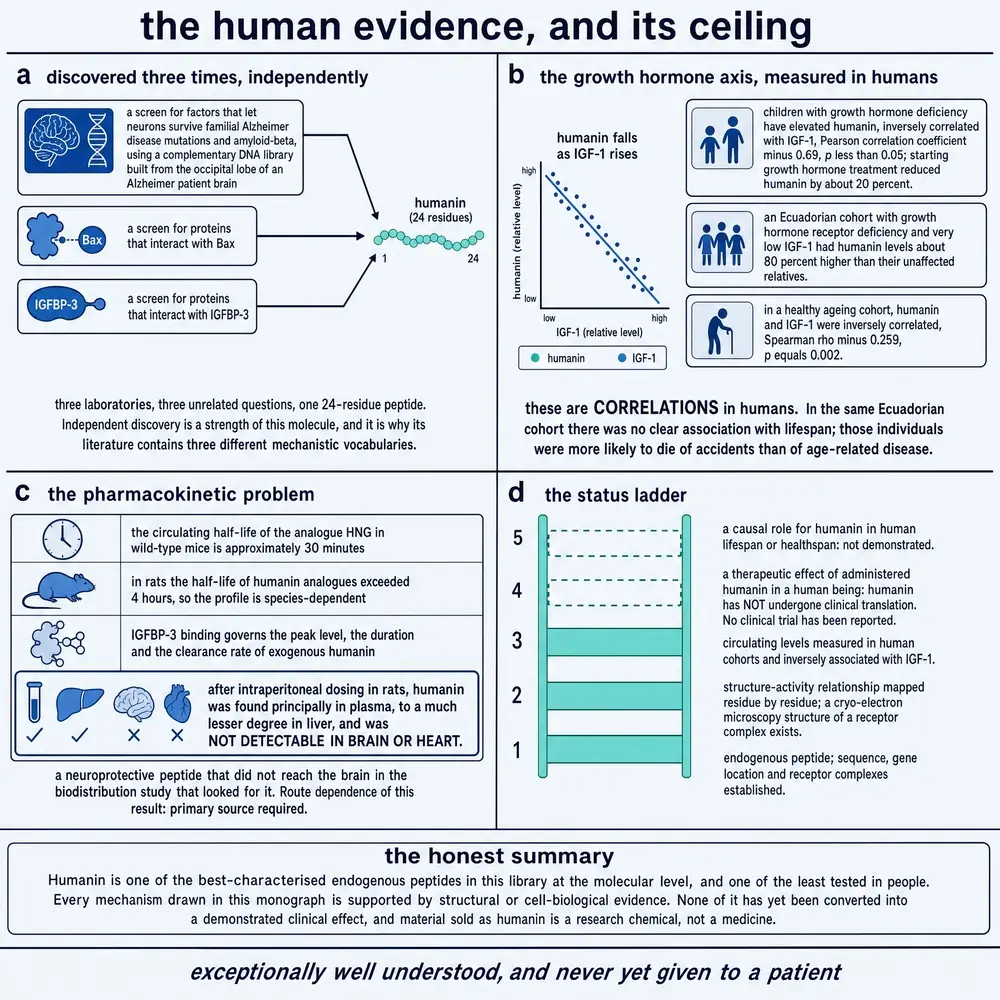
The growth hormone finding is the most mechanistically interesting and the smallest. In children being evaluated for short stature, plasma humanin was uncorrelated with peak stimulated growth hormone but strongly negatively correlated with IGF-I (r = −0.69). Treating them with recombinant growth hormone lowered humanin by about a fifth. And in an Ecuadorian cohort with growth hormone receptor deficiency — Laron syndrome, a condition associated with striking protection from cancer and diabetes — humanin was 80 per cent higher than in matched relatives (Lee et al., 2014). Mouse data in the same paper align: long-lived growth-hormone-deficient dwarfs have more humanin, short-lived growth-hormone-transgenics less.
The Laron result is the one most often quoted, and it is six people against six, on an assay whose own authors flag long-term drift, in a paper whose senior authors declare stockholding in a company founded on this peptide class. It is a genuinely interesting finding and it is not a robust one.
The cross-species work is more persuasive than any of the human cohorts. Humanin overexpression extends lifespan in C. elegans, dependent on the FOXO transcription factor daf-16; transgenic mice show overlapping phenotypes; middle-aged mice treated twice weekly with the analogue showed improved metabolic measures and lower inflammatory markers; and humanin levels are stable across the lifespan of the naked mole-rat, a species with negligible senescence (Yen et al., 2020). Whether any of that transfers to people is exactly what the human data above fail to establish.
15The biomarker record
Read with the assay caveat attached, the human observational literature describes a molecule that moves with illness in almost every system examined — usually downward, sometimes upward, occasionally both.
Cardiovascular. In 327 patients, humanin fell stepwise from controls through unstable angina to myocardial infarction. In 40 patients undergoing coronary function testing, those with endothelial dysfunction had lower humanin than those without. Against that, plaque tissue shows the opposite direction, with more humanin in the unstable lesions.
Diabetes and pregnancy. In 225 subjects, serum humanin was lower in type 2 diabetes and correlated negatively with glycated haemoglobin, glucose and triglycerides. It is lower in gestational diabetes. In polycystic ovary syndrome the result is unusually informative: serum humanin was significantly lower in 40 patients than in 40 matched controls, while humanin measured in their vastus lateralis muscle showed no difference at all — a dissociation the authors read as evidence that the circulating fall is not of muscle origin.
Kidney. In haemodialysis patients followed for two years, the relationship with cardiovascular mortality is U-shaped: both the lowest and the highest humanin concentrations carried elevated risk. Adding humanin to five established risk models improved discrimination.
Lung. In stable chronic obstructive pulmonary disease, humanin was higher than in smokers without disease, and high humanin independently predicted malnutrition, worse walking distance, and future exacerbations — severe ones with a hazard ratio of 3.4. This is a well-conducted prospective study in which more of the protective peptide predicted worse outcomes.
Mitochondrial disease is where a widely repeated statement inverts. The claim that humanin is “decreased in MELAS” traces to a paper whose MELAS data are cybrid cell lines and mitochondrial-DNA-depleted cells, not patients. In actual patient tissue the finding is the reverse: humanin is increased in MELAS skeletal muscle, staining strongly in every ragged-red fibre, and is expressed in the muscle of patients with chronic progressive external ophthalmoplegia. A peptide induced by mitochondrial crisis is what those biopsies show.
Cancer is contradictory. Two studies report humanin elevated in breast cancer; a third reports it decreased. In prostate cancer, higher humanin was associated with lower risk in European-American men and the association was null in African-American men, with a significant interaction.
Alzheimer’s disease deserves its own paragraph, because the contradiction there is usually presented as brain-versus-blood biology and is actually a disparity in evidence quality. The claim that humanin is elevated in the Alzheimer’s brain rests on one brain, one immunoblot, no quantification, in 2002. The claim that it is reduced in cerebrospinal fluid rests on seven subjects, in a paper whose text and figure legend disagree about how those seven were split. The only claim with a real cohort behind it is that plasma humanin is lower in Alzheimer’s disease, in 120 patients — and a 2025 study designed to test it found that transcript levels differed while plasma protein concentrations did not discriminate Alzheimer’s from mild cognitive impairment. Circulating humanin is not an established Alzheimer’s biomarker.
16Two letters in the mitochondrial genome
The strongest human evidence about humanin is not pharmacological. It is genetic, and it is strong precisely because nobody administered anything.
rs2854128 sits at the very end of the humanin reading frame. It does not change any amino acid; it changes the stop codon from TAA to TAG. Carriers have about 14 per cent less circulating humanin. The variant is present in roughly 42 per cent of Caucasian-Americans and is much rarer in other groups. Tested against cognitive scores in 10,158 participants of a national ageing study, it showed a significant association, with a race interaction: among African-American carriers the effect corresponded to about two years of additional cognitive age, against roughly two months in Caucasian-American carriers (Yen et al., 2018).
P3S is a change of one base at position 2,639 that turns the third residue from proline to serine. It is present in about 12 per cent of a cohort of 146 female centenarians, principally Ashkenazi, against under 0.2 per cent in other populations. Within those centenarians it was strongly enriched among the ones carrying APOE4, the major genetic risk factor for Alzheimer’s disease — six of twenty APOE4 carriers had it, against about seven per cent of the rest. Humanin P3S binds APOE4 far more tightly than wild-type humanin does: dissociation constants of 0.69 nM against 12.7 nM by surface plasmon resonance, which is the single most rigorous binding measurement in the humanin literature. In mice carrying both APP/PS1 and human APOE4, the P3S peptide reduced brain amyloid more than wild-type humanin did (Miller et al., 2024).
The published commentary on that paper supplies the counterweight, and this monograph gives it equal space. P3S is a haplogroup marker: it defines mitochondrial haplogroup N1b, and by the commentator’s arithmetic roughly 9.5 million non-Ashkenazi people carry it — in whom no benefit has been demonstrated. The same commentary points out that S14G-humanin, the most potent synthetic analogue in the whole literature, occurs naturally in haplogroup U6a7a1a, and that nobody has shown carrying it is advantageous either (Logan, 2024).
Set against these two positives is a clean, well-powered negative that is rarely cited. When all thirteen nuclear humanin-like gene regions were looked up in a large coronary artery disease meta-analysis, no association survived correction for multiple testing, and no significant expression quantitative-trait loci were found.
17Nobody has taken it
The following is reported as a registry finding and nothing more. Every search below was run directly against the registry on 3 August 2026, not taken from any paper’s summary.
ClinicalTrials.gov returns seven records for humanin. Every one of them measures humanin as a biomarker or an outcome; none administers it. Three are observational studies of acute kidney injury or cardiac surgery. Two are interventional, but their interventions are anaesthetic agents and an exercise programme respectively, with humanin as the endpoint. One is an observational study in polycystic ovary syndrome and one a long-running study of cerebral palsy muscle biopsies.
Searching for colivelin returns zero records. Searching for S14G-humanin returns zero records. The European Union Clinical Trials Register returns zero for humanin, for colivelin and for MOTS-c. The single relevant record on the ISRCTN registry is observational, with humanin as an assayed outcome.
No human being has been given humanin, S14G-humanin or colivelin in a registered clinical trial anywhere, at any time. Twenty-five years after the discovery, with a preclinical literature spanning more than a dozen organ systems, the compound has never entered a first-in-human study.
There is a completed Phase 1 trial that is frequently associated with this field: 88 subjects, healthy volunteers and patients with fatty liver disease, sponsored by a company founded on mitochondrial-derived peptides, with no results posted to the registry. The drug in that trial was CB4211, an analogue of MOTS-c — not of humanin. The company’s own release describes it as such. It is not humanin’s clinical record and must not be reported as such.
Two further facts complete the regulatory picture. Humanin has no marketing authorisation anywhere, for any indication — searches of the United States drug label and approval databases return no match. And one completed study that did measure it, a 106-patient investigation of humanin in atrial tissue and plasma after cardiac surgery, closed in 2019 and has never posted results; no corresponding publication could be located.
Finally, the residual gaps in this search are stated rather than glossed. The World Health Organization’s international registry portal could not be queried directly because its search interface has been retired; its three largest constituent registries were queried individually instead. And the United States orphan-drug designation database did not return an interpretable result for the query submitted, so orphan status is recorded here as unverified, not as absent.
18Sold, unstudied, and forming amyloid
The facts in this section are commercial and regulatory. None of them is evidence of efficacy or of safety, and none is presented as such.
Humanin and S14G-humanin are available for purchase. Two distinct channels exist and they should not be confused. The first is the ordinary laboratory-reagent trade: humanin analogues appear in the catalogues of established chemical suppliers, sold to research institutions as reagents, which is unremarkable and is how most of the studies in this document were done. The second is the research-chemical market, where the same peptides are offered as lyophilised powder in five- and ten-milligram vials, labelled “research use only” and “not for human consumption”, alongside websites that index them together with dosing content.
Set that against what has been established. There is no approved indication in any jurisdiction. There is no completed trial of any phase in which the compound was given to a person. There is no published human pharmacokinetic data — no measurement of what happens to injected humanin in a human body over time, at all. There is no published human safety or tolerability data. The only pharmacokinetic work in the corpus is in rodents.
And there is no formal toxicology. Across the whole preclinical literature — hundreds of animal studies over twenty-five years — this corpus contains no dedicated safety-pharmacology study and no repeat-dose toxicology study for humanin or any analogue. The reassuring observations that exist are real but they are incidental: that the analogue did not worsen dystrophic muscle in one study, that it did not prolong bleeding time in another, that it did not blunt an anti-inflammatory drug’s effect in a third. Each is a secondary endpoint inside an experiment designed to measure something else.
To this must be added the structural finding from 2025 described in section 06: humanin forms amyloid-like β-sheet fibrils, and the residues required for fibrillation are the residues required for secretion. What that implies for a peptide injected repeatedly into a living body has not been investigated by anyone.
The gap between the commercial availability of this molecule and the evidence base underneath it is, on the numbers assembled here, among the widest in the peptide market.
19What would have to be true
It is easy to end a document like this with a list of promising directions. More useful is to state what would actually have to be established, and in what order, for the claims made about humanin to become claims about people.
First, the measurement. The single cheapest and most consequential experiment the field has not done is to quantify endogenous circulating humanin by mass spectrometry, in a way that distinguishes the mitochondrial twenty-four-residue product from the twenty-one-residue form and from the one-residue-different peptides encoded by the nuclear paralogues. Until that exists, every human observational finding in section 15 rests on an uncalibrated proxy, and the thirtyfold disagreement between platforms cannot be adjudicated. A head-to-head comparison of the commercial kits on shared samples would cost very little and would resolve a great deal.
Second, the origin. The clean genetic demonstration that humanin is translated inside mitochondria was done in the rat because the rat lacks the confounding nuclear copies. The equivalent experiment in human cells — mitochondrial-DNA depletion, cytoplasmic translation blockade, and a readout that can tell the mitochondrial product from the nuclear ones — has never been performed. Everything the compound is named for depends on its answer.
Third, the receptor. Four accounts coexist, partitioned by experimental system rather than by evidence. What would settle it is not another system: it is one system in which the candidate receptors are all present and are knocked out in turn, with a measured binding constant attached. The field has no dissociation constant for humanin at any surface receptor, which is an unusual position for a molecule twenty-five years old.
Fourth, the tumour question. The reassuring chemoprotection data and the adverse tumour-promotion data used different malignancies, and no study has tested them against each other. The two facts that recur in the adverse literature — that chemotherapy induces humanin in tumour cells, and that silencing it restores chemosensitivity — are not incidental findings but a coherent mechanism, and they were reported by groups with no stake in the peptide as a drug. Any programme contemplating systemic administration to humans has to address this first, and the field’s own reviewers have said so in print.
Fifth, which animal findings are most likely to travel. On the evidence assembled here, the cardiac work is the strongest candidate: it has a plateaued dose–response, a mechanism, and a large-animal replication. It also carries the clearest warning, because that replication failed at seventy-five minutes of ischaemia and produced no change in circulating injury markers in either arm. The metabolic work is mechanistically the most interesting — a peripherally injected peptide acting on the liver through the hypothalamus and the vagus — and is the least likely to be reproduced by simply giving the peptide to a person, precisely because the route matters. The neurological work is the largest and the least secure, resting heavily on models of familial disease and containing a total failure in the one model of sporadic disease that was tried.
And what would refute the framing. If mass spectrometry fails to detect endogenous humanin in human plasma at the concentrations the immunoassays report, the entire human biomarker literature becomes a literature about something else. If the nuclear paralogues turn out to be the translated source, humanin remains a real and interesting peptide but stops being a mitochondrial-derived one, and the conceptual claim it founded — that mitochondria signal outward — would have to be re-established on MOTS-c and the others, half of which fail the same evolutionary test humanin passes.
Humanin is a genuine biological discovery, made by an unusual and clever experiment, whose principal legacy is conceptual: it is the reason the field takes seriously the idea that the mitochondrion writes messages to the rest of the cell. Its cytoprotective activity in cultured cells is robust and reproducible across many laboratories. Its activity in animals is broad and, in places, quantitatively convincing.
What it is not, on the evidence assembled here, is a therapy. It has no human data of any interventional kind, no pharmacokinetics, no toxicology, no validated assay, no measured receptor affinity, and an unresolved signal that it helps tumours survive chemotherapy. The distance between those two paragraphs is the subject of this document.
This monograph describes published research. It does not recommend human use of humanin or of any analogue, and it specifies no dose, route or schedule for any person. Every quantity reported in it is a parameter of a study that was carried out, given with the species, the population and the duration attached. No human interventional data exist for this compound; there is therefore no human dose to report, and none is implied by any animal figure quoted here.
20References
Generated from verified NCBI records rather than from recall. Author lists, journal names, volumes, pages and identifiers are taken from the PubMed record for each identifier, not written out by hand. The build refuses to run if any cited identifier fails to resolve.
Two retracted papers were excluded by a gate. This compound's harvest contains two primary papers that were retracted in 2016 and are still indexed as live records, both concerning S14G-humanin — the analogue that carries most of the animal literature. Nothing in the index links a retracted paper to its notice automatically. Their identifiers are held in a barred list that stage 05 checks before it will emit a reference list at all.
- Alser M, Ramanjaneya M, Rizwana Anwardeen N, Donati F, Botrè F, Jerobin J, et al.. The Effect of Chronic Endurance Exercise on Serum Levels of MOTS-c and Humanin in Professional Athletes. Rev Cardiovasc Med. 2022;23(5):181.
PMID 39077591 · doi:10.31083/j.rcm2305181 · PMC11273660 - Amado CA, Martín-Audera P, Agüero J, Lavín BA, Guerra AR, Muñoz P, et al.. Associations between serum mitokine levels and outcomes in stable COPD: an observational prospective study. Sci Rep. 2022;12(1):17315.
PMID 36243733 · doi:10.1038/s41598-022-21757-5 · PMC9569360 - Amado CA, Martín-Audera P, Agüero J, Ferrer-Pargada D, Josa Laorden B, Boucle D, et al.. Alterations in circulating mitochondrial signals at hospital admission for COPD exacerbation. Chron Respir Dis. 2023;20:14799731231220058.
PMID 38112134 · doi:10.1177/14799731231220058 · PMC10734331 - Amman AM, Wolfe V, Piraino G, Ziady A, Zingarelli B. Humanin-G Ameliorates Hemorrhage-Induced Acute Lung Injury in Mice Through AMPKα1-Dependent and -Independent Mechanisms. Biomedicines. 2024;12(11).
PMID 39595179 · doi:10.3390/biomedicines12112615 · PMC11592305 - Arakawa T, Niikura T, Kita Y. The biological activity of Humanin analogs correlates with structure stabilities in solution. Int J Biol Macromol. 2011;49(1):93-7.
PMID 21510972 · doi:10.1016/j.ijbiomac.2011.04.003 - Arisaka F, Arakawa T, Niikura T, Kita Y. Active form of neuroprotective Humanin, HN, and inactive analog, S7A-HN, are monomeric and disordered in aqueous phosphate solution at pH 6.0; No correlation of solution structure with activity. Protein Pept Lett. 2009;16(2):132-7.
PMID 19200035 · doi:10.2174/092986609787316225 - Arneson D, Zhang G, Ahn IS, Ying Z, Diamante G, Cely I, et al.. Systems spatiotemporal dynamics of traumatic brain injury at single-cell resolution reveals humanin as a therapeutic target. Cell Mol Life Sci. 2022;79(9):480.
PMID 35951114 · doi:10.1007/s00018-022-04495-9 · PMC9372016 - Bachar AR, Scheffer L, Schroeder AS, Nakamura HK, Cobb LJ, Oh YK, et al.. Humanin is expressed in human vascular walls and has a cytoprotective effect against oxidized LDL-induced oxidative stress. Cardiovasc Res. 2010;88(2):360-6.
PMID 20562421 · doi:10.1093/cvr/cvq191 · PMC2952532 - Benaki D, Zikos C, Evangelou A, Livaniou E, Vlassi M, Mikros E, et al.. Solution structure of humanin, a peptide against Alzheimer's disease-related neurotoxicity. Biochem Biophys Res Commun. 2005;329(1):152-60.
PMID 15721287 · doi:10.1016/j.bbrc.2005.01.100 - Benaki D, Zikos C, Evangelou A, Livaniou E, Vlassi M, Mikros E, et al.. Solution structure of Ser14Gly-humanin, a potent rescue factor against neuronal cell death in Alzheimer's disease. Biochem Biophys Res Commun. 2006;349(2):634-42.
PMID 16945331 · doi:10.1016/j.bbrc.2006.08.087 - Bodzioch M, Lapicka-Bodzioch K, Zapala B, Kamysz W, Kiec-Wilk B, Dembinska-Kiec A. Evidence for potential functionality of nuclearly-encoded humanin isoforms. Genomics. 2009;94(4):247-56.
PMID 19477263 · doi:10.1016/j.ygeno.2009.05.006 - Bolignano D, Greco M, Pugliese S, Duni A, Presta P, Errante A, et al.. Circulating Humanin Improves the Prognostic Accuracy of Cardiovascular Risk Models in Chronic Hemodialysis Patients. Cardiorenal Med. 2026;16(1):179-189.
PMID 41849628 · doi:10.1159/000551517 · PMC13193688 - Bolignano D, Greco M, Presta P, Duni A, Zicarelli M, Mercuri S, et al.. Unbalanced circulating Humanin levels and cardiovascular risk in chronic hemodialysis patients: a pilot, prospective study. J Nephrol. 2024;37(7):1863-1870.
PMID 39102184 · doi:10.1007/s40620-024-02032-4 · PMC11519124 - Cai H, Cao P, Sun W, Shao W, Li R, Wang L, et al.. Circulating humanin is lower in coronary artery disease and is a prognostic biomarker for major cardiac events in humans. Biochim Biophys Acta Gen Subj. 2022;1866(1):130010.
PMID 34525397 · doi:10.1016/j.bbagen.2021.130010 - Caricasole A, Bruno V, Cappuccio I, Melchiorri D, Copani A, Nicoletti F. A novel rat gene encoding a Humanin-like peptide endowed with broad neuroprotective activity. FASEB J. 2002;16(10):1331-3.
PMID 12154011 · doi:10.1096/fj.02-0018fje - Celvin B, Zaman F, Aulin C, Sävendahl L. Humanin prevents undesired apoptosis of chondrocytes without interfering with the anti-inflammatory effect of dexamethasone in collagen-induced arthritis. Clin Exp Rheumatol. 2020;38(1):129-135.
PMID 31172921 - Chai GS, Duan DX, Ma RH, Shen JY, Li HL, Ma ZW, et al.. Humanin attenuates Alzheimer-like cognitive deficits and pathological changes induced by amyloid β-peptide in rats. Neurosci Bull. 2014;30(6):923-935.
PMID 25391447 · doi:10.1007/s12264-014-1479-3 · PMC5562564 - Chen J, Sun M, Zhang X, Miao Z, Chua BH, Hamdy RC, et al.. Increased oligodendrogenesis by humanin promotes axonal remyelination and neurological recovery in hypoxic/ischemic brains. Hippocampus. 2015;25(1):62-71.
PMID 25139533 · doi:10.1002/hipo.22350 - Cheng J, Li M, Motta E, Barci D, Song W, Zhou D, et al.. Myeloid cells coordinately induce glioma cell-intrinsic and cell-extrinsic pathways for chemoresistance via GP130 signaling. Cell Rep Med. 2024;5(8):101658.
PMID 39053460 · doi:10.1016/j.xcrm.2024.101658 · PMC11384956 - Chiba T, Yamada M, Hashimoto Y, Sato M, Sasabe J, Kita Y, et al.. Development of a femtomolar-acting humanin derivative named colivelin by attaching activity-dependent neurotrophic factor to its N terminus: characterization of colivelin-mediated neuroprotection against Alzheimer's disease-relevant insults in vitro and in vivo. J Neurosci. 2005;25(44):10252-61.
PMID 16267233 · doi:10.1523/JNEUROSCI.3348-05.2005 · PMC6725789 - Chiba T, Yamada M, Sasabe J, Terashita K, Aiso S, Matsuoka M, et al.. Colivelin prolongs survival of an ALS model mouse. Biochem Biophys Res Commun. 2006;343(3):793-8.
PMID 16564029 · doi:10.1016/j.bbrc.2006.02.184 - Chin YP, Keni J, Wan J, Mehta H, Anene F, Jia Y, et al.. Pharmacokinetics and tissue distribution of humanin and its analogues in male rodents. Endocrinology. 2013;154(10):3739-44.
PMID 23836030 · doi:10.1210/en.2012-2004 · PMC3776863 - Conte M, Ostan R, Fabbri C, Santoro A, Guidarelli G, Vitale G, et al.. Human Aging and Longevity Are Characterized by High Levels of Mitokines. J Gerontol A Biol Sci Med Sci. 2019;74(5):600-607.
PMID 29955888 · doi:10.1093/gerona/gly153 - Conte M, Sabbatinelli J, Chiariello A, Martucci M, Santoro A, Monti D, et al.. Disease-specific plasma levels of mitokines FGF21, GDF15, and Humanin in type II diabetes and Alzheimer's disease in comparison with healthy aging. Geroscience. 2021;43(2):985-1001.
PMID 33131010 · doi:10.1007/s11357-020-00287-w · PMC8110619 - Eltermaa M, Jakobson M, Utt M, Kõks S, Mägi R, Starkopf J. Genetic variants in humanin nuclear isoform gene regions show no association with coronary artery disease. BMC Res Notes. 2019;12(1):759.
PMID 31753007 · doi:10.1186/s13104-019-4807-x · PMC6873426 - Emser SV, Spielvogel CP, Millesi E, Steinborn R. Mitochondrial polymorphism m.3017C>T of SHLP6 relates to heterothermy. Front Physiol. 2023;14:1207620.
PMID 37675281 · doi:10.3389/fphys.2023.1207620 · PMC10478271 - Eriksson E, Wickström M, Perup LS, Johnsen JI, Eksborg S, Kogner P, et al.. Protective role of humanin on bortezomib-induced bone growth impairment in anticancer treatment. J Natl Cancer Inst. 2014;106(3):djt459.
PMID 24586107 · doi:10.1093/jnci/djt459 - Gong Z, Tasset I, Diaz A, Anguiano J, Tas E, Cui L, et al.. Humanin is an endogenous activator of chaperone-mediated autophagy. J Cell Biol. 2018;217(2):635-647.
PMID 29187525 · doi:10.1083/jcb.201606095 · PMC5800795 - Gong Z, Su K, Cui L, Tas E, Zhang T, Dong HH, et al.. Central effects of humanin on hepatic triglyceride secretion. Am J Physiol Endocrinol Metab. 2015;309(3):E283-92.
PMID 26058861 · doi:10.1152/ajpendo.00043.2015 · PMC4525112 - Gottardo MF, Pidre ML, Zuccato C, Asad AS, Imsen M, Jaita G, et al.. Baculovirus-based gene silencing of Humanin for the treatment of pituitary tumors. Apoptosis. 2018;23(2):143-151.
PMID 29352443 · doi:10.1007/s10495-018-1444-0 - Gruschus JM, Morris DL, Tjandra N. Evidence of natural selection in the mitochondrial-derived peptides humanin and SHLP6. Sci Rep. 2023;13(1):14110.
PMID 37644144 · doi:10.1038/s41598-023-41053-0 · PMC10465549 - Guo B, Zhai D, Cabezas E, Welsh K, Nouraini S, Satterthwait AC, et al.. Humanin peptide suppresses apoptosis by interfering with Bax activation. Nature. 2003;423(6938):456-61.
PMID 12732850 · doi:10.1038/nature01627 - Ha CP, Hua TNM, Vo VTA, Om J, Han S, Cha SK, et al.. Humanin activates integrin αV-TGFβ axis and leads to glioblastoma progression. Cell Death Dis. 2024;15(6):464.
PMID 38942749 · doi:10.1038/s41419-024-06790-8 · PMC11213926 - Harada M, Habata Y, Hosoya M, Nishi K, Fujii R, Kobayashi M, et al.. N-Formylated humanin activates both formyl peptide receptor-like 1 and 2. Biochem Biophys Res Commun. 2004;324(1):255-61.
PMID 15465011 · doi:10.1016/j.bbrc.2004.09.046 - Hashimoto Y, Niikura T, Tajima H, Yasukawa T, Sudo H, Ito Y, et al.. A rescue factor abolishing neuronal cell death by a wide spectrum of familial Alzheimer's disease genes and Abeta. Proc Natl Acad Sci U S A. 2001;98(11):6336-41.
PMID 11371646 · doi:10.1073/pnas.101133498 · PMC33469 - Hashimoto Y, Ito Y, Niikura T, Shao Z, Hata M, Oyama F, et al.. Mechanisms of neuroprotection by a novel rescue factor humanin from Swedish mutant amyloid precursor protein. Biochem Biophys Res Commun. 2001;283(2):460-8.
PMID 11327724 · doi:10.1006/bbrc.2001.4765 - Hashimoto Y, Niikura T, Ito Y, Sudo H, Hata M, Arakawa E, et al.. Detailed characterization of neuroprotection by a rescue factor humanin against various Alzheimer's disease-relevant insults. J Neurosci. 2001;21(23):9235-45.
PMID 11717357 · doi:10.1523/JNEUROSCI.21-23-09235.2001 · PMC6763898 - Hashimoto Y, Suzuki H, Aiso S, Niikura T, Nishimoto I, Matsuoka M. Involvement of tyrosine kinases and STAT3 in Humanin-mediated neuroprotection. Life Sci. 2005;77(24):3092-104.
PMID 16005025 · doi:10.1016/j.lfs.2005.03.031 - Hashimoto Y, Kurita M, Aiso S, Nishimoto I, Matsuoka M. Humanin inhibits neuronal cell death by interacting with a cytokine receptor complex or complexes involving CNTF receptor alpha/WSX-1/gp130. Mol Biol Cell. 2009;20(12):2864-73.
PMID 19386761 · doi:10.1091/mbc.e09-02-0168 · PMC2695794 - Hashimoto Y, Kurita M, Matsuoka M. Identification of soluble WSX-1 not as a dominant-negative but as an alternative functional subunit of a receptor for an anti-Alzheimer's disease rescue factor Humanin. Biochem Biophys Res Commun. 2009;389(1):95-9.
PMID 19703422 · doi:10.1016/j.bbrc.2009.08.095 - Hashimoto Y, Nawa M, Kurita M, Tokizawa M, Iwamatsu A, Matsuoka M. Secreted calmodulin-like skin protein inhibits neuronal death in cell-based Alzheimer's disease models via the heterotrimeric Humanin receptor. Cell Death Dis. 2013;4(3):e555.
PMID 23519124 · doi:10.1038/cddis.2013.80 · PMC3615737 - Hashimoto Y, Terashita K, Niikura T, Yamagishi Y, Ishizaka M, Kanekura K, et al.. Humanin antagonists: mutants that interfere with dimerization inhibit neuroprotection by Humanin. Eur J Neurosci. 2004;19(9):2356-64.
PMID 15128389 · doi:10.1111/j.0953-816X.2004.03298.x - Hekım MG, Ozcan S, Yur M, Yıldırım N, Ozcan M. Exploring the potential of humanin as a biomarker for early breast cancer detection: a study of serum levels and diagnostic performance. Biomarkers. 2023;28(6):555-561.
PMID 37552125 · doi:10.1080/1354750X.2023.2246700 - Hoang PT, Park P, Cobb LJ, Paharkova-Vatchkova V, Hakimi M, Cohen P, et al.. The neurosurvival factor Humanin inhibits beta-cell apoptosis via signal transducer and activator of transcription 3 activation and delays and ameliorates diabetes in nonobese diabetic mice. Metabolism. 2010;59(3):343-9.
PMID 19800083 · doi:10.1016/j.metabol.2009.08.001 · PMC2932671 - Horwath O, Corell L, Wan J, Hjalmarsson E, Starck J, Reitzner SM, et al.. Circulating mitochondrial-derived microproteins at rest and in response to an acute bout of endurance exercise in individuals with cerebral palsy. Exp Physiol. 2026;111(8):3772-3783.
PMID 42349896 · doi:10.1113/EP093298 · PMC13394186 - Hosseini MH, Baghalishahi M, Moshrefi M, Pinto-Fraga J, Khajeh S, Hosseini NS, et al.. Preserving brain health in aging: structural and biochemical benefits of water based resistance training, a randomized controlled trial. BMC Geriatr. 2026;26(1).
PMID 41975304 · doi:10.1186/s12877-026-07413-x · PMC13107886 - Huang J, Zhang D, Zou L, Liu H, Xia W, Zhu C, et al.. S14G-Humanin ameliorates ovarian dysfunction in a cyclophosphamide-induced premature ovarian insufficiency mouse model. Mol Hum Reprod. 2025;31(3).
PMID 40811024 · doi:10.1093/molehr/gaaf042 - Huang J, Feng Q, Zou L, Liu Y, Bao M, Xia W, et al.. [Gly14]-humanin exerts a protective effect against D-galactose-induced primary ovarian insufficiency in mice. Reprod Biomed Online. 2024;48(2):103330.
PMID 38163419 · doi:10.1016/j.rbmo.2023.103330 - Hudson G, Nalls M, Evans JR, Breen DP, Winder-Rhodes S, Morrison KE, et al.. Two-stage association study and meta-analysis of mitochondrial DNA variants in Parkinson disease. Neurology. 2013;80(22):2042-8.
PMID 23645593 · doi:10.1212/WNL.0b013e318294b434 · PMC3716399 - Ikonen M, Liu B, Hashimoto Y, Ma L, Lee KW, Niikura T, et al.. Interaction between the Alzheimer's survival peptide humanin and insulin-like growth factor-binding protein 3 regulates cell survival and apoptosis. Proc Natl Acad Sci U S A. 2003;100(22):13042-7.
PMID 14561895 · doi:10.1073/pnas.2135111100 · PMC240741 - Jia Y, Swerdloff RS, Lue Y, Dai-Ju J, Surampudi P, Cohen P, et al.. The IL-27 component EBI-3 and its receptor subunit IL-27Rα are essential for the cytoprotective action of humanin on male germ cells†. Biol Reprod. 2021;104(3):717-730.
PMID 33330922 · doi:10.1093/biolre/ioaa225 · PMC8527998 - Jia Y, Lue YH, Swerdloff R, Lee KW, Cobb LJ, Cohen P, et al.. The cytoprotective peptide humanin is induced and neutralizes Bax after pro-apoptotic stress in the rat testis. Andrology. 2013;1(4):651-9.
PMID 23686888 · doi:10.1111/j.2047-2927.2013.00091.x · PMC3696635 - Jia Y, Ohanyan A, Lue YH, Swerdloff RS, Liu PY, Cohen P, et al.. The effects of humanin and its analogues on male germ cell apoptosis induced by chemotherapeutic drugs. Apoptosis. 2015;20(4):551-61.
PMID 25666707 · doi:10.1007/s10495-015-1105-5 · PMC4351164 - Jia Y, Lue Y, Swerdloff RS, Lasky JL, Panosyan EH, Dai-Ju J, et al.. The humanin analogue (HNG) prevents temozolomide-induced male germ cell apoptosis and other adverse effects in severe combined immuno-deficiency (SCID) mice bearing human medulloblastoma. Exp Mol Pathol. 2019;109:42-50.
PMID 31085184 · doi:10.1016/j.yexmp.2019.104261 · PMC6680022 - Jung JE, Sun G, Bautista Garrido J, Obertas L, Mobley AS, Ting SM, et al.. The Mitochondria-Derived Peptide Humanin Improves Recovery from Intracerebral Hemorrhage: Implication of Mitochondria Transfer and Microglia Phenotype Change. J Neurosci. 2020;40(10):2154-2165.
PMID 31980585 · doi:10.1523/JNEUROSCI.2212-19.2020 · PMC7055138 - Kal S, Mahata S, Jati S, Mahata SK. Mitochondrial-derived peptides: Antidiabetic functions and evolutionary perspectives. Peptides. 2024;172:171147.
PMID 38160808 · doi:10.1016/j.peptides.2023.171147 · PMC10838678 - Kang N, Kim KW, Shin DM. Humanin suppresses receptor activator of nuclear factor-κB ligand-induced osteoclast differentiation via AMP-activated protein kinase activation. Korean J Physiol Pharmacol. 2019;23(5):411-417.
PMID 31496878 · doi:10.4196/kjpp.2019.23.5.411 · PMC6717796 - Karachaliou CE, Livaniou E. Neuroprotective Action of Humanin and Humanin Analogues: Research Findings and Perspectives. Biology (Basel). 2023;12(12).
PMID 38132360 · doi:10.3390/biology12121534 · PMC10740898 - Kariya S, Hirano M, Furiya Y, Sugie K, Ueno S. Humanin detected in skeletal muscles of MELAS patients: a possible new therapeutic agent. Acta Neuropathol. 2005;109(4):367-72.
PMID 15759134 · doi:10.1007/s00401-004-0965-5 - Kavak AG, Karslioglu I, Saracaloglu A, Demiryürek S, Demiryürek AT. Impact of Radiation Therapy on Serum Humanin and MOTS-c Levels in Patients with Lung or Breast Cancer. Curr Radiopharm. 2024;17(3):229-237.
PMID 38314601 · doi:10.2174/0118744710254730231114181358 - Kim J, Choi JW, Namkung J. Expression Profile of Mouse Gm20594, Nuclear-Encoded Humanin-Like Gene. J Lifestyle Med. 2021;11(1):13-22.
PMID 33763338 · doi:10.15280/jlm.2021.11.1.13 · PMC7957044 - Kim SJ, Mehta HH, Wan J, Kuehnemann C, Chen J, Hu JF, et al.. Mitochondrial peptides modulate mitochondrial function during cellular senescence. Aging (Albany NY). 2018;10(6):1239-1256.
PMID 29886458 · doi:10.18632/aging.101463 · PMC6046248 - Kim SJ, Devgan A, Miller B, Lee SM, Kumagai H, Wilson KA, et al.. Humanin-induced autophagy plays important roles in skeletal muscle function and lifespan extension. Biochim Biophys Acta Gen Subj. 2022;1866(1):130017.
PMID 34624450 · doi:10.1016/j.bbagen.2021.130017 · PMC8595716 - Kim SM, Kang JI, Yoon HS, Choi YK, Go JS, Oh SK, et al.. HNG, A Humanin Analogue, Promotes Hair Growth by Inhibiting Anagen-to-Catagen Transition. Int J Mol Sci. 2020;21(12).
PMID 32604799 · doi:10.3390/ijms21124553 · PMC7348781 - Kin T, Sugie K, Hirano M, Goto YI, Nishino I, Ueno S. Humanin expression in skeletal muscles of patients with chronic progressive external ophthalmoplegia. J Hum Genet. 2006;51(6):555-558.
PMID 16639504 · doi:10.1007/s10038-006-0397-2 - Kuliawat R, Klein L, Gong Z, Nicoletta-Gentile M, Nemkal A, Cui L, et al.. Potent humanin analog increases glucose-stimulated insulin secretion through enhanced metabolism in the β cell. FASEB J. 2013;27(12):4890-8.
PMID 23995290 · doi:10.1096/fj.13-231092 · PMC3834779 - Kumfu S, Charununtakorn ST, Jaiwongkam T, Chattipakorn N, Chattipakorn SC. Humanin prevents brain mitochondrial dysfunction in a cardiac ischaemia-reperfusion injury model. Exp Physiol. 2016;101(6):697-707.
PMID 27059110 · doi:10.1113/EP085749 - Kumfu S, Charununtakorn ST, Jaiwongkam T, Chattipakorn N, Chattipakorn SC. Humanin Exerts Neuroprotection During Cardiac Ischemia-Reperfusion Injury. J Alzheimers Dis. 2018;61(4):1343-1353.
PMID 29376862 · doi:10.3233/JAD-170708 - Kutuk IS, Akin S, Demirel H, Mumusoglu S, Ciftci T, Yildiz BO. Assessment of mitochondrial peptide humanin in women with polycystic ovary syndrome: serum and skeletal muscle profile. J Endocrinol Invest. 2026;49(3):635-642.
PMID 41186863 · doi:10.1007/s40618-025-02747-6 - Lee C, Wan J, Miyazaki B, Fang Y, Guevara-Aguirre J, Yen K, et al.. IGF-I regulates the age-dependent signaling peptide humanin. Aging Cell. 2014;13(5):958-61.
PMID 25040290 · doi:10.1111/acel.12243 · PMC4172517 - Lee C, Yen K, Cohen P. Humanin: a harbinger of mitochondrial-derived peptides?. Trends Endocrinol Metab. 2013;24(5):222-8.
PMID 23402768 · doi:10.1016/j.tem.2013.01.005 · PMC3641182 - Lin B, Schneider K, Ozgul M, Ianopol N, Seiler MJ. Neuroprotective effect of intraperitoneal Humanin-G in retinal degeneration of Royal College of Surgeons rats. Sci Rep. 2026.
PMID 42471450 · doi:10.1038/s41598-026-62834-3 - Liu C, Gao Y. Berberine targets the STAT3 signaling pathway to improve cognitive impairment in chronic cerebral hypoperfusion rats. AIMS Neurosci. 2026;13(1):119-136.
PMID 42004572 · doi:10.3934/Neuroscience.2026005 · PMC13086593 - Liu C, Gidlund EK, Witasp A, Qureshi AR, Söderberg M, Thorell A, et al.. Reduced skeletal muscle expression of mitochondrial-derived peptides humanin and MOTS-C and Nrf2 in chronic kidney disease. Am J Physiol Renal Physiol. 2019;317(5):F1122-F1131.
PMID 31432706 · doi:10.1152/ajprenal.00202.2019 - Liu Y, Hu C, Luo S, Han Y, Li L, Wei L, et al.. S14G-humanin (HNG) ameliorates diabetic nephropathy tubular injury by inhibiting Z-DNA/ZBP1-mediated necroptosis. Eur J Pharmacol. 2026;1031:179175.
PMID 42492881 · doi:10.1016/j.ejphar.2026.179175 - Logan IS. Humanin P3S, haplogroup N1b and the risk of Alzheimer's disease. Aging Cell. 2024;23(7):e14207.
PMID 38757793 · doi:10.1111/acel.14207 · PMC11258445 - Lu Y, Bartoszek EM, Cortada M, Bodmer D, Levano Huaman S. Mitochondrial-derived peptides, HNG and SHLP3, protect cochlear hair cells against gentamicin. Cell Death Discov. 2024;10(1):445.
PMID 39433756 · doi:10.1038/s41420-024-02215-9 · PMC11493991 - Luciano F, Zhai D, Zhu X, Bailly-Maitre B, Ricci JE, Satterthwait AC, et al.. Cytoprotective peptide humanin binds and inhibits proapoptotic Bcl-2/Bax family protein BimEL. J Biol Chem. 2005;280(16):15825-35.
PMID 15661735 · doi:10.1074/jbc.M413062200 - Lue Y, Swerdloff R, Liu Q, Mehta H, Hikim AS, Lee KW, et al.. Opposing roles of insulin-like growth factor binding protein 3 and humanin in the regulation of testicular germ cell apoptosis. Endocrinology. 2010;151(1):350-7.
PMID 19952275 · doi:10.1210/en.2009-0577 - Lue Y, Swerdloff R, Wan J, Xiao J, French S, Atienza V, et al.. The Potent Humanin Analogue (HNG) Protects Germ Cells and Leucocytes While Enhancing Chemotherapy-Induced Suppression of Cancer Metastases in Male Mice. Endocrinology. 2015;156(12):4511-21.
PMID 26384090 · doi:10.1210/en.2015-1542 · PMC4655208 - Lue Y, Gao C, Swerdloff R, Hoang J, Avetisyan R, Jia Y, et al.. Humanin analog enhances the protective effect of dexrazoxane against doxorubicin-induced cardiotoxicity. Am J Physiol Heart Circ Physiol. 2018;315(3):H634-H643.
PMID 29775411 · doi:10.1152/ajpheart.00155.2018 · PMC6734085 - Ma Y, Li S, Wei X, Huang J, Lai M, Wang N, et al.. Comparison of serum concentrations of humanin in women with and without gestational diabetes mellitus. Gynecol Endocrinol. 2018;34(12):1064-1067.
PMID 29909696 · doi:10.1080/09513590.2018.1482869 - Mamiya T, Ukai M. [Gly(14)]-Humanin improved the learning and memory impairment induced by scopolamine in vivo. Br J Pharmacol. 2001;134(8):1597-9.
PMID 11739234 · doi:10.1038/sj.bjp.0704429 · PMC1572908 - Maximov V, Martynenko A, Hunsmann G, Tarantul V. Mitochondrial 16S rRNA gene encodes a functional peptide, a potential drug for Alzheimer's disease and target for cancer therapy. Med Hypotheses. 2002;59(6):670-3.
PMID 12445508 · doi:10.1016/s0306-9877(02)00223-2 - Mendelsohn AR, Larrick JW. Mitochondrial-Derived Peptides Exacerbate Senescence. Rejuvenation Res. 2018;21(4):369-373.
PMID 30058454 · doi:10.1089/rej.2018.2114 - Miller B, Kim SJ, Cao K, Mehta HH, Thumaty N, Kumagai H, et al.. Humanin variant P3S is associated with longevity in APOE4 carriers and resists APOE4-induced brain pathology. Aging Cell. 2024;23(7):e14153.
PMID 38520065 · doi:10.1111/acel.14153 · PMC11258485 - Miller B, Wan J. Assay Development and Measurement of the Aging Biomarker Humanin. Methods Mol Biol. 2020;2144:201-209.
PMID 32410037 · doi:10.1007/978-1-0716-0592-9_18 - Miller B, Kim SJ, Kumagai H, Yen K, Cohen P. Mitochondria-derived peptides in aging and healthspan. J Clin Invest. 2022;132(9).
PMID 35499074 · doi:10.1172/JCI158449 · PMC9057581 - Moin H, Shafi R, Ishtiaq A, Liaquat A, Majeed S, Zaidi NN. Effectiveness of analog of Humanin in ameliorating streptozotocin-induced diabetic nephropathy in Sprague Dawley rats. Peptides. 2023;165:171014.
PMID 37119975 · doi:10.1016/j.peptides.2023.171014 - Moreno Ayala MA, Gottardo MF, Zuccato CF, Pidre ML, Nicola Candia AJ, Asad AS, et al.. Humanin Promotes Tumor Progression in Experimental Triple Negative Breast Cancer. Sci Rep. 2020;10(1):8542.
PMID 32444831 · doi:10.1038/s41598-020-65381-7 · PMC7244539 - Morris DL, Kastner DW, Johnson S, Strub MP, He Y, Bleck CKE, et al.. Humanin induces conformational changes in the apoptosis regulator BAX and sequesters it into fibers, preventing mitochondrial outer-membrane permeabilization. J Biol Chem. 2019;294(50):19055-19065.
PMID 31690630 · doi:10.1074/jbc.RA119.011297 · PMC6916494 - Morris DL, Johnson S, Bleck CKE, Lee DY, Tjandra N. Humanin selectively prevents the activation of pro-apoptotic protein BID by sequestering it into fibers. J Biol Chem. 2020;295(52):18226-18238.
PMID 33106313 · doi:10.1074/jbc.RA120.013023 · PMC11843584 - Morris DL, Nyenhuis SB, Gruschus JM, Nyenhuis DA, Puja R, Hinshaw JE, et al.. Humanin variants aggregate to produce different fibril morphologies. J Biol Chem. 2025;301(7):110403.
PMID 40543583 · doi:10.1016/j.jbc.2025.110403 · PMC12281593 - Mottaghi-Dastjerdi N, Soltany-Rezaee-Rad M, Sepehrizadeh Z, Roshandel G, Ebrahimifard F, Setayesh N. Genome expression analysis by suppression subtractive hybridization identified overexpression of Humanin, a target gene in gastric cancer chemoresistance. Daru. 2014;22(1):14.
PMID 24401285 · doi:10.1186/2008-2231-22-14 · PMC3896685 - Muzumdar RH, Huffman DM, Calvert JW, Jha S, Weinberg Y, Cui L, et al.. Acute humanin therapy attenuates myocardial ischemia and reperfusion injury in mice. Arterioscler Thromb Vasc Biol. 2010;30(10):1940-8.
PMID 20651283 · doi:10.1161/ATVBAHA.110.205997 · PMC2941397 - Muzumdar RH, Huffman DM, Atzmon G, Buettner C, Cobb LJ, Fishman S, et al.. Humanin: a novel central regulator of peripheral insulin action. PLoS One. 2009;4(7):e6334.
PMID 19623253 · doi:10.1371/journal.pone.0006334 · PMC2709436 - Negintaji K, Zarifkar A, Ghasemi R, Moosavi M. Humanin Does Not Protect Against STZ-Induced Spatial Memory Impairment. J Mol Neurosci. 2015;56(2):290-8.
PMID 25744099 · doi:10.1007/s12031-015-0531-8 - Niikura T, Chiba T, Aiso S, Matsuoka M, Nishimoto I. Humanin: after the discovery. Mol Neurobiol. 2004;30(3):327-40.
PMID 15655255 · doi:10.1385/MN:30:3:327 - Niikura T, Tajima H, Kita Y. Neuronal cell death in Alzheimer's disease and a neuroprotective factor, humanin. Curr Neuropharmacol. 2006;4(2):139-47.
PMID 18615127 · doi:10.2174/157015906776359577 · PMC2430668 - Niikura T, Sidahmed E, Hirata-Fukae C, Aisen PS, Matsuoka Y. A humanin derivative reduces amyloid beta accumulation and ameliorates memory deficit in triple transgenic mice. PLoS One. 2011;6(1):e16259.
PMID 21264226 · doi:10.1371/journal.pone.0016259 · PMC3022031 - Nishimoto I, Okamoto T, Matsuura Y, Takahashi S, Okamoto T, Murayama Y, et al.. Alzheimer amyloid protein precursor complexes with brain GTP-binding protein G(o). Nature. 1993;362(6415):75-9.
PMID 8446172 · doi:10.1038/362075a0 - Nishimoto I, Matsuoka M, niikura T. Unravelling the role of Humanin. Trends Mol Med. 2004;10(3):102-5.
PMID 15106598 · doi:10.1016/j.molmed.2004.01.001 - Njomen E, Evans HG, Gedara SH, Heyl DL. Humanin Peptide Binds to Insulin-Like Growth Factor-Binding Protein 3 (IGFBP3) and Regulates Its Interaction with Importin-β. Protein Pept Lett. 2015;22(10):869-76.
PMID 26216267 · doi:10.2174/0929866522666150728114955 - Oh YK, Bachar AR, Zacharias DG, Kim SG, Wan J, Cobb LJ, et al.. Humanin preserves endothelial function and prevents atherosclerotic plaque progression in hypercholesterolemic ApoE deficient mice. Atherosclerosis. 2011;219(1):65-73.
PMID 21763658 · doi:10.1016/j.atherosclerosis.2011.06.038 · PMC3885346 - Paharkova V, Alvarez G, Nakamura H, Cohen P, Lee KW. Rat Humanin is encoded and translated in mitochondria and is localized to the mitochondrial compartment where it regulates ROS production. Mol Cell Endocrinol. 2015;413:96-100.
PMID 26116236 · doi:10.1016/j.mce.2015.06.015 - Peña Agudelo JA, Pidre ML, Garcia Fallit M, Pérez Küper M, Zuccato C, Nicola Candia AJ, et al.. Mitochondrial Peptide Humanin Facilitates Chemoresistance in Glioblastoma Cells. Cancers (Basel). 2023;15(16).
PMID 37627089 · doi:10.3390/cancers15164061 · PMC10452904 - Ramanjaneya M, Bettahi I, Jerobin J, Chandra P, Abi Khalil C, Skarulis M, et al.. Mitochondrial-Derived Peptides Are Down Regulated in Diabetes Subjects. Front Endocrinol (Lausanne). 2019;10:331.
PMID 31214116 · doi:10.3389/fendo.2019.00331 · PMC6554664 - Ramirez-Torres A, Reagan AL, Howard LE, Wiggins E, Vidal AC, Wan J, et al.. Racial differences in circulating mitochondria-derived peptides may contribute to prostate cancer health disparities. Prostate. 2022;82(13):1248-1257.
PMID 35789022 · doi:10.1002/pros.24398 · PMC9388542 - Ren L, Li Q, You T, Zhao X, Xu X, Tang C, et al.. Humanin analogue, HNG, inhibits platelet activation and thrombus formation by stabilizing platelet microtubules. J Cell Mol Med. 2020;24(8):4773-4783.
PMID 32174022 · doi:10.1111/jcmm.15151 · PMC7176859 - Rodríguez-Esparragón F, Cazorla-Rivero SE, Torrealba E, Cánovas-Molina Á, González-Hernández AN, Martín-Alfaro R, et al.. Insights into the Biomarker Potential of Humanin and Mots-c Expression and Telomere Length in Alzheimer's Disease. Int J Mol Sci. 2025;26(22).
PMID 41303353 · doi:10.3390/ijms262210866 · PMC12652385 - Rossini L, Hashimoto Y, Suzuki H, Kurita M, Gianfriddo M, Scali C, et al.. VSTM2L is a novel secreted antagonist of the neuroprotective peptide Humanin. FASEB J. 2011;25(6):1983-2000.
PMID 21393573 · doi:10.1096/fj.10-163535 - Ruiz D, Santibañez M, Lavín BA, Berja A, Montalban C, Vazquez LA. Evolution of Mitochondrially Derived Peptides Humanin and MOTSc, and Changes in Insulin Sensitivity during Early Gestation in Women with and without Gestational Diabetes. J Clin Med. 2022;11(11).
PMID 35683389 · doi:10.3390/jcm11113003 · PMC9181699 - Saracaloglu A, Mete AÖ, Ucar DF, Demiryürek S, Erbagcı E, Demiryürek AT. Evaluation of Serum Humanin and MOTS-c Peptide Levels in Patients with COVID-19 and Healthy Subjects. Curr Protein Pept Sci. 2023;24(3):277-283.
PMID 36799414 · doi:10.2174/1389203724666230217101202 - Shahzaib M, Aprile D, Laporta G, Galderisi U, Colonna G. Network Topology and Interactomic Analysis Reveal the Regulatory Framework of the Humanin Protein Family (MTRNR2Lx Class). Biomolecules. 2026;16(7).
PMID 42509775 · doi:10.3390/biom16070981 · PMC13406609 - Sharp TE, Gong Z, Scarborough A, Goetzman ES, Ali MJ, Spaletra P, et al.. Efficacy of a Novel Mitochondrial-Derived Peptide in a Porcine Model of Myocardial Ischemia/Reperfusion Injury. JACC Basic Transl Sci. 2020;5(7):699-714.
PMID 32760857 · doi:10.1016/j.jacbts.2020.04.015 · PMC7393416 - Surampudi P, Chang I, Lue Y, Doumit T, Jia Y, Atienza V, et al.. Humanin protects against chemotherapy-induced stage-specific male germ cell apoptosis in rats. Andrology. 2015;3(3):582-589.
PMID 25891800 · doi:10.1111/andr.12036 · PMC5034733 - Tajima H, Niikura T, Hashimoto Y, Ito Y, Kita Y, Terashita K, et al.. Evidence for in vivo production of Humanin peptide, a neuroprotective factor against Alzheimer's disease-related insults. Neurosci Lett. 2002;324(3):227-31.
PMID 12009529 · doi:10.1016/s0304-3940(02)00199-4 - Tajima H, Kawasumi M, Chiba T, Yamada M, Yamashita K, Nawa M, et al.. A humanin derivative, S14G-HN, prevents amyloid-beta-induced memory impairment in mice. J Neurosci Res. 2005;79(5):714-23.
PMID 15678515 · doi:10.1002/jnr.20391 - Takeshita Y, Hashimoto Y, Nawa M, Uchino H, Matsuoka M. SH3-binding protein 5 mediates the neuroprotective effect of the secreted bioactive peptide humanin by inhibiting c-Jun NH2-terminal kinase. J Biol Chem. 2013;288(34):24691-704.
PMID 23861391 · doi:10.1074/jbc.M113.469692 · PMC3750166 - Terashita K, Hashimoto Y, Niikura T, Tajima H, Yamagishi Y, Ishizaka M, et al.. Two serine residues distinctly regulate the rescue function of Humanin, an inhibiting factor of Alzheimer's disease-related neurotoxicity: functional potentiation by isomerization and dimerization. J Neurochem. 2003;85(6):1521-38.
PMID 12787071 · doi:10.1046/j.1471-4159.2003.01797.x - Thummasorn S, Shinlapawittayatorn K, Chattipakorn SC, Chattipakorn N. High-dose Humanin analogue applied during ischemia exerts cardioprotection against ischemia/reperfusion injury by reducing mitochondrial dysfunction. Cardiovasc Ther. 2017;35(5).
PMID 28726291 · doi:10.1111/1755-5922.12289 - Urban C, Hayes HV, Piraino G, Wolfe V, Lahni P, O'Connor M, et al.. Colivelin, a synthetic derivative of humanin, ameliorates endothelial injury and glycocalyx shedding after sepsis in mice. Front Immunol. 2022;13:984298.
PMID 36119052 · doi:10.3389/fimmu.2022.984298 · PMC9478210 - Voigt A, Jelinek HF. Humanin: a mitochondrial signaling peptide as a biomarker for impaired fasting glucose-related oxidative stress. Physiol Rep. 2016;4(9).
PMID 27173674 · doi:10.14814/phy2.12796 · PMC4873641 - von Walden F, Fernandez-Gonzalo R, Norrbom J, Emanuelsson EB, Figueiredo VC, Gidlund EK, et al.. Acute endurance exercise stimulates circulating levels of mitochondrial-derived peptides in humans. J Appl Physiol (1985). 2021;131(3):1035-1042.
PMID 34351816 · doi:10.1152/japplphysiol.00706.2019 · PMC12854548 - Wagner ML, Ammann A, Piraino G, Wolfe V, O'Connor M, Lahni P, et al.. PROTECTIVE EFFECTS OF HUMANIN-G IN HEMORRHAGIC SHOCK IN FEMALE MICE VIA AMPKα1-INDEPENDENT MECHANISMS. Shock. 2023;60(1):64-74.
PMID 37079467 · doi:10.1097/SHK.0000000000002134 · PMC10523894 - Wang Y, Li N, Zeng Z, Tang L, Zhao S, Zhou F, et al.. Humanin regulates oxidative stress in the ovaries of polycystic ovary syndrome patients via the Keap1/Nrf2 pathway. Mol Hum Reprod. 2021;27(2).
PMID 33337472 · doi:10.1093/molehr/gaaa081 - Wang ZJ, Han WN, Yang GZ, Yuan L, Liu XJ, Li QS, et al.. The neuroprotection of Rattin against amyloid β peptide in spatial memory and synaptic plasticity of rats. Hippocampus. 2014;24(1):44-53.
PMID 23996574 · doi:10.1002/hipo.22202 - Widmer RJ, Flammer AJ, Herrmann J, Rodriguez-Porcel M, Wan J, Cohen P, et al.. Circulating humanin levels are associated with preserved coronary endothelial function. Am J Physiol Heart Circ Physiol. 2013;304(3):H393-7.
PMID 23220334 · doi:10.1152/ajpheart.00765.2012 · PMC3774506 - Woodhead JST, D'Souza RF, Hedges CP, Wan J, Berridge MV, Cameron-Smith D, et al.. High-intensity interval exercise increases humanin, a mitochondrial encoded peptide, in the plasma and muscle of men. J Appl Physiol (1985). 2020;128(5):1346-1354.
PMID 32271093 · doi:10.1152/japplphysiol.00032.2020 · PMC7717117 - Wu M, Shi H, He Y, Yuan L, Qu X, Zhang J, et al.. Colivelin Ameliorates Impairments in Cognitive Behaviors and Synaptic Plasticity in APP/PS1 Transgenic Mice. J Alzheimers Dis. 2017;59(3):1067-1078.
PMID 28731445 · doi:10.3233/JAD-170307 - Wu MN, Zhou LW, Wang ZJ, Han WN, Zhang J, Liu XJ, et al.. Colivelin ameliorates amyloid β peptide-induced impairments in spatial memory, synaptic plasticity, and calcium homeostasis in rats. Hippocampus. 2015;25(3):363-72.
PMID 25332198 · doi:10.1002/hipo.22378 - Wu Y, Zhang H, Guan L, Jia X, Wang M. S14G-humanin alleviates acute lung injury by inhibiting the activation of NF-κB. Aging (Albany NY). 2023;15(23):13865-13875.
PMID 38054825 · doi:10.18632/aging.205267 · PMC10756097 - Xu X, Chua CC, Gao J, Hamdy RC, Chua BH. Humanin is a novel neuroprotective agent against stroke. Stroke. 2006;37(10):2613-9.
PMID 16960089 · doi:10.1161/01.STR.0000242772.94277.1f - Xu X, Chua CC, Gao J, Chua KW, Wang H, Hamdy RC, et al.. Neuroprotective effect of humanin on cerebral ischemia/reperfusion injury is mediated by a PI3K/Akt pathway. Brain Res. 2008;1227:12-8.
PMID 18590709 · doi:10.1016/j.brainres.2008.06.018 · PMC2575816 - Xu X, Chua KW, Chua CC, Liu CF, Hamdy RC, Chua BH. Synergistic protective effects of humanin and necrostatin-1 on hypoxia and ischemia/reperfusion injury. Brain Res. 2010;1355:189-94.
PMID 20682300 · doi:10.1016/j.brainres.2010.07.080 · PMC3412340 - Yamada M, Chiba T, Sasabe J, Terashita K, Aiso S, Matsuoka M. Nasal Colivelin treatment ameliorates memory impairment related to Alzheimer's disease. Neuropsychopharmacology. 2008;33(8):2020-32.
PMID 17928813 · doi:10.1038/sj.npp.1301591 - Yamagishi Y, Hashimoto Y, Niikura T, Nishimoto I. Identification of essential amino acids in Humanin, a neuroprotective factor against Alzheimer's disease-relevant insults. Peptides. 2003;24(4):585-95.
PMID 12860203 · doi:10.1016/s0196-9781(03)00106-2 - Yen K, Lee C, Mehta H, Cohen P. The emerging role of the mitochondrial-derived peptide humanin in stress resistance. J Mol Endocrinol. 2013;50(1):R11-9.
PMID 23239898 · doi:10.1530/JME-12-0203 · PMC3705736 - Yen K, Mehta HH, Kim SJ, Lue Y, Hoang J, Guerrero N, et al.. The mitochondrial derived peptide humanin is a regulator of lifespan and healthspan. Aging (Albany NY). 2020;12(12):11185-11199.
PMID 32575074 · doi:10.18632/aging.103534 · PMC7343442 - Yen K, Wan J, Mehta HH, Miller B, Christensen A, Levine ME, et al.. Humanin Prevents Age-Related Cognitive Decline in Mice and is Associated with Improved Cognitive Age in Humans. Sci Rep. 2018;8(1):14212.
PMID 30242290 · doi:10.1038/s41598-018-32616-7 · PMC6154958 - Ying G, Iribarren P, Zhou Y, Gong W, Zhang N, Yu ZX, et al.. Humanin, a newly identified neuroprotective factor, uses the G protein-coupled formylpeptide receptor-like-1 as a functional receptor. J Immunol. 2004;172(11):7078-85.
PMID 15153530 · doi:10.4049/jimmunol.172.11.7078 - Yuan L, Han WN, Li SF, Liu XJ, Wu MN, Qi JS. [[Gly14]-humanin protects against Aβ₃₁₋₃₅-induced impairment of spatial learning and memory in rats]. Sheng Li Xue Bao. 2012;64(6):625-32.
PMID 23258324 - Zacharias DG, Kim SG, Massat AE, Bachar AR, Oh YK, Herrmann J, et al.. Humanin, a cytoprotective peptide, is expressed in carotid atherosclerotic [corrected] plaques in humans. PLoS One. 2012;7(2):e31065.
PMID 22328926 · doi:10.1371/journal.pone.0031065 · PMC3273477 - Zaman F, Zhao Y, Celvin B, Mehta HH, Wan J, Chrysis D, et al.. Humanin is a novel regulator of Hedgehog signaling and prevents glucocorticoid-induced bone growth impairment. FASEB J. 2019;33(4):4962-4974.
PMID 30657335 · doi:10.1096/fj.201801741R · PMC6988867 - Zhai D, Luciano F, Zhu X, Guo B, Satterthwait AC, Reed JC. Humanin binds and nullifies Bid activity by blocking its activation of Bax and Bak. J Biol Chem. 2005;280(16):15815-24.
PMID 15661737 · doi:10.1074/jbc.M411902200 - Zhang W, Zhang W, Li Z, Hao J, Zhang Z, Liu L, et al.. S14G-humanin improves cognitive deficits and reduces amyloid pathology in the middle-aged APPswe/PS1dE9 mice. Pharmacol Biochem Behav. 2012;100(3):361-9.
PMID 21993310 · doi:10.1016/j.pbb.2011.09.012 - Zhloba AA, Subbotina TF, Molchan NS, Polushin YS. [The level of circulating humanin in patients with ischemic heart disease.]. Klin Lab Diagn. 2018;63(8):466-470.
PMID 30726649 · doi:10.18821/0869-2084-2018-63-8-466-470 - Zhu Y, Lin X, Zong X, Han S, Wang M, Su Y, et al.. Structural basis of FPR2 in recognition of Aβ42 and neuroprotection by humanin. Nat Commun. 2022;13(1):1775.
PMID 35365641 · doi:10.1038/s41467-022-29361-x · PMC8976073 - Zuccato CF, Asad AS, Nicola Candia AJ, Gottardo MF, Moreno Ayala MA, Theas MS, et al.. Mitochondrial-derived peptide humanin as therapeutic target in cancer and degenerative diseases. Expert Opin Ther Targets. 2019;23(2):117-126.
PMID 30582721 · doi:10.1080/14728222.2019.1559300
Sources without a PubMed record
Sequence databases, structural databases, trial registries and the mitochondrial reference genome have no PubMed record and are listed separately, so that the generated list above remains wholly machine-verified.
- Matsuoka M (2004). Obituary: Ikuo Nishimoto (1956-2003). Trends in Molecular Medicine. 10(3):105.
doi:10.1016/j.molmed.2004.01.013
Not indexed in PubMed. Verified independently against Crossref and Semantic Scholar. Appears in the same issue as, and on the closing page of, Nishimoto's own posthumous humanin review (PMID 15106598, pp. 102-105). - UniProt Consortium (2026). UniProtKB Q8IVG9 (HUNIN_HUMAN) - Humanin, gene MT-RNR2; and P0CJ68-P0CJ77, S4R3Y5, P0DMP1, S4R3P1 (MTRNR2L1-MTRNR2L13). UniProt Knowledgebase. retrieved 3 August 2026.
Source of the 24-residue sequence and of the standing caution that the physiologically active humanin peptide may be encoded by a nuclear paralogue. - National Center for Biotechnology Information (2026). Homo sapiens mitochondrion, complete genome (revised Cambridge Reference Sequence), NC_012920.1. GenBank. MT-RNR2 at m.1671-3229; humanin ORF derived at m.2633-2707.
The ORF coordinates, the 24-residue and 21-residue translations, and the codon arithmetic for the S14G and P3S variants were computed directly from this record for this monograph rather than taken from a secondary source. - US National Library of Medicine (2026). ClinicalTrials.gov registry, searched for humanin, MTRNR2, colivelin, S14G-humanin and mitochondrial-derived peptide. ClinicalTrials.gov. searched 3 August 2026 via API v2.
Seven records for humanin, all observational or measuring humanin as an outcome; zero for colivelin; zero for S14G-humanin. NCT03998514 (CohBar, n=88, completed, no results posted) administered CB4211, an analogue of MOTS-c, not of humanin. - European Medicines Agency (2026). EU Clinical Trials Register, searched for humanin, colivelin, MOTS-c and CB4211. clinicaltrialsregister.eu. searched 3 August 2026; zero records for each term.
Phase 1 trials conducted solely in adults outside an agreed paediatric investigation plan are not publicly displayed on this register; the zero result is qualified accordingly. - RCSB Protein Data Bank (2026). Structures 1Y32, 2GD3, 5GIW and 7WVX - humanin and analogues by solution NMR, and FPR2 with N-formyl-humanin by cryo-electron microscopy. RCSB PDB. retrieved 3 August 2026.
Three of the four are synthetic peptide in 30% trifluoroethanol.
21How this document was assembled
The corpus was built against project 05, the Therapeutic Peptide Research Library. For this compound the interesting part of the arithmetic is how much had to be discarded, and why — because the name humanin looks like the safest retrieval key in this series and is not.
Four ways to retrieve the wrong document. The first is an overloaded abbreviation. HNG is the standard short form of S14G-humanin, so admitting it looks obviously correct; measured against the 823-record indexed surface, 113 records — 14.5 per cent — were admitted on that string alone and not one was about this peptide. They were papers on hybrid nanogenerators, human nasal gland cells, human neutrophil gelatinase, helium-rich natural gas, home nasogastric tubes, handgrip exercise and hydrogel poultry nutrition. Matching without regard to case made it worse, admitting HnG from a 1975 paper on mortality in Leghorn chickens. Removing the abbreviation cost nothing at all: every genuine S14G-humanin paper in the harvest names humanin in full somewhere in its title or abstract. The external check is the one that matters — humanin was described in 2001, and after the correction the earliest surviving record in the corpus is the 2001 paper itself.
The second is the reverse case: the right molecule in the wrong role. Colivelin is sold as an off-the-shelf STAT3 activator, and across pharmacology it is used as a positive-control reagent rather than studied. Of 176 records naming it, 161 never name humanin at all — work on evodiamine, berberine, coptisine, asiaticoside, tofacitinib and several cancers, in which the peptide appears once, in a methods sentence, to switch a pathway back on. Those 161 are genuinely about the molecule in the sense that they use it; they are not documents about it. The remedy is corroboration that is specific to the humanin family, because the obvious corroborators — STAT3, JAK2, ERK — are exactly the pathway the reagent is being used to probe, and would have admitted every one of them.
The third is a property of the corpus rather than of the molecule. Biomedical prose is saturated with the bigram human in — human in vitro, human in vivo, in human in-vivo studies — and wherever text extraction, de-hyphenation or column reflow drops that space, the token humanin appears with clean word boundaries on both sides. No lookbehind can help, because the left-hand boundary is real; the discriminator is the word that follows.
The fourth is an embedded superstring, and here it denotes different molecules entirely. Humanin-like peptide, small humanin-like peptide and the thirteen nuclear paralogues MTRNR2L1 to MTRNR2L13 all contain this compound's name in full. One of them, MTRNR2L12, has acquired an independent literature in which humanin is never the subject.
The numbers. Every file with a document extension in the project's stores was opened — 47,292 of them — and its extracted text searched with the same matcher used everywhere else in the pipeline, so that the sweep and the harvest cannot drift apart. 402 files were admitted. Of those, only 141 were peer-reviewed scientific full texts. 157 were vendor and commercial pages, 56 were this project's own earlier monographs and internal write-ups, 39 were reference sources, and 9 were bibliographic search caches. Reporting the larger number as a corpus would be true and useless.
That last category is worth one sentence of its own, because it was mis-classified at first. A source is normally classified by the store it came from; two files in a peer-reviewed store turned out to be saved database query results holding 500 and 261 bibliographic records, and between them they contributed 1,150 phantom printed pages — seventeen per cent of the apparent reading corpus. One of them was a cache for MOTS-c, the sibling compound, which names humanin throughout and therefore passed the identity gate legitimately. A container can hold more than one kind of thing, so the classifier now tests the shape of the file as well as its location.
The external harvest. A scoped PubMed query returned 823 records, of which 525 survived the identity screen — a rejection rate of thirty-six per cent on a compound whose name has no dictionary homograph. Because PubMed indexes only titles, abstracts and subject headings, a second route searched PubMed Central's full text and returned 1663 matches, of which 1352 were invisible to the first route. Stage 03 fetched the union: 1712 documents.
The far-side screen. Of those 1712 fetched documents, 537 never named the compound in their retrieved body at all — they had been returned because they cite a paper about it. 7 were about a sibling mitochondrial-derived peptide and 2 named something else by the same string. 690 mentioned it below the substantive-use threshold and were counted but not read, and 19 yielded no body text at all. That last group is not a retrieval failure: a substantial number of the deposited records in this field carry the abstract only, and several of the papers most central to the mechanism are among them.
The reading corpus is therefore 494 unique scientific full texts, approximately 8,109 printed-page equivalents. It is a union keyed on identifier rather than a sum: 104 documents were present in both the local library and the external harvest and are counted once.
What was derived rather than cited. The humanin reading frame, its position in the mitochondrial genome, the twenty-four-residue and twenty-one-residue translations, the molecular formulae and masses, and the codon arithmetic for the S14G and P3S variants were computed for this document directly from the revised Cambridge Reference Sequence, and checked against the corresponding sequence-database record. They are stated here as derivations, not attributed to any paper.
What was checked against the instrument rather than a summary. Every trial-registry and regulatory statement in section 17 was obtained by querying the registry directly on the compilation date. Where a registry could not be queried — the World Health Organization's international portal has retired its search interface — the fact is stated rather than converted into a negative finding, and the three largest constituent registries were queried individually instead.
22Evidence handling
Findings are labelled by the kind of study that produced them, in the sentence that reports them, and the label is taken from the source record rather than inferred. Six tiers are used: human interventional, human observational, animal in vivo, animal or human in vitro, in silico, and review or commentary. Registry and market facts are their own category and are never converted into outcome claims.
For this compound one of those tiers is empty. There is no human interventional evidence of any kind. That is not a gap in this document's retrieval; it is a fact about the compound, established in section 17 by direct registry search, and it is the reason no human dose appears anywhere in these pages.
Six molecules are kept apart throughout. Humanin, S14G-humanin, HNGF6A, AGA-(C8R)HNG17, colivelin and rattin differ in potency across roughly nine orders of magnitude, and a result obtained with one is never reported as a result about another. The same separation is enforced against MOTS-c and the small humanin-like peptides, which share this compound's reviews, its laboratories and much of its conceptual frame, and which are the subject of their own work in this series.
Conflicts are presented as conflicts. Where the literature disagrees — on whether circulating humanin rises or falls with age, on which receptor mediates its effects, on whether it is elevated or reduced in breast cancer, on whether it protects tumours or spares only normal tissue — both sides are given with their sample sizes and their methods, and where one side is substantially better evidenced that is said plainly rather than left for the reader to infer. Where a widely repeated claim rests on weaker evidence than its circulation suggests, the evidence is stated: an unlabelled figure panel, a single immunoblot, seven subjects, six subjects per group.
Negative and failed studies appear beside the positive ones, in the same section, not quarantined into a late paragraph. The failure of the analogue in the streptozotocin model sits in the neurological section; the collapse of the porcine infarct result at seventy-five minutes sits beside the result it qualifies; the absence of any change in circulating troponin sits with both.
Values that could not be verified are named as unverified rather than quietly omitted. Where a number appears in secondary sources but could not be traced to a primary record — the cause of the discoverer's death, the demographics of the patient whose tissue produced the founding library, a dissociation constant that is described as high affinity but never measured — the document says so.
Continue exploring