
FOXO4-DRI a peptide built backwards, to kill the cells that will not die
In 2017 a laboratory in Rotterdam published a molecule that did something no drug had done before: it persuaded worn-out cells to commit suicide, left healthy ones alone, and made old mice visibly better rather than merely slower to decline. Nine years later the mechanism has been confirmed in atomic detail and reproduced in a dozen tissues — and the compound has still never been given to a human being in a registered trial. This document sets out what was found, who found it, how well it has held up, and precisely where the evidence stops.
Every finding here is labelled, in the sentence that reports it, by the kind of study that produced it — a measurement in a dish, an experiment in mice, a structure solved by nuclear magnetic resonance. These are different kinds of claim and the difference is never left to the reader to infer. Where a result comes from cultured cells, the text says so; it is never phrased so as to imply that the same thing happens in a person.
Concentrations, doses and schedules appear only as the parameters of a published experiment, always with the species, route and duration attached. Nothing in this document is a recommendation to administer this compound to any person, and no human dose, route or schedule is stated or implied anywhere in it. Adverse findings, failures to replicate and outright contradictions are reported next to the results they bear on, not collected into a disclaimer at the end.
01A molecule with a job description
Most drugs are found. Somebody screens a library of ten thousand compounds against a target, or notices that a soil bacterium kills the culture growing next to it, or observes that patients taking a blood-pressure medicine grow unexpected hair. The molecule comes first and the explanation comes afterwards.
FOXO4-DRI was not found. It was specified. A group led by Peter de Keizer at Erasmus University Medical Center in Rotterdam worked out why a particular population of damaged cells was refusing to die, identified the single protein–protein handshake that was keeping them alive, and then wrote down the sequence of a molecule that would break that handshake and nothing else. The peptide they described in Cell in March 2017 is the physical expression of a hypothesis (Baar et al., 2017a).
That origin explains almost everything interesting about the compound, including its weaknesses. A designed molecule inherits the accuracy of the model it was designed from. If the model is right, the molecule is precise in a way that a screening hit rarely is. If the model is incomplete — if the handshake it breaks also matters somewhere the designers were not looking — the molecule will be precisely wrong, and the failure will show up somewhere unexpected. Both of those things have since happened, and Part Four and Part Five of this document are largely about them.
This first Part describes what the molecule is: which protein it was carved from, the chemical trick that makes it survive contact with a living cell, and what the finished construct actually consists of.
02FOXO4, and why this protein and not its siblings
The FOXO family are transcription factors — proteins that bind DNA and decide which genes get read. Mammals carry four of them: FOXO1, FOXO3, FOXO4 and FOXO6. They share a common ancestor and a common structural signature, a DNA-binding module called the Forkhead domain, and they overlap heavily in what they do. All four sit downstream of insulin and growth-factor signalling, all four respond to oxidative stress, and the family as a whole has been tied to lifespan in organisms from nematodes to humans.
Human FOXO4 is 505 amino acids long (UniProt P98177). Reading from the amino terminus, it carries an unstructured N-terminal region, the Forkhead DNA-binding domain at roughly residues 86–208, signals that control its movement into and out of the nucleus, and a transactivation domain at the far end that recruits the machinery of transcription (Asadi et al., 2025; Bourgeois et al., 2025).
The choice of FOXO4 over its siblings was deliberate, and the reasoning is worth following because it is the single decision the compound's safety case rests on. FOXO1 and FOXO3 are load-bearing. They are required for normal development, for cell differentiation and for tumour suppression, and an animal without them is in serious trouble. FOXO4 is not like that. Baar and colleagues noted two facts about it: that it is only marginally expressed in most tissues, and that mice lacking it entirely do not show a striking phenotype (Baar et al., 2017a). A protein that a mouse can live without is a far more attractive thing to interfere with than one it cannot.
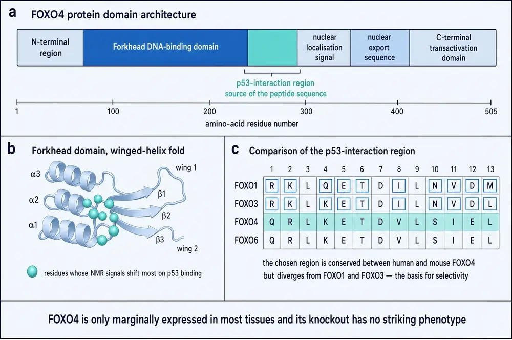
Having chosen the protein, the designers then had to choose a stretch of it — a region involved in the interaction they wanted to break, conserved between mouse and human so that the mouse experiments would mean something, and different enough from the same region in FOXO1 and FOXO3 that a molecule mimicking it would not blunder into those proteins instead.
The stretch they settled on lies in that N-terminal part of the Forkhead domain. Mapping it precisely is possible from the published sequence, and doing so is a useful check on the whole design: read the peptide back into ordinary orientation and it corresponds, residue for residue, to positions 99 to 124 of human FOXO4. The molecule is a literal excerpt from the protein it was built to displace.
03The mirror trick
There is an obvious problem with using a fragment of a protein as a drug. Bodies are full of proteases — enzymes whose entire function is to find peptide bonds and cut them. A 26-residue peptide injected into a mouse has a short and uneventful career.
The solution used here is old, elegant, and depends on a fact about chemistry that has no obvious reason to be useful. Amino acids are chiral: each one exists in two mirror-image forms, conventionally L and D, which cannot be superimposed on one another any more than a left hand can be laid onto a right. Life on Earth builds proteins almost exclusively from L-amino acids, and proteases — being themselves made of L-amino acids, and having evolved to cut L-backbones — are stereospecific. Hand them a peptide built entirely from D-amino acids and they do not recognise it as food.
Simply mirroring a peptide, however, destroys it functionally as well as chemically: the side chains that do the actual binding end up pointing the wrong way. The retro-inverso construction is the fix. Build the molecule from D-amino acids and reverse the order of the sequence, and the two inversions cancel where it matters. The backbone now runs in the opposite direction, and its hydrogen-bond donors and acceptors are swapped — but the side chains, read left to right in space, sit approximately where they sat in the original. The topology that does the binding survives; the chemistry that invited digestion does not.
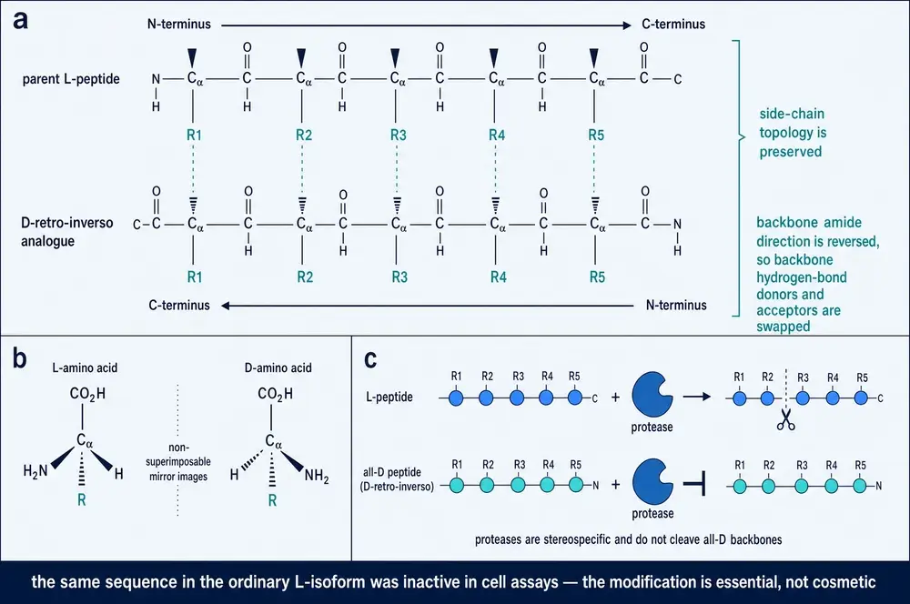
The last point in that caption deserves emphasis, because it is the kind of control that separates a real result from a hopeful one. The same 26 residues, in the same order, synthesised as an ordinary L-peptide, did nothing to senescent cells. The stereochemistry is not a formulation convenience bolted on to improve shelf life. It is load-bearing.
The team also had a precedent to point at. D-retro-inverso peptides were not exotic in 2017: one had gone through a double-blind, randomised, placebo-controlled Phase IIb trial and another through a Phase I trial in solid tumours, both reported as tolerated (Baar et al., 2017, citing Suckfüll et al., 2014 and Warso et al., 2013). Those trials involved entirely different peptides addressing entirely different targets. They establish that the chemistry can be taken into people; they say nothing whatever about this molecule.
04The molecule itself
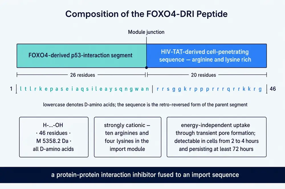
The finished construct is two things welded together. The first 26 positions are the FOXO4 excerpt described above — the part that does the work. The remaining 20 are a cell-penetrating sequence derived from HIV-TAT, a short, intensely positively charged stretch that carries cargo across cell membranes. Without it the peptide would be a competent inhibitor of an interaction it could never reach, since the target sits inside the nucleus and peptides do not ordinarily cross membranes at all.
Read the sequence in the figure and the division is visible without any chemistry: the first stretch is a mixed, ordinary-looking peptide sequence, while the tail is a run of r and k — ten arginines and four lysines in twenty positions. That is the import module, and its charge is the mechanism. It does not bind a receptor or use a transporter; it interacts with the membrane directly and forms transient pores, which is why uptake is energy-independent.
The molecule as a whole is 46 residues and 5358.2 daltons. That is large for something called a peptide and small for something called a protein, and it sits in an awkward pharmacological middle: too big to behave like a conventional small-molecule drug, too small and too charged to behave like an antibody. Section 21 returns to what that means in practice, which is that almost nothing is known about where this compound goes in a body or how long it stays there.
05The cell that stops but does not leave
A cell that suffers damage it cannot repair has, in principle, two decent options. It can die, which is tidy and is what apoptosis is for. Or it can stop dividing permanently, which prevents it passing broken DNA to its descendants and is the reason senescence exists as a tumour-suppressive mechanism. Both options protect the organism.
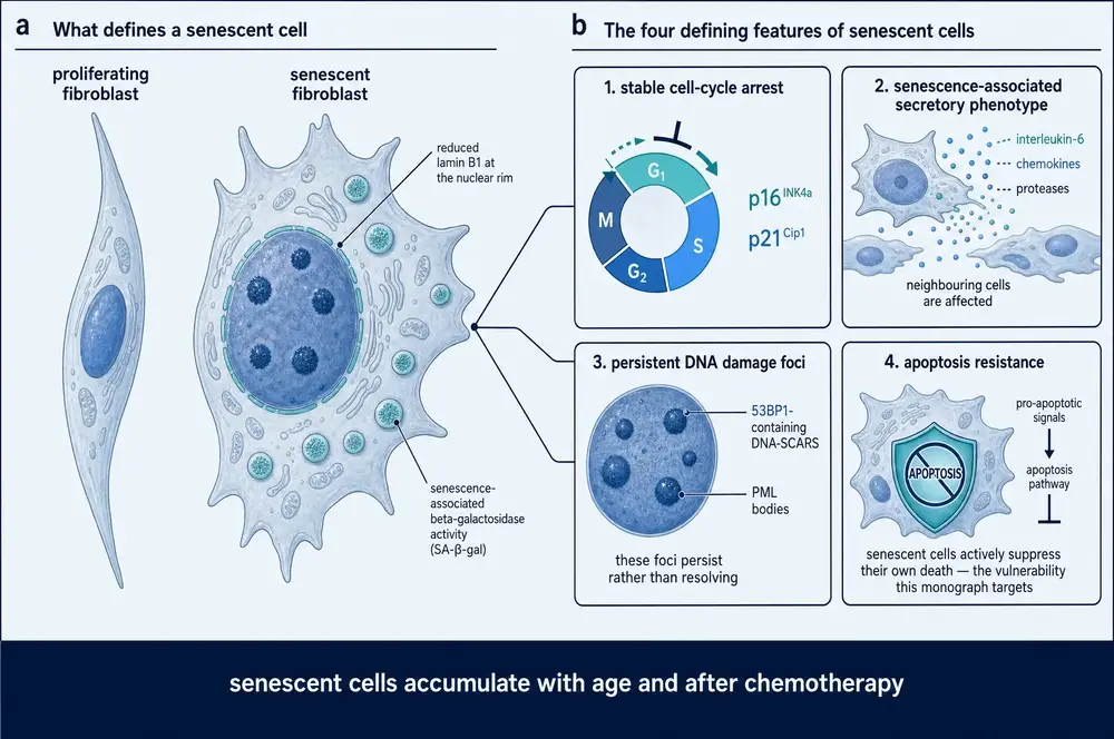
The difficulty is what happens next. A cell that dies is cleared and replaced. A cell that becomes senescent stays where it is, sometimes for the rest of the animal's life, and it is not inert. It swells. Its lysosomes proliferate, which is why the standard stain for senescence detects β-galactosidase activity at pH 6. Lamin B1 falls at the nuclear rim. It bolts the cell cycle shut using the inhibitors p16INK4a and p21Cip1. And it begins to secrete — interleukin-6, chemokines, matrix-degrading proteases — a programme collectively named the senescence-associated secretory phenotype, or SASP.
The SASP is the reason senescent cells are a problem rather than merely a curiosity. A senescent cell exports inflammation to its neighbours. A small number of them can degrade the function of a tissue far out of proportion to their abundance, and they accumulate with age and after chemotherapy. This is the observation the entire senolytic field is built on: that clearing these cells from mice — originally by genetic tricks rather than drugs — delays multiple features of ageing at once.
06Apoptosis resistance is the vulnerability
The fourth feature in that figure is the one that matters here, and it is the least intuitive. Senescent cells are not passively surviving. They are actively suppressing their own death, and they are doing it while carrying signals that ought to kill them.
Baar and colleagues established this directly rather than assuming it. In irradiated human IMR90 fibroblasts — a standard lung fibroblast line, made senescent by ionising radiation — they found the pro-apoptotic proteins PUMA and BIM elevated and the anti-apoptotic guardian BCL-2 reduced. In their own phrasing, senescent cells appear primed to undergo apoptosis, but the execution of the death program is restrained (Baar et al., 2017a).
That framing is the whole strategy. If a senescent cell were simply robust, killing it would require force, and force is not selective. But a cell holding its foot on the brake of a running engine can be killed by releasing the brake — and the brake is a thing that healthy cells are not using. Selectivity comes free, provided the brake is correctly identified.
So the question became: what is the brake? The team went looking among transcription factors previously linked to apoptosis — STAT1, STAT2 and STAT4, RELB, NF-κB, TP53 and FOXO4. Several could be eliminated on prior evidence: interfering with JAK-STAT signalling was known not to affect senescent cell viability, and the same group had previously seen comparable results for NF-κB and p53 inhibition. That left the one candidate nobody had examined in this role, which was FOXO4.
The elimination step above is doing more work than it appears to. FOXO4 was not the strongest hit in an unbiased screen; it was the survivor of a short candidate list after the alternatives were ruled out on the basis of previously published work, some of it the same group's. That is a legitimate way to arrive at a hypothesis, and the experiments that followed test the hypothesis properly. It is worth stating plainly, though, because it bears on a question Part Five returns to: whether FOXO4 is the mechanism of senescent-cell apoptosis resistance, or one of several.
07The nuclear complex that keeps the cell alive
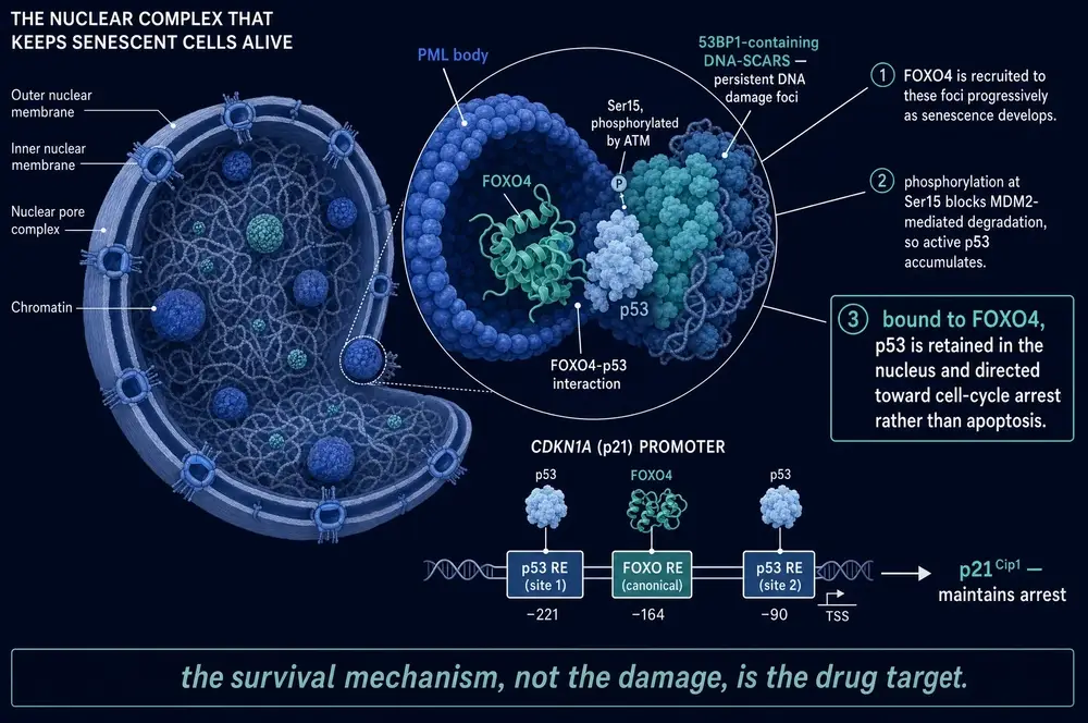
What FOXO4 does in a senescent cell is hold on to p53.
p53 is the most-studied tumour suppressor in biology and its reputation is as an executioner: DNA damage activates it, and activated p53 either arrests the cell cycle or triggers apoptosis. Which of the two it chooses is context-dependent, and in a senescent cell the choice has already been made and locked in. The mechanism is spatial.
As senescence develops, promyelocytic leukaemia (PML) bodies in the nucleus fuse with persistent DNA-damage foci containing 53BP1 — structures known as DNA-SCARS, which unlike ordinary damage foci never resolve. FOXO4 is progressively recruited into these structures. Inside them it binds p53 that has been phosphorylated on serine 15 by the damage-response kinase ATM, a modification that blocks MDM2-mediated degradation so that active p53 accumulates instead of being cleared away.
Bound to FOXO4, that p53 is held in the nucleus. It is not inactive — it is transcriptionally engaged, and the promoter of CDKN1A, the gene encoding p21Cip1, contains a canonical FOXO response element flanked by two p53 binding sites, an arrangement that lets the pair sustain the arrest programme together. What that p53 is not doing is the other thing p53 can do, which is leave the nucleus and kill the cell from the mitochondria.
This is the complete target definition, and it is unusually clean. A protein–protein interaction, in a defined subnuclear compartment, present in senescent cells and largely absent from healthy ones, holding a known executioner in a place where it cannot execute. Break the interaction and the prediction is specific: p53 should leave the nucleus, reach the mitochondria, and kill the cell — and only in cells where the complex existed in the first place.
Part Three is the test of that prediction.
08Rotterdam, and the road to 2017
Peter de Keizer had been circling this problem for seven years before the peptide existed.
In 2010, working on how cancer-causing mutations force cells into permanent arrest, he and colleagues showed that the BRAF V600E oncogene — the mutation found in most melanomas — drives senescence through FOXO4. The route ran through reactive oxygen species and JNK-mediated phosphorylation of FOXO4, and it produced arrest via p21Cip1 rather than through the more familiar p16INK4a. The paper closed on a framing that reads, in retrospect, like a statement of intent: that FOXO proteins mediate a trade-off between cancer and ageing (de Keizer et al., 2010).
That is the intellectual thread. FOXO4 was already, in this laboratory's hands, the protein that connected oncogene-induced arrest to p21 and therefore to ageing. What changed between 2010 and 2017 was the field around it. Between those dates, work elsewhere — most influentially from Judith Campisi's group and from the Mayo Clinic — established that senescent cells could be cleared from mice genetically, and that doing so delayed multiple age-related pathologies. The proof of principle existed. What did not exist was a molecule.
The 2017 paper is the meeting of those two lines. Its author list reads like a map of the problem: Marjolein Baar as first author, with de Keizer as senior author at Erasmus MC in Rotterdam; Tobias Madl's group supplying the nuclear magnetic resonance that proved the peptide competes for p53; Jan Hoeijmakers, whose long work on DNA-repair-deficient mice supplied the XpdTTD/TTD fast-ageing model; and Judith Campisi, whose laboratory had defined much of what a senescent cell is (Baar et al., 2017a).
It is worth recording that the discoverers were more restrained about the result than the coverage of it was. In a commentary published later the same year, Baar, van Willigenburg and de Keizer set out the finding and then immediately noted that more research was needed to establish its translational potential — and, pointedly, whether removing senescent cells is even safe (Baar et al., 2017b). Section 17 shows that this was not routine throat-clearing.
09Competition, measured
The first thing to establish was that the peptide does the one thing it was designed to do: compete with FOXO4 for p53.
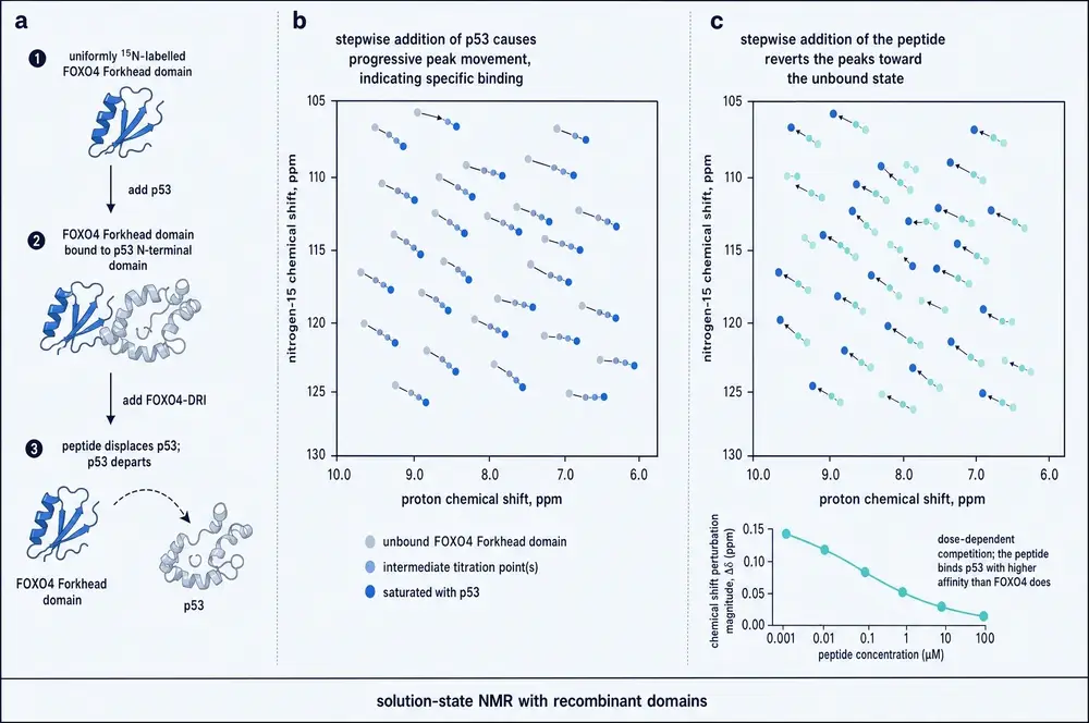
This was tested by nuclear magnetic resonance, which is the right instrument for the question because it reports on individual atoms rather than on bulk binding. A uniformly 15N-labelled FOXO4 Forkhead domain gives a two-dimensional spectrum in which each amide in the protein produces its own cross peak. Add a binding partner and the peaks belonging to residues at the interface move — a chemical shift perturbation. Which peaks move tells you where the contact is; how far they move tells you how much is bound.
Titrating in the N-terminal domain of p53 (residues 1–312) produced progressive, specific shifts, confirming the interaction. Then, adding the peptide to that preformed complex drove the same peaks back toward their unbound positions — dose-dependently, and to a degree indicating that the peptide engages p53 with higher affinity than FOXO4 itself does (Baar et al., 2017a).
Two things follow from this experiment that are easy to miss. The first is that the peptide binds p53, not FOXO4 — it is a mimic of the FOXO4 surface, so it occupies the site on p53 that FOXO4 would otherwise occupy. The second is that it does so better than the native protein, which is not a given for a fragment removed from its structural context, and which is largely what makes the strategy work at achievable concentrations.
10What happens inside the cell
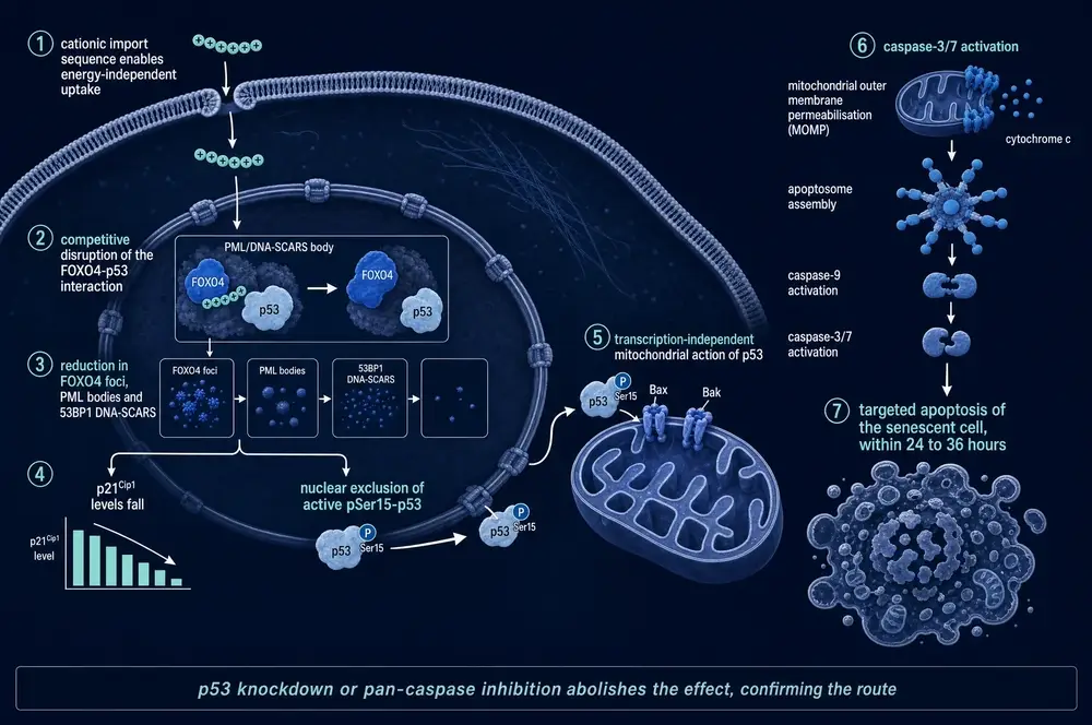
Displacing a protein in a test tube is not the same as killing a cell, and the chain between the two was established step by step.
Uptake first: using an antibody against the HIV-TAT tag, the peptide was detectable inside cells 2–4 hours after administration and remained detectable for at least 72 hours. Then the structures: the peptide reduced the number of senescence-induced FOXO4 foci, PML bodies and 53BP1 DNA-SCARS — while leaving small 53BP1 foci unaffected, which is the control that shows it is dispersing the senescence-associated assemblies specifically rather than flattening DNA-damage signalling in general.
Then the consequences. p21Cip1 fell. Active serine-15 phosphorylated p53 accumulated and was excluded from the nucleus. And cells died — on a timescale of 24 to 36 hours, by real-time density measurement (Baar et al., 2017a).
11Selectivity, and the controls that matter
A compound that kills senescent cells is only interesting if it spares the others, and this is where the founding paper is at its strongest — not because the selectivity figure is large, but because of what was done to rule out the alternative explanations.
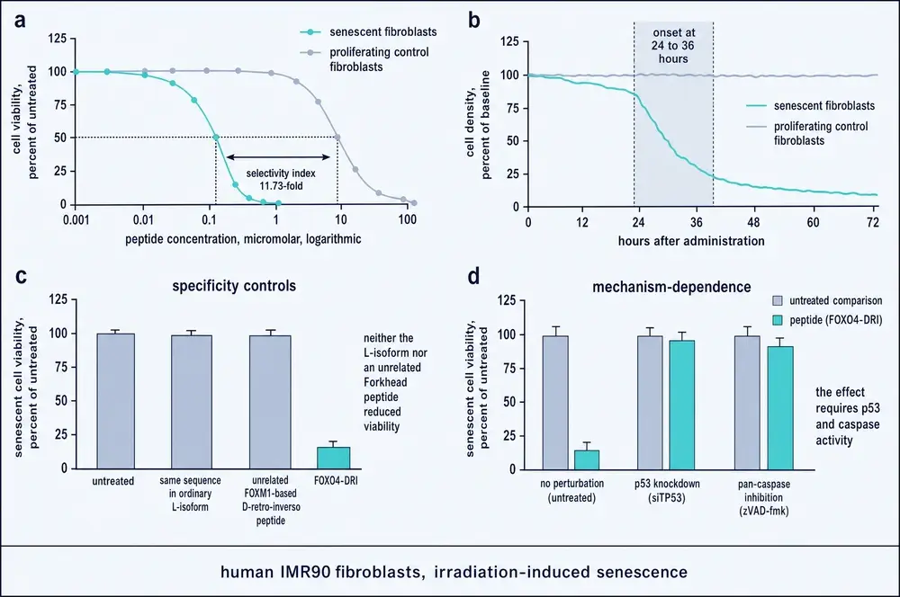
The headline number is a selectivity index of 11.73-fold: senescent IMR90 fibroblasts lost viability at roughly an order of magnitude lower concentration than proliferating controls. The index is defined as the ratio of the half-maximal effect concentrations between the two populations, and it is a ratio rather than an absolute margin — a distinction that matters when extrapolating, and one this document returns to.
The controls are the substantive part. Two of them are decisive:
- The same sequence as an ordinary L-peptide did nothing. This eliminates the possibility that the effect comes from the sequence's charge, its bulk, or its cell-penetrating tail, and locates it in the stereochemistry-dependent binding.
- An unrelated D-retro-inverso peptide based on FOXM1 — a different Forkhead protein, built the same way — also did nothing. This eliminates the possibility that D-retro-inverso peptides are simply toxic to senescent cells as a class.
Two further experiments tested the mechanism rather than the selectivity. Stable knockdown of p53 abolished the killing. So did pan-caspase inhibition. Together they establish that the compound works through p53 and through caspase-mediated intrinsic apoptosis, and not through some non-specific membrane toxicity that the cationic tail might plausibly have caused (Baar et al., 2017a).
12Three mouse models, and a claim about restoration
The animal work is where the paper made its reputation, and its design carries an argument.
Three models were used. In the first, mice were given doxorubicin, an anthracycline chemotherapy that induces senescence throughout the body and produces a characteristic syndrome of malaise, liver and kidney injury. In the second, XpdTTD/TTD mice — carrying a DNA-repair defect that produces accelerated ageing — were treated as they declined. In the third, ordinary mice were allowed to grow genuinely old.
Across all three, intermittent dosing improved the readouts: body condition and liver and kidney injury markers in the chemotoxicity model; fur density, renal function measured by blood urea nitrogen and creatinine, and spontaneous activity in both the fast-ageing and the naturally aged animals. The treatment was reported as well tolerated under the conditions used (Baar et al., 2017a).
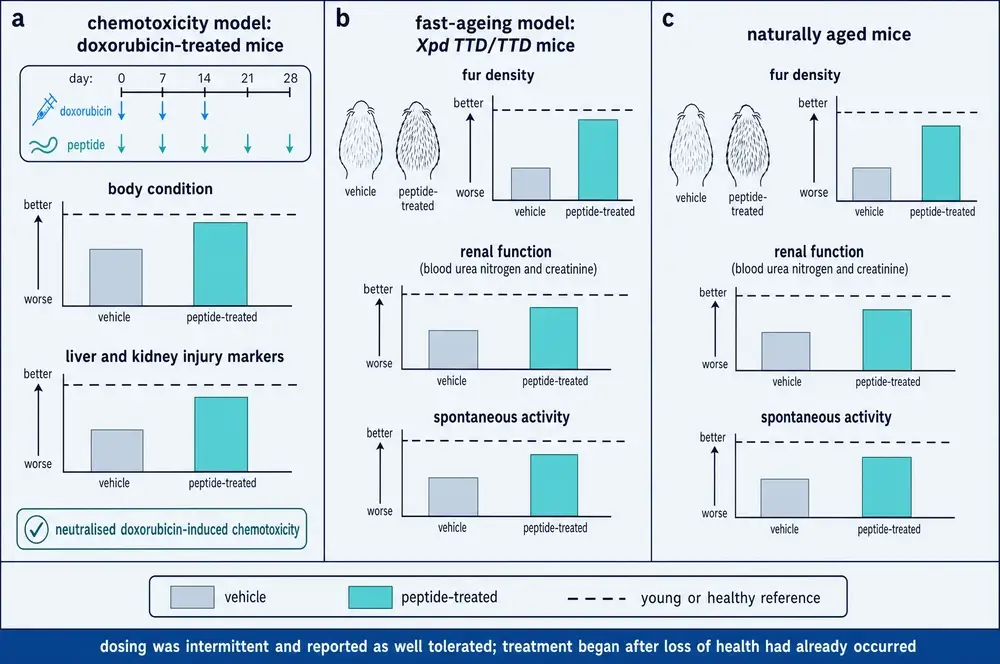
The design argument is in the timing. Earlier senescent-cell-clearance experiments had largely been preventive: clear the cells as they arise and the animal ages more slowly. Here, treatment began after health had already been lost. That is a different and much stronger claim — not that decline can be postponed, but that some of it can be reversed — and it is the reason the paper landed as hard as it did. A commentary in the same issue of Cell framed the result as rejuvenation by therapeutic elimination of senescent cells (Krimpenfort & Berns, 2017).
Three cautions belong here rather than later. The readouts are gross physiological measures — how much fur, how much movement, what the blood chemistry looks like — and not survival; the paper does not demonstrate extended lifespan. The XpdTTD/TTD model is a DNA-repair mutant, and its resemblance to ordinary human ageing is a matter of ongoing argument rather than a settled fact. And every result in this section is from mice.
13The structure got clearer, and more complicated
The 2017 paper proved competition without solving a structure. The NMR showed that peaks moved; it did not show what the complex looked like. Filling that in took most of a decade, and the picture that emerged is messier than the original schematic — in a way that matters for anyone trying to improve on the molecule.
Three findings define the current state. First, the interaction is not a single tidy contact. Mandal and colleagues showed that while the contact between the p53 transactivation domain and the FOXO4 Forkhead domain is essential to the complex's stability, additional contacts — involving FOXO4's N-terminal segment, p53's C-terminal regulatory domain, and the DNA-binding domains of both proteins — help stabilise it. They also established a functional consequence the original model did not predict: forming the complex blocks p53 from binding DNA, without impairing FOXO4's own DNA binding (Mandal et al., 2022).
Second, the interaction is structurally plastic rather than fixed. Kohoutova and colleagues found the contacts highly heterogeneous: the p53 transactivation domain engages the N-terminal helical bundle of the FOXO4 Forkhead domain but retains substantial flexibility within the complex, and simulations driven by the NMR data suggest multiple binding modes rather than one (Kohoutova et al., 2025).
Third — and this is the most useful result for drug design — Bourgeois and colleagues solved solution NMR structural models of the p53 transactivation domain bound both to the FOXO4 Forkhead domain and to the peptide itself. Two things came out of it. The peptide and its target are both intrinsically disordered, and they form a transiently folded complex — a structure that exists but does not sit still. And the cationic cell-permeability tail, which had been treated as an inert delivery module, turns out to contribute to the interaction itself. They also found that phosphorylating p53 increases its affinity for both FOXO4 and the peptide, which supplies a mechanistic reason for the selectivity: the senescent cell is precisely where phosphorylated p53 accumulates (Bourgeois et al., 2025).
That last point is worth pausing on. The design assumed a clean division of labour — a targeting module and a delivery module. The structural work says the division is not clean. Anyone shortening the tail to reduce charge is also altering the binding surface.
14Successor peptides
If the mechanism is real, better molecules should be derivable from it, and two groups have derived them.
Le and colleagues used molecular modelling to design a series of peptides mimicking FOXO4, and characterised one, ES2, in detail. It disrupts FOXO4–p53 foci, activates p53-mediated apoptosis, and — unlike the parent compound — binds FOXO4 preferentially over p53. In mouse work it eliminated senescent human cancer cells; intratumoural delivery of ES2 with a BRAF inhibitor increased apoptosis and conferred a survival advantage in melanoma models; and repeated systemic delivery to older mice reduced senescent cell numbers in the liver with what the authors describe as minimal toxicity (Le et al., 2021). A commentary in the same journal placed this work in the context of the senolytic field's shift from broad anti-apoptotic inhibitors toward precise interface disruption (Tripathi et al., 2021).
Kang and colleagues took the structural route. Using NMR to identify which part of the p53 transactivation domain matters — hydrophobic contacts — they designed an optimised inhibitor with reduced negative charge, attached a cationic cell-penetrating peptide, and produced CPP-CAND. It is shorter than the parent compound, built from ordinary L-amino acids, and reported as more selective; it disrupted nuclear FOXO4–p53 foci and induced caspase-dependent apoptosis in senescent cancer cells produced by several different chemotherapies (Kang et al., 2025).
The strategic significance of CPP-CAND is that it is an L-peptide. If a conventional peptide can be made to work, the D-retro-inverso construction — expensive to synthesise, and unusual enough to complicate regulatory paths — may turn out to have been a step on the way rather than a destination.
15Tissue by tissue
The most persuasive thing about the FOXO4-DRI literature is not any single follow-up study. It is that independent groups, working on unrelated organs with unrelated motivations, kept picking the compound up as a tool and kept getting a version of the same answer.
| Tissue / setting | Model | Principal finding | Source |
|---|---|---|---|
| Testis — Leydig cells | Senescent TM3 cells; naturally aged mice | Selective apoptosis of senescent Leydig cells; improved testicular microenvironment and age-related testosterone insufficiency | Zhang et al., 2020 |
| Testis — spermatogenesis | Aged mice | Reduced SASP secretion from Leydig cells; improved sperm quality and spermatogenesis | Li et al., 2024 |
| Cartilage | Human chondrocytes expanded in vitro | Removed over half the cells at high passage while sparing low-passage cells; reduced senescence markers, but did not improve chondrogenic potential | Huang et al., 2021 |
| Lung — fibrosis | Bleomycin-induced mouse model | Reduced senescent cells and SASP; attenuated collagen deposition, comparable to pirfenidone; preferentially killed TGF-β-induced myofibroblasts | Han et al., 2022 |
| Lung — fibrosis (independent) | Bleomycin-induced mouse model | Milder pathology and less collagen; redistributed nuclear p53 and reduced total extracellular matrix protein content | Liu et al., 2023 |
| Lung — cancer and radiotherapy | NSCLC cells and mouse models | Killed senescence-like cancer-associated fibroblasts, radiosensitising tumours while reducing radiation-induced pulmonary fibrosis | Meng et al., 2021 |
| Skin — keloid | Human keloid organ cultures and fibroblasts | Promoted apoptosis and reduced G0/G1 fraction, with nuclear exclusion of pSer15-p53 — the closest approach to human tissue in this corpus | Kong et al., 2025 |
| Vasculature — endothelium | Aged and progeroid mice; oxygen-glucose-deprived endothelial cells | Selective apoptosis of senescent endothelial cells via p53/BCL-2/caspase-3; improved vascular function | Hu et al., 2025 |
| Mesenchymal stem cells | Human umbilical cord MSCs | FOXO4 suppression (by shRNA, not the peptide) promoted apoptosis of senescent cells and reduced IL-6 — independent support for the target rather than the drug | Wu et al., 2020 |
Read that table for its shape rather than its contents. Nine settings, at least seven independent groups, three continents, and two of the entries — the bleomycin lung-fibrosis studies from Han and from Liu — are straightforward independent replications of one another. The mechanism travels. Wherever a tissue accumulates senescent cells and someone applies this compound, senescent cells die and something measurable improves.
Two entries are more informative than the rest. The chondrocyte study is the only one in which the compound did what it was supposed to do and the downstream benefit did not follow: it removed senescent cells and reduced senescence markers, but the cartilage those cells went on to make was not better for it (Huang et al., 2021). Removing senescent cells is not automatically the same as restoring function. And the keloid work is the nearest this literature comes to a human system — not a trial, but human tissue in organ culture rather than a mouse (Kong et al., 2025).
16Upstream: why FOXO4 is there in the first place
A separate line of work has been asking not what FOXO4 does in senescent cells but why there is so much of it, and the answer arrived in 2026.
Sun and colleagues showed that FOXO4 protein is stabilised in senescent cells by reduced proteasomal degradation, and identified the machinery: the deubiquitinase USP37 binds FOXO4 directly and strips K48-linked polyubiquitin chains from it. Upstream of that, persistent DNA-damage signalling activates the kinase CHK2, which phosphorylates USP37 and strengthens its binding to FOXO4. Removing USP37 destabilises FOXO4 and sensitises senescent cells to apoptosis — an effect rescued by re-expressing FOXO4 (Sun et al., 2026).
This is significant for two reasons. It supplies the missing link between the damage that causes senescence and the survival programme that sustains it: the same persistent DNA-damage response that will not resolve is what keeps FOXO4 elevated. And it independently validates the target by a completely different route — genetic rather than pharmacological, upstream rather than at the interface. When knocking down the enzyme that stabilises a protein reproduces the effect of a drug that blocks that protein's interaction, the target is probably real.
Related work has been mapping FOXO4's regulation more broadly. A 2026 structured review classified the FOXO4 evidence base by how directly it supports FOXO4 specifically, as opposed to being extrapolated from FOXO1 or FOXO3, and found the strongest FOXO4-specific support for three axes: cysteine-dependent redox sensing, stress-regulated nuclear trafficking, and FOXO4–p53-mediated survival of senescent cells. Notably, several antioxidant functions routinely attributed to FOXO4 were found to rest on evidence from its paralogues rather than on direct measurement (Mateescu et al., 2026). The third of those axes is the one this monograph is about, and it is the best-supported.
17The result that points the other way
In 2023, a study in Circulation reported that eliminating senescent cells can promote pulmonary hypertension.
Born and colleagues examined senescence markers in patients with pulmonary arterial hypertension and found them elevated: p16, p21 and the DNA-damage markers γ-H2AX and 53BP1, with vascular cells co-staining for several at once. So far this is the standard senolytic rationale — disease tissue is full of senescent cells, so clear them.
They then cleared them, four different ways: a suicide-gene construct driven by the p16 promoter, the BCL-2-family inhibitor ABT263, genetic inactivation of p16, and FOXO4-DRI. In mouse models of pulmonary hypertension, elimination of senescent cells made the disease worse rather than better (Born et al., 2023).
It would be easy to set this aside as one contrary finding among many positive ones. Three features make that unsafe. The effect was reproduced across four mechanistically unrelated methods of clearance, including two genetic ones, so it is a property of removing the cells rather than an off-target effect of any compound. Roughly 30 per cent of lung senescent cells in these animals were pulmonary endothelial cells — a population whose loss is plausibly harmful. And the finding is directionally consistent with what senescence is for: these cells arise as a protective response, and in some tissues they may still be performing that function when a drug removes them.
The correct reading is not that senolytics are dangerous, nor that this study is wrong. It is that the benefit of clearing senescent cells is tissue- and context-dependent, and that the compound's selectivity for senescent cells — which is its principal virtue — provides no protection whatever against the possibility that those cells were needed.
Set against this, the same period produced the field's most enthusiastic claims. A 2026 review reports that in aged mammalian models the compound reduces senescent cell accumulation, restores cerebral blood flow and blood-brain-barrier integrity, reverses hippocampal atrophy and improves cognition, and that in Alzheimer's and tauopathy models it clears amyloid-β and pathological tau (Alameen et al., 2026). Those are substantial claims and they are made in a review rather than a primary report; this monograph has not been able to trace each of them to a primary source within its corpus, and they should be treated as the review's summary rather than as established results. The same review notes preliminary human work on “FOXO4-axis modulators” — but the example it gives is high-dose fisetin, a flavonoid, not this peptide. That distinction is easy to lose and important to keep.
18Where it sits among the senolytics
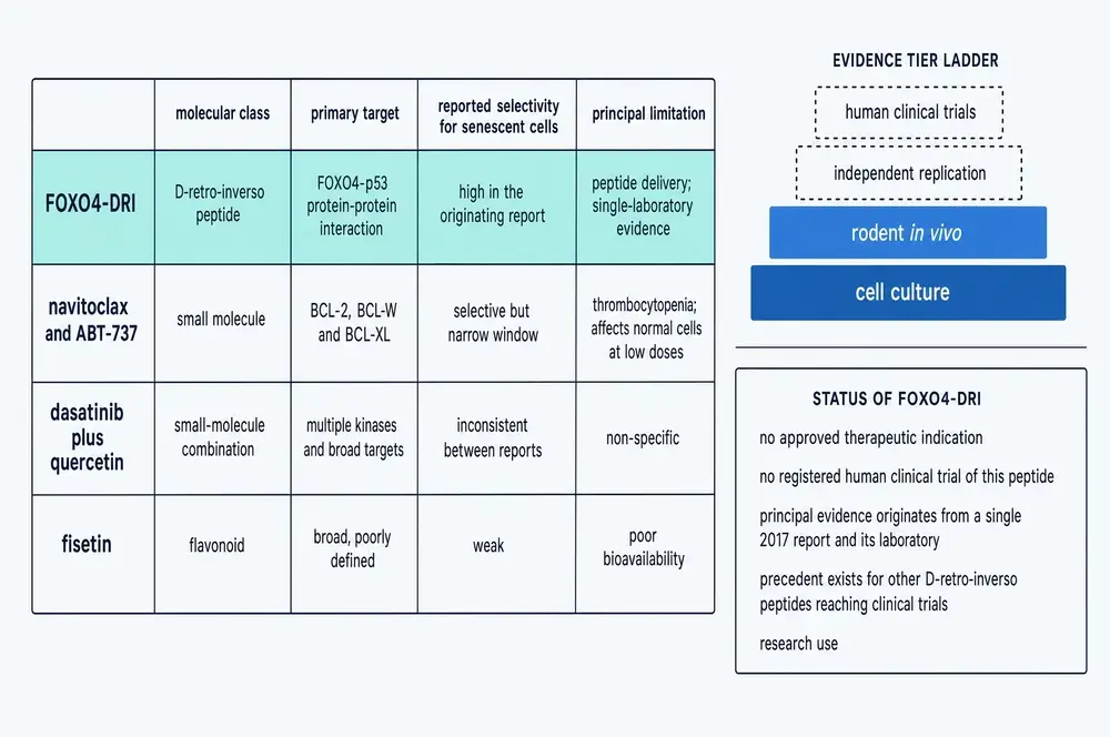
FOXO4-DRI is not the only compound that kills senescent cells, and comparing it with the alternatives clarifies both what is distinctive about it and what is unproven.
The other senolytics in common use work by blunter means. Navitoclax and ABT-737 inhibit the anti-apoptotic BCL-2 family — BCL-2, BCL-W and BCL-XL — which senescent cells lean on to stay alive. They are effective and they are selective, but the window is narrow: BCL-XL inhibition causes thrombocytopenia, because platelets depend on it too. The combination of dasatinib and quercetin, the most widely studied senolytic regimen, hits multiple kinases and a broad range of targets, and its selectivity varies between reports. Fisetin, a flavonoid, is weak and poorly bioavailable.
Against that field, this compound's distinguishing feature is the nature of its target. It is the only entry that works by blocking a defined protein–protein interaction rather than by inhibiting an anti-apoptotic protein or by broad-spectrum activity. That is what makes its mechanism legible — and it is also why the failure mode described in section 17 is instructive, because a precisely selective compound is still only as safe as the assumption that the cells it selects for should be removed.
19The shape of the literature
One useful way to judge a compound's evidence base is to look at the literature's shape rather than its contents — how much there is, when it appeared, and what kind of documents it consists of.
Two features of that chart matter. The first is that the sustained activity after 2017 is real — this is not a compound that produced one striking paper and was forgotten. The second is that the total is small. Seventy-three indexed records across a quarter of a century is a modest literature by the standards of a serious drug candidate, and a substantial share of it is review.
The body-text sweep sharpens that point. Searching the full-text database rather than titles and abstracts found 363 articles that name this compound somewhere in their text. Classifying each by how substantively it uses the compound gives the real distribution: 8 papers name it in their Methods, that is, actually ran experiments with it; 5 more are experimental papers that discuss it; 65 are reviews that discuss it substantively; and 191 — slightly over half — mention it once or twice in passing, typically in a table of senolytic agents. The compound is talked about an order of magnitude more often than it is used.
20What is solid, and what is thin
Weighing this evidence base means separating claims that rest on measurements from claims that rest on inference, and being explicit about which kind of system produced each.
What is solid. The molecular mechanism is as well established as almost anything in this field. The interaction exists, its structure has been solved by three independent groups, the peptide competes for it, and the downstream requirement for p53 and for caspases has been demonstrated by knockdown and by inhibition. The senolytic effect in cell culture is robust and has been reproduced in at least nine tissue settings by independent laboratories. The target has been validated a second time by a completely different route, through the CHK2–USP37 stabilisation axis.
What is thin. Everything downstream of the mouse. There is no pharmacokinetic profile — no half-life, no clearance figure, no tissue-distribution study for this compound anywhere in this corpus. The in-vivo benefit is measured on gross functional readouts over weeks, not on survival. The selectivity index of 11.73-fold is a ratio between two cultured cell populations under one senescence-induction protocol, and there is no reason to assume it transfers to a whole animal with many cell types and many routes of senescence. And the founding in-vivo result — the restoration claim that made the compound's reputation — has not been repeated in its original form by an independent laboratory.
What is contested. Whether clearing senescent cells is beneficial in every tissue. Section 17 sets out the case that it is not.
21Delivery, and the pharmacology that does not exist
The practical obstacle to this compound is not its mechanism. It is that it is a 46-residue, highly cationic peptide.
Peptides of that size are not orally available; they are digested. The D-retro-inverso construction solves proteolysis but not absorption, and it solves nothing at all about distribution. Cationic cell-penetrating sequences enter cells efficiently, but they enter all cells efficiently — the selectivity of this compound is generated after entry, by whether the target complex is present, not by where the peptide goes. That is an elegant design and a demanding one, because it means the whole body is exposed and the margin depends entirely on the intracellular discrimination holding up.
The successor peptides described in section 14 are attempts on exactly this problem — shorter, less charged, ordinary L-amino acids — and that is the clearest signal of where the field thinks the difficulty lies.
It should be said plainly that the absence of pharmacokinetic data is not a gap this monograph failed to find. It is a gap in the literature. A compound with nine years of publication history, an established mechanism and reproducible activity across a dozen tissues has never, so far as this corpus shows, had its disposition in a living body characterised.
Nothing in this document recommends the use of FOXO4-DRI in any person. No human dose, route, schedule or duration is stated or implied anywhere in it, and none can be derived from what is reported here. Every quantitative parameter given is a description of a published experiment in cells, in tissue, or in mice.
As of 1 August 2026, this compound has no approved therapeutic indication in any jurisdiction, and a direct query of ClinicalTrials.gov returns no registered human clinical trial of it under any of its designations. The human safety of removing senescent cells from any tissue is unestablished, and at least one line of evidence indicates that in the pulmonary vasculature it is actively harmful. This monograph describes published research. It is not medical advice.
22What would change the picture
It is worth ending on what is missing rather than on what is known, because the list is short and specific.
A pharmacokinetic study would change the most. Half-life, clearance and tissue distribution in any species would convert an interesting mechanism into a candidate that could be reasoned about. An independent replication of the restoration experiment — the three-model in-vivo work of 2017, repeated in another laboratory — would settle whether the founding result is robust or a single institution's finding. A lifespan study would test the claim the field is actually interested in, as opposed to the functional proxies that stand in for it. And a systematic map of which tissues benefit from senescent-cell clearance and which are harmed by it would tell us whether the pulmonary hypertension result is a special case or the first of several.
None of those is exotic. All of them are ordinary next steps that have not been taken in nine years, and the reason they have not is the most informative fact in this document: the compound is easy to buy, easy to use as a laboratory tool, and has no owner with an interest in developing it. It has been adopted as a reagent rather than pursued as a drug.
23References
Generated from verified NCBI records rather than from recall. Author lists, journal names, volumes, pages and identifiers are taken from the PubMed record for each citation, and the build refuses to run if any identifier fails to resolve. This safeguard exists because it has been needed: in two earlier monographs in this series, reference lists drafted from memory contained identifiers that pointed at real but unrelated papers. It was needed again here — three citations in the first draft of this document named the wrong paper, and the generator caught all three.
- Alameen AAM, Al-Kuraishy HM, Fawzy MN, Batiha GE. Targeting the FOXO4-p53 axis by retro-inverso peptide senolytic agents: a pharmacological strategy to mitigate brain aging and cognitive decline. Naunyn-Schmiedeberg's archives of pharmacology. 2026;399(10):14659-14676.
PMID 42024235 · doi:10.1007/s00210-026-05309-6 - Asadi Y, Moundounga RK, Chakroborty A, Pokokiri A, Wang H. FOXOs and their roles in acute and chronic neurological disorders. Front Mol Biosci. 2025;12:1538472.
PMID 40260403 · doi:10.3389/fmolb.2025.1538472 · PMC12010098 - Baar MP, Brandt RMC, Putavet DA, Klein JDD, Derks KWJ, Bourgeois BRM, et al.. Targeted Apoptosis of Senescent Cells Restores Tissue Homeostasis in Response to Chemotoxicity and Aging. Cell. 2017a;169(1):132-147.e16.
PMID 28340339 · doi:10.1016/j.cell.2017.02.031 · PMC5556182 - Baar MP, Van Willigenburg H, de Keizer PLJ. Maintenance and repair of an aging life cycle. Oncotarget. 2017b;8(50):86985-86986.
PMID 29152057 · doi:10.18632/oncotarget.18046 · PMC5675609 - Born E, Lipskaia L, Breau M, Houssaini A, Beaulieu D, Marcos E, et al.. Eliminating Senescent Cells Can Promote Pulmonary Hypertension Development and Progression. Circulation. 2023;147(8):650-666.
PMID 36515093 · doi:10.1161/CIRCULATIONAHA.122.058794 - Bourgeois B, Madl T. Regulation of cellular senescence via the FOXO4-p53 axis. FEBS letters. 2018;592(12):2083-2097.
PMID 29683489 · doi:10.1002/1873-3468.13057 · PMC6033032 - Bourgeois B, Spreitzer E, Platero-Rochart D, Paar M, Zhou Q, Usluer S, et al.. The disordered p53 transactivation domain is the target of FOXO4 and the senolytic compound FOXO4-DRI. Nature communications. 2025;16(1):5672.
PMID 40593617 · doi:10.1038/s41467-025-60844-9 · PMC12216184 - de Keizer PL, Packer LM, Szypowska AA, Riedl-Polderman PE, van den Broek NJ, de Bruin A, et al.. Activation of forkhead box O transcription factors by oncogenic BRAF promotes p21cip1-dependent senescence. Cancer research. 2010;70(21):8526-36.
PMID 20959475 · doi:10.1158/0008-5472.CAN-10-1563 · PMC2989643 - Han X, Yuan T, Zhang J, Shi Y, Li D, Dong Y, et al.. FOXO4 peptide targets myofibroblast ameliorates bleomycin-induced pulmonary fibrosis in mice through ECM-receptor interaction pathway. Journal of cellular and molecular medicine. 2022;26(11):3269-3280.
PMID 35510614 · doi:10.1111/jcmm.17333 · PMC9170815 - Hu Z, Li F, Hu C, Shan Q, Tang Z, Jiang M, et al.. FOXO4-DRI regulates endothelial cell senescence via the P53 signaling pathway. Frontiers in bioengineering and biotechnology. 2025;13:1729166.
PMID 41625068 · doi:10.3389/fbioe.2025.1729166 · PMC12852416 - Huang Y, He Y, Makarcyzk MJ, Lin H. Senolytic Peptide FOXO4-DRI Selectively Removes Senescent Cells From in vitro Expanded Human Chondrocytes. Frontiers in bioengineering and biotechnology. 2021;9:677576.
PMID 33996787 · doi:10.3389/fbioe.2021.677576 · PMC8116695 - Kang D, Lim Y, Ahn D, Lee J, Park CJ. Peptide Inhibitors Targeting FOXO4-p53 Interactions and Inducing Senescent Cancer Cell-specific Apoptosis. Journal of medicinal chemistry. 2025;68(15):15683-15694.
PMID 40739602 · doi:10.1021/acs.jmedchem.5c00537 · PMC12363517 - Kohoutova K, Srb P, Obsilova V, Veverka V, Obsil T. Structural plasticity of the FOXO-DBD:p53-TAD interaction. Nature communications. 2025;16(1):4907.
PMID 40425537 · doi:10.1038/s41467-025-59106-5 · PMC12117093 - Kong YX, Li ZS, Liu YB, Pan B, Fu X, Xiao R, et al.. FOXO4-DRI induces keloid senescent fibroblast apoptosis by promoting nuclear exclusion of upregulated p53-serine 15 phosphorylation. Communications biology. 2025;8(1):299.
PMID 39994346 · doi:10.1038/s42003-025-07738-0 · PMC11850796 - Krimpenfort P, Berns A. Rejuvenation by Therapeutic Elimination of Senescent Cells. Cell. 2017;169(1):3-5.
PMID 28340347 · doi:10.1016/j.cell.2017.03.014 - Le HH, Cinaroglu SS, Manalo EC, Ors A, Gomes MM, Duan Sahbaz B, et al.. Molecular modelling of the FOXO4-TP53 interaction to design senolytic peptides for the elimination of senescent cancer cells. EBioMedicine. 2021;73:103646.
PMID 34689087 · doi:10.1016/j.ebiom.2021.103646 · PMC8546421 - Li Y, Zhang C, Cheng H, Lv L, Zhu X, Ma M, et al.. FOXO4-DRI improves spermatogenesis in aged mice through reducing senescence-associated secretory phenotype secretion from Leydig cells. Experimental gerontology. 2024;195:112522.
PMID 39025385 · doi:10.1016/j.exger.2024.112522 - Liu Y, Hou Q, Wang R, Liu Y, Cheng Z. FOXO4-D-Retro-Inverso targets extracellular matrix production in fibroblasts and ameliorates bleomycin-induced pulmonary fibrosis in mice. Naunyn-Schmiedeberg's archives of pharmacology. 2023;396(10):2393-2403.
PMID 37074394 · doi:10.1007/s00210-023-02452-2 - Mandal R, Kohoutova K, Petrvalska O, Horvath M, Srb P, Veverka V, et al.. FOXO4 interacts with p53 TAD and CRD and inhibits its binding to DNA. Protein science : a publication of the Protein Society. 2022;31(5):e4287.
PMID 35481640 · doi:10.1002/pro.4287 · PMC8994487 - Mateescu DM, Gavrilescu DM, Marinescu AR, Rosca O, Lazureanu VE, Ilie AC, et al.. FOXO4 as a Redox-Sensitive Regulator of Antioxidant Defense and Cellular Senescence: Cysteine-Based Signaling, p53 Interaction, and Therapeutic Targeting. Antioxidants (Basel, Switzerland). 2026;15(7).
PMID 42510573 · doi:10.3390/antiox15070842 · PMC13404206 - Meng J, Li Y, Wan C, Sun Y, Dai X, Huang J, et al.. Targeting senescence-like fibroblasts radiosensitizes non-small cell lung cancer and reduces radiation-induced pulmonary fibrosis. JCI insight. 2021;6(23).
PMID 34877934 · doi:10.1172/jci.insight.146334 · PMC8675198 - Suckfuell M, Lisowska G, Domka W, Kabacinska A, Morawski K, Bodlaj R, et al.. Efficacy and safety of AM-111 in the treatment of acute sensorineural hearing loss: a double-blind, randomized, placebo-controlled phase II study. Otol Neurotol. 2014;35(8):1317-26.
PMID 24979398 · doi:10.1097/MAO.0000000000000466 - Sun J, Geng A, Song Z, Chen L, Zhang ZN, Jiang Y, et al.. CHK2-USP37 axis stabilizes FOXO4 to sustain senescence and evade apoptosis. Proceedings of the National Academy of Sciences of the United States of America. 2026;123(16):e2526252123.
PMID 41980094 · doi:10.1073/pnas.2526252123 · PMC13099602 - Tripathi U, Chaib S, Gerdes EOW, Hogan KA, Zhu Y. Development of a novel senolytic by precise disruption of FOXO4-p53 complex. EBioMedicine. 2021;74:103693.
PMID 34768086 · doi:10.1016/j.ebiom.2021.103693 · PMC8601985 - Warso MA, Richards JM, Mehta D, Christov K, Schaeffer C, Rae Bressler L, et al.. A first-in-class, first-in-human, phase I trial of p28, a non-HDM2-mediated peptide inhibitor of p53 ubiquitination in patients with advanced solid tumours. Br J Cancer. 2013;108(5):1061-70.
PMID 23449360 · doi:10.1038/bjc.2013.74 · PMC3619084 - Wu PP, Hu WL, Yin CC, Fei JW. [FOXO4 maintains senescence in human umbilical cord mesenchymal stem cells by repressing apoptosis]. Sheng li xue bao : [Acta physiologica Sinica]. 2020;72(4):426-432.
PMID 32820304 - Zhang C, Xie Y, Chen H, Lv L, Yao J, Zhang M, et al.. FOXO4-DRI alleviates age-related testosterone secretion insufficiency by targeting senescent Leydig cells in aged mice. Aging. 2020;12(2):1272-1284.
PMID 31959736 · doi:10.18632/aging.102682 · PMC7053614
24How this document was assembled
The corpus was built by a six-stage pipeline against project 05, the Therapeutic Peptide Research Library. Every file with a document extension in the project's document stores was opened and its extracted text searched for explicit mentions of FOXO4-DRI under any of its designations — the hyphenated form, the spelled-out D-retro-inverso form, and the vendor name proxofim. Matches on FOXO4 alone, or on senolytic or senescence, were recorded but never counted on their own, because the overwhelming majority of such documents concern the biology rather than this compound. That sweep opened 45,807 files and returned 10 matches.
Classifying those by kind of source is the step that matters. Only 5 were peer-reviewed scientific full texts. The other 5 were vendor catalogue material, an archived web capture and an internal working document — none of which is evidence about the compound. The local library is simply thin here, and the substance of this monograph comes from the external layer.
The pipeline queried NCBI directly by two routes. A PubMed harvest returned 73 indexed records spanning 2002 to 2026, of which 46 carried a PMCID and were fetched as full text. PubMed indexes titles, abstracts and MeSH terms only, so a second route searched PubMed Central's full text: that returned 363 articles naming the compound somewhere in their body. Those were not admitted wholesale. Each was fetched and classified by how substantively it uses the compound, with primary-versus-secondary decided by document structure — a real Methods section naming the peptide — rather than by the title, because a title regex was tried first and mislabelled several reviews as experimental work.
| Class | What it means | Count |
|---|---|---|
| Primary, tool use | Names the peptide in its Methods; ran experiments with it | 8 |
| Primary, context | Experimental paper discussing it outside Methods | 5 |
| Review, substantive | Three or more mentions, real discussion | 65 |
| Passing mention | One or two mentions, typically a row in a table of senolytics | 191 |
| No mention in body | Matched the index but not the retrieved text | 60 |
Only the first three classes entered the reading corpus. Merging the local and fetched sets and counting the union once — keyed by PMCID, so the 3 documents present in both stores are not double-counted — gives the corpus this monograph is written from: 126 scientific full texts, roughly 1,725 printed-page equivalents. Summing the two stores instead of keying them would have reported 1,772 pages.
| Stage | What it does | Result |
|---|---|---|
| 01b | Targeted scan of the project's document stores | 45,807 files opened |
| 02 | PubMed E-utilities harvest, complete publication record | 73 records |
| 02b | PubMed Central full-text sweep | 363 articles |
| 03 / 03b | Full-text retrieval and substantive-use screening | 126 retained |
| 04 | Classification and keyed-union inventory | 1,725 pages |
| 05 | Reference list generation from verified records | 27 citations |
| 06 | Assembly of this document | 12 figures |
Three traps are worth recording, because each produced a wrong result during this build before it was caught. A PubMed article record contains reference and comment lists full of identifier nodes belonging to other papers, so every lookup is scoped to the article's own subtree. A corpus size must be counted as a keyed union rather than a sum, or documents held in two stores are counted twice. And the ten commissioned plates were treated as evidence to be checked rather than as decoration: every value printed on them was verified against the corpus, which corrected one domain boundary the originating paper prints as a transposition typo, identified two annotations the evidence base does not carry, and established the peptide's exact position in its parent protein.
25Evidence handling
Findings in this document are labelled by the kind of study that produced them. Structures solved by nuclear magnetic resonance, measurements in cultured cells, experiments in mice and observations in human tissue held in organ culture are different kinds of claim, and the difference is stated in the sentence that reports the result rather than left to the reader to infer. Animal and in-vitro findings are never phrased so as to imply a human outcome, and the species is named every time. There is no human trial evidence for this compound to label.
Recency is weighted, but not blindly. A newer finding is given precedence over an older one unless a preponderance of evidence contradicts it. Here that rule mostly favours the newer work: the structural picture of the FOXO4–p53 interaction published between 2022 and 2025 supersedes the simpler model in the 2017 report, and the 2026 finding that FOXO4 is stabilised by a CHK2–USP37 axis is treated as current. It cuts the other way for the 2026 review layer, whose broadest claims — reversal of hippocampal atrophy, clearance of amyloid and tau — are reported as the review's summary because they could not be traced to primary work within this corpus. Recency of publication is not recency of evidence.
Where evidence conflicts, both sides are given, and the reason one does or does not supersede the other is stated. The clearest instance is section 17: a 2023 report that clearing senescent cells worsens pulmonary hypertension is not discounted for being outnumbered by positive findings, because it was reproduced across four mechanistically unrelated clearance methods, and because it bears on a question the positive findings do not address. Where a widely repeated claim is not supported by the primary record, it is named as unsupported rather than quietly omitted.

Continue exploring