
GHK‑Cu A wrapper for one atom of copper, and the fifty years spent arguing about what it does
GHK‑Cu is three amino acids holding a single copper ion, and almost everything difficult about it follows from the fact that the copper is the interesting part. Copper runs the enzyme that gives collagen its tensile strength, the enzyme that disposes of superoxide, and the last step of respiration — and loose in solution it is one of the most efficient generators of oxidative damage in biology. Any organism that needs copper has to solve the problem of moving it without letting it react on the way. This molecule is one of the answers, and it is one the body builds for itself, out of its own torn collagen, at the site of an injury. What that fact does and does not license is the subject of this document.
01A difference between young blood and old blood
In the early 1970s a doctoral student at the University of California, San Francisco was running an experiment that sounds, today, like something from a much more recent decade. Loren Pickart was keeping slices of human liver alive in culture and bathing them in human plasma. The plasma came from two groups of donors: people aged roughly twenty to twenty-five, and people aged roughly fifty to seventy. The question was whether the older tissue behaved differently depending on whose blood it was sitting in.
It did. Liver tissue from older donors, incubated in plasma from younger ones, shifted its protein output toward a younger pattern — specifically, it made less fibrinogen, the clotting protein whose production climbs with age and with inflammation (Pickart & Thaler, 1973). Something in young plasma was carrying an instruction, and the instruction was legible to old tissue.
This is worth pausing on, because the idea has since been rediscovered several times with much more publicity. The proposition that circulating factors carry age information, and that young circulation can partially reinstruct old tissue, is now associated with parabiosis experiments and with plasma-exchange trials. Pickart was working the same seam in 1973, with liver slices and a fibrinogen assay, and the substance he pulled out of it is the subject of this monograph.
The first two publications, both in 1973, describe the isolated activity and then confirm it with material made from scratch: a tripeptide, synthesised in the laboratory, reproduced what the plasma fraction did (Pickart & Thaler, 1973; Pickart et al., 1973). That is the right way to close such a loop, and it closed early.
There is a detail in those first papers that later accounts tend to leave out, and it deserves to be here rather than buried. The titles say the tripeptide prolongs survival of normal liver cells and stimulates growth in neoplastic liver. It made healthy hepatocytes last longer and it made hepatoma cells grow. Contemporary work described the compound’s effects across cultured systems as ranging from stimulation of growth and differentiation to outright toxicity, depending on the system (Pickart et al., 1980). A molecule that begins its published life as a hepatoma growth stimulant, and is now sold as an anti-ageing agent, has had an interesting career, and the reader is entitled to know that.
02Three letters
Identifying the active substance took four more years. In 1977 the sequence was published: glycyl‑L‑histidyl‑L‑lysine, three residues, no more (Schlesinger et al., 1977). Glycine, histidine, lysine — Gly‑His‑Lys, or GHK.
Three amino acids is very small. For scale, insulin has fifty-one, and the shortest peptide hormones in the human body run to nine or ten. A tripeptide is barely a molecule with a shape; it has no folding to speak of, no secondary structure, nothing that looks like a binding surface. That the activity survived reduction to three residues was a genuine surprise, and it created an obvious puzzle. What could a fragment that small possibly be doing?
The answer, when it came, was that the peptide was not doing it.
03The metal was the point
The clue arrived as a nuisance. Purifying the tripeptide out of plasma was inefficient, and the losses were traced to metals: copper and iron were co-isolating with the peptide and interfering at several steps of the procedure. Stripping them out with a chelating resin raised recovery of the labelled peptide from plasma eight-fold (Pickart et al., 1979). The metals were not contaminants of the preparation. They were attached to the thing being prepared.
Follow-up work established that the tripeptide associates with copper, cobalt, iron, molybdenum, manganese, nickel and zinc at physiological pH, and has no affinity at all for calcium, potassium or sodium — and that its effects on cultured hepatoma cells required the metal to be present (Pickart & Thaler, 1980). Then, in 1980, a paper in Nature put the case together: the tripeptide readily forms complexes with copper(II) and enhances the uptake of copper into cultured cells (Pickart et al., 1980). The same paper noted a structural resemblance the authors found suggestive — that the copper transport sites on albumin and alpha‑fetoprotein also bind the metal at a histidine sitting next to a basic residue.
04Names, and why they matter
Few compounds of this size carry so many names, and the differences between them are not cosmetic.
| Name | What it denotes | Where it is used |
|---|---|---|
| GHK | the free tripeptide, no metal | chemistry and much of the animal literature |
| GHK‑Cu | the copper complex | most biology, most commerce |
| Copper tripeptide‑1 | the INCI name of the cosmetic ingredient | ingredient lists, regulatory filings |
| Tripeptide‑1 | the INCI name of the metal-free peptide | ingredient lists |
| Prezatide copper acetate | the pharmaceutical name of the same complex | the 1990s drug-development programme |
| Iamin, PC1020 | trade and code names of that drug candidate | the clinical trials in section 18 |
| Palmitoyl tripeptide‑1 | GHK with a fatty acid attached, no copper | a different cosmetic ingredient |
Three of these distinctions cause real confusion in the literature. The first is GHK against GHK‑Cu. Several of the most-cited animal studies used the uncomplexed peptide and said so; at least one set of authors stated explicitly that their experiment could not establish whether the copper complex would have worked better (Zhou et al., 2017). Reviews then cite those results as evidence about GHK‑Cu. This document keeps them apart.
The second is palmitoyl tripeptide‑1, which is neither the copper complex nor a source of copper. It appears in cosmetic literature under names that look adjacent to GHK‑Cu and is sometimes swept into the same evidence pile. It is a different ingredient.
The third is stoichiometry, and it is the least discussed and possibly the most consequential. The Cosmetic Ingredient Review’s own safety assessment identifies copper tripeptide‑1 with the formula Cu2+[Gly‑His‑Lys]2− — that is, two peptides per copper — and one published toxicity study records that its commercially supplied GHK‑Cu reagent had a stated peptide-to-copper ratio of 2:1 (Li et al., 2016). Much of the biological literature treats GHK‑Cu as a 1:1 complex. Both species exist and both are described in the chemistry. Material sold and studied under one name is therefore not guaranteed to be the same substance from one paper to the next.
A practical corollary follows for anyone reading a certificate of analysis. The free peptide has the molecular formula C14H24N6O4 and an average mass of 340.38 Da. The 1:1 copper complex is C14H23CuN6O4+ at 402.92 Da. A document describing material as the copper complex while reporting a mass of 340 is describing either the free peptide or a paperwork error.
05The copper problem
To see why a copper carrier would be worth having, it helps to look at what copper is for. Three enzymes make the case on their own.
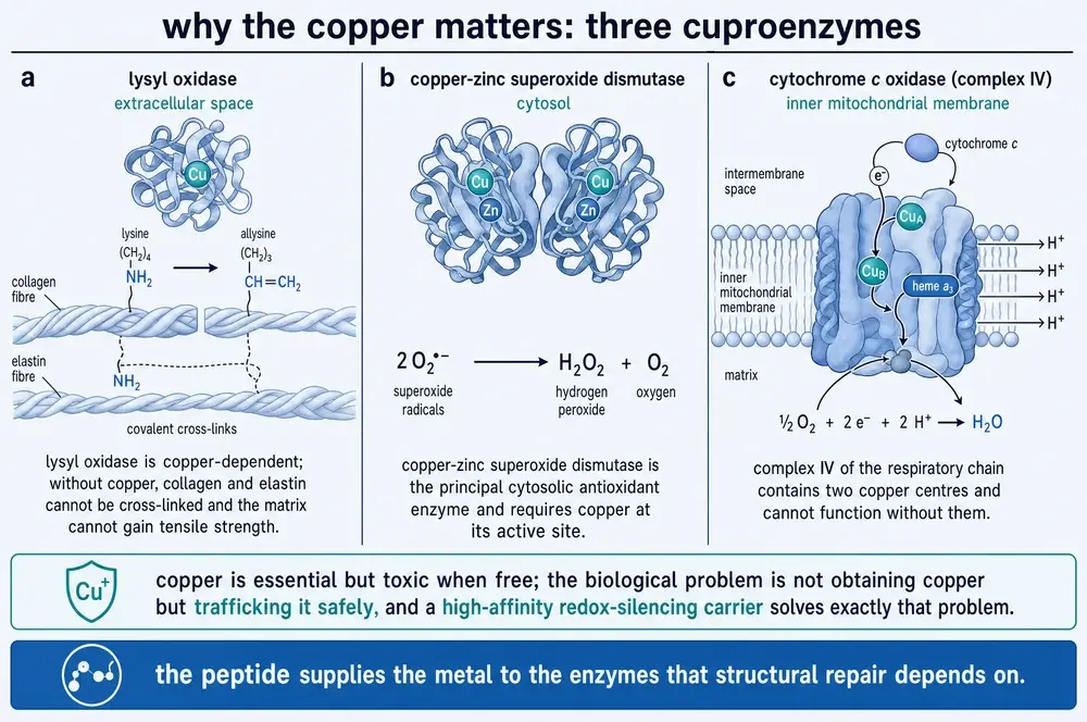
Lysyl oxidase works outside the cell, on collagen and elastin fibres that have already been secreted. It converts particular lysine side chains into a reactive aldehyde, and those aldehydes then form the covalent cross-links that turn a heap of protein strands into a material with tensile strength. Lysyl oxidase is a copper enzyme. Without copper, connective tissue can be synthesised but cannot be made strong.
Copper–zinc superoxide dismutase is the principal antioxidant enzyme of the cytosol. It takes superoxide, the radical that leaks continuously from respiration, and converts it to hydrogen peroxide and oxygen. Its active site is a copper ion.
Cytochrome c oxidase is complex IV of the respiratory chain, the enzyme that finally hands electrons to oxygen and makes water. It contains two copper centres and does not work without them.
So copper is not optional. Neither is it safe. A free cupric ion in the presence of a reducing agent and oxygen will cycle between its two oxidation states and generate hydroxyl radicals, which attack essentially anything nearby. The body’s response to this is to make sure there is almost no free copper anywhere: plasma copper is held on ceruloplasmin and albumin, and inside cells it is passed hand to hand by dedicated chaperone proteins. The concentration of genuinely unbound copper ion in a cell is often quoted as less than one atom per cell.
The biological problem, then, is not obtaining copper. It is trafficking copper. A small, soluble, high-affinity carrier that holds the metal in a non-damaging form and can hand it off is a solution to a real problem, and that is what GHK‑Cu appears to be. The chemistry that follows in Part Two is about how well it does that job, and it is the part of this molecule’s story that is not seriously contested.
06The coordination sphere
The copper sits in a square-planar arrangement held by three nitrogen atoms that the peptide supplies, one from each of the first two residues plus one from the histidine side chain. In order along the chain they are: the free α‑amino nitrogen at the glycine end; the nitrogen of the glycine–histidine amide bond, which loses its proton to make the bond; and one of the two nitrogens in the histidine imidazole ring. The fourth equatorial position is left open, and a water molecule sits loosely above the plane, completing a square pyramid.
That arrangement was established by a convergence of methods across the 1980s — X‑ray crystallography, electron paramagnetic resonance, X‑ray absorption and nuclear magnetic resonance — and it has been confirmed again since, most recently at 1.3 Å resolution in crystal structures where the tripeptide was fused to unrelated proteins purely as a copper-binding handle for solving their structures (Mehr et al., 2020). Nobody argues about it.
Two refinements matter for reading the rest of this document. First, the solid-state and solution structures are not the same. The crystal form is polymeric, with copper pairs bridged through oxygen and the fourth site occupied by a carboxylate from a neighbouring complex; in solution that polymer does not exist, and the complex is a discrete 1:1 species (Freedman et al., 1982). Second, the fourth site is not usually empty. It is an open coordination position on a copper ion in a solution full of imidazole rings, and it takes one whenever it can — from a second molecule of the tripeptide, from free histidine, or from a histidine side chain on a protein. What circulates is often not the bare complex but a ternary one (Bossak‑Ahmad et al., 2020).
07How tightly, and tighter than what
The comparison that appears in almost every account of this molecule is that the tripeptide binds copper with a stability constant of log K 16.44, against 16.2 for the high-affinity copper site at the N‑terminus of serum albumin — from which it is concluded that the two are essentially equipotent and that the peptide can take copper directly from albumin. Those figures are reported consistently across the discoverer’s reviews (Pickart et al., 2012; Pickart et al., 2015; Pickart & Margolina, 2018) and attributed there to a 1981 study by Lau and Sarkar.
The figures are not wrong. What is usually missing is the rest of that study’s result. Lau and Sarkar measured the distribution of copper directly by equilibrium dialysis, and found that at equal molar concentrations of albumin and peptide, about 42 per cent of the copper sat on the peptide — consistent with near-equal affinity. But at the concentrations that actually obtain in plasma, where albumin is present in vast excess over any low-molecular-weight ligand, only about 6 per cent of plasma copper is associated with low-molecular-weight components at all (Lau & Sarkar, 1981). Per molecule the two carriers are comparable. In the copper budget of blood they are not, because there is a great deal more albumin than there is tripeptide.
The practical upshot is that the tripeptide is a good copper carrier and a minor one. It will hold copper against almost everything except a dedicated transport protein, it will exchange with albumin readily in either direction, and it is present in plasma at concentrations three to four orders of magnitude below albumin’s. Any claim that it materially redistributes the body’s copper needs to reckon with that arithmetic.
08Redox silencing, and where it stops
The safety case for putting copper into a cream rests on one claim: that copper bound in this coordination sphere is redox-silenced. Loose copper cycles between Cu(II) and Cu(I) and generates hydroxyl radicals; bound copper, according to the claim, does not, and so can be delivered without doing oxidative damage. The claim is repeated in essentially every review of the compound.
It is a real finding, and it is narrower than the way it is usually stated. Three independent lines of recent chemistry mark out where it holds and where it fails.
Against ascorbate, it holds
The classic pro-oxidant test is whether a copper species will catalyse the oxidation of ascorbate. Bound in the tripeptide, copper is inert against physiological levels of ascorbate. Better than inert, in fact: adding the tripeptide to a system in which copper is bound to amyloid‑β and merrily oxidising ascorbate slows that oxidation in a dose-dependent way, and at 30 µM — an excess over the amyloid peptide — brings the rate almost to zero (Tosto et al., 2023). The tripeptide wins the copper away from the damaging site and holds it quietly. This is the strongest form of the redox-silencing claim and it is well supported.
Against glutathione, it does not
Glutathione is the strongest reductant that naturally occurs in human plasma, and it is present inside cells at millimolar concentration. A 2021 study followed what happens when the two meet, in water at pH 7.4 and 37 °C, and the answer is: a great deal (Ufnalska et al., 2021). The copper is reduced — not slowly, but within the mixing time of the experiment, reaching about 60 per cent conversion at a 2.2-fold excess of glutathione and complete conversion at four- to six-fold. The reduction proceeds through a short-lived thiolate intermediate, and the stable product is not a reduced form of the complex at all: it is a Cu(I)4(glutathione)6 cluster, with the tripeptide released intact and chemically unmodified. Under aerobic conditions the copper is then re-oxidised and re-captured by the peptide, and the cycle can be run again.
Two details make this more than a curiosity. Glutathione is consumed in excess of what the stoichiometry requires, which means the copper–peptide complex is catalysing additional glutathione oxidation rather than simply being reduced by it; and the re-oxidation step proceeds through superoxide. Cyclic voltammetry independently confirms that the complex is electrochemically active. The authors’ own framing is not that the complex is inert but that it is a clean delivery vehicle for Cu(I) — the oxidation state that cellular copper importers actually accept — with the peptide surviving the transaction undamaged.
And it has measurable oxidase activity of its own
In 2026 a group characterising the complex as a catalyst reported that it behaves as a laccase mimic: it oxidises phenols and catecholamines using molecular oxygen, through a Cu(II)/Cu(I) cycle with superoxide as a trapped intermediate, and on several kinetic measures it outperforms the fungal enzyme it was compared against (Chen et al., 2026). Taken at face value that is awkward for the redox-silencing claim, and the paper does not attempt to reconcile the two.
The reconciliation is in the conditions. That assay runs at 80 °C, at catalyst concentrations in the millimolar range, against millimolar phenolic substrate, in buffer. Human plasma concentrations of the tripeptide are sub-micromolar and body temperature is 37 °C. No activity data at 37 °C are reported. Most tellingly, the paper’s own interference panel shows that serum albumin substantially suppresses the catalytic activity — and albumin is precisely the protein the complex is supposed to be sharing copper with in blood.
09Cut out of collagen
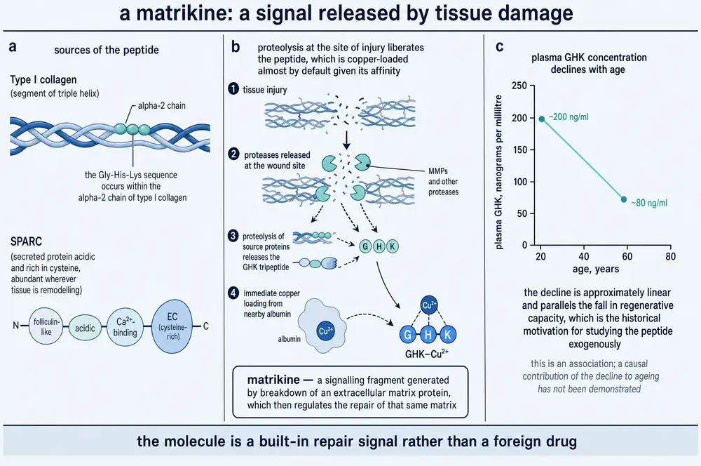
The most elegant fact about this molecule is where it comes from. The sequence Gly‑His‑Lys is not manufactured as a signal. It occurs inside larger proteins, and it is released when those proteins are cut up — which is exactly what happens when tissue is damaged and proteases are released at the wound. The injury generates its own repair signal out of the material that was injured. Molecules that work this way are called matrikines, and this is the textbook example.
Two source proteins are named in the literature. Both were checked against the reference protein database for this monograph rather than taken from review text, and both hold up — with additional detail the reviews do not carry.
| Protein | Accession | Length | Gly‑His‑Lys at | Note |
|---|---|---|---|---|
| Collagen type I, α2 chain | P08123 | 1,366 | 943–945 | a standard collagen Gly‑X‑Y triplet |
| SPARC / osteonectin | P09486 | 303 | 138–140 | abundant wherever tissue is remodelling |
| Collagen type I, α1 chain | P02452 | 1,464 | absent | the sequence is specific to the α2 chain |
| Serum albumin | P02768 | 609 | absent | its copper site is the different motif DAHK |
The two negatives are as informative as the two positives. The α1 chain of type I collagen — two of the three strands in every type I fibre — does not contain the sequence at all, so release depends on degrading the minority chain. And albumin does not contain it either. Albumin’s high-affinity copper site is its own N‑terminal sequence Asp‑Ala‑His‑Lys, with the copper co-ordinated at the histidine in third position. The two motifs are structurally analogous — which is what the 1980 Nature paper noticed — but they are not the same sequence, and a reader who takes “albumin carries GHK” from a review will have the chemistry wrong.
One further piece of chemistry belongs here rather than later, because it changes what “GHK‑Cu” means in skin specifically. The stratum corneum contains millimolar concentrations of cis-urocanic acid, a histidine breakdown product that is part of the skin’s natural moisturising factor — and urocanic acid carries an imidazole ring, which is exactly what the complex’s open fourth site wants. Measured directly, it binds with a conditional constant of 540 M−1 at pH 7.4. Feeding real stratum-corneum concentrations into the speciation model, the authors calculate that about 46 per cent of peptide-bound copper in skin would exist as the ternary complex rather than the binary one (Bossak‑Ahmad et al., 2020). Whatever is doing the work in a cream is, on that arithmetic, about half something the cell-culture experiments never tested.
10The dermal matrix
The second substantial body of work on this compound did not come from the laboratory that discovered it. Through the late 1980s and the 1990s a group at Reims — François-Xavier Maquart, Jacques-Paul Borel, Anne Siméon, Yanusz Wegrowski and colleagues — worked out what the copper complex does to fibroblasts, and their results are the foundation of every cosmetic claim made for the molecule since.
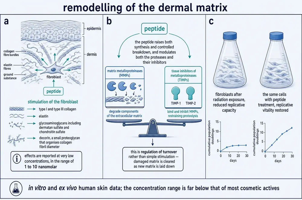
The first paper set the concentration range that still defines the field. Collagen synthesis by cultured fibroblasts began to rise somewhere between 10−12 and 10−11 M, peaked at 10−9 M — one nanomolar — and did so without any change in cell number, so this was more collagen per cell rather than more cells (Maquart et al., 1988). Sulfated glycosaminoglycan synthesis followed the same shape, maximal at 10−9 to 10−8 M and falling back toward control at higher concentrations (Wegrowski et al., 1992). A dose–response that rises and then falls is characteristic of a signalling molecule rather than a nutrient, and it is worth noting how low these concentrations are: a nanomolar effect is two to three orders of magnitude below where most cosmetic actives are studied.
The Reims group then established what turned out to be the conceptually important result. The complex does not simply push matrix production upward. It raises the matrix metalloproteinases — the enzymes that degrade matrix — alongside their tissue inhibitors TIMP‑1 and TIMP‑2 (Siméon et al., 1999; Siméon et al., 2000a). Old material is cleared while new material is laid down. That is a description of turnover, and it is a materially different claim from “increases collagen”: a tissue that only accumulates collagen is a scar.
Within that turnover the changes are selective rather than uniform. Dermatan sulfate and chondroitin sulfate accumulate; hyaluronic acid does not. Decorin — the small proteoglycan that controls how thick a collagen fibril grows — goes up, while its relative biglycan goes down (Siméon et al., 2000b). Selectivity of that kind is hard to explain as a general stimulatory effect and is the best evidence in the literature that something specific is being signalled.
11Wounds in living animals
The single strongest experiment in this literature is a 1993 study in the Journal of Clinical Investigation, and it deserves to be described properly because it is the one most often gestured at and least often read (Maquart et al., 1993).
Stainless-steel mesh cylinders were implanted under the skin of rats. Such a chamber fills with granulation tissue over weeks, and its contents can be removed and weighed and assayed — it converts wound healing into a measurable quantity of new tissue. Chambers received repeated injections of either saline or the copper complex at several concentrations. What came out of the treated chambers weighed more, and contained more DNA, more total protein, more collagen and more glycosaminoglycan, in a concentration-dependent way. The increase in collagen was twice the increase in non-collagen protein, so this was not simply more tissue. Messenger RNA for type I and type III collagen rose; messenger RNA for transforming growth factor β did not, which argues against the effect being mediated by the obvious fibrotic pathway. And a control tripeptide of similar size, glutamyl‑histidyl‑proline, had no significant effect.
That is a properly controlled in-vivo experiment with a specificity control, a dose–response and a mechanistic negative. It is a study in rats. Nothing in it is a statement about people.
12Lungs, and the first real target
The most active current research on this compound is not in skin. Since about 2016 several groups, largely in China and largely independent of one another and of the original laboratory, have tested it in models of lung injury. This body of work is the most methodologically modern in the field, and one paper in it produced something the previous forty years had not: a molecular target.
| Model | Species | Dose and route, as reported | Principal result |
|---|---|---|---|
| Endotoxin acute lung injury | mouse, C57BL/6 | 1 or 10 µg/g, intraperitoneal, ×3 over 3 d | reduced lung injury score, neutrophils, myeloperoxidase; blocked NF-κB p65 and p38 activation |
| Cigarette-smoke emphysema | mouse, C57BL/6J | 0.2, 2 or 20 µg/g, intraperitoneal, alternate days, 12 wk | less alveolar enlargement at the two higher doses; Nrf2 up, Keap1 down; low dose ineffective on most endpoints |
| Bleomycin fibrosis (GHK, not GHK‑Cu) | mouse, C57BL/6 | 2.6, 26 or 260 µg/mL/d, intraperitoneal, d4–21 | less collagen deposition; TGF-β1/Smad2/3 suppressed dose-dependently |
| Silica-induced fibrosis | mouse; plus human observation | 2 or 20 mg/kg, intraperitoneal | cytokines and hydroxyproline normalised; binds peroxiredoxin 6 directly |
| Smoke-induced muscle wasting | mouse, C57BL/6J | 0.2 or 2 mg/kg, intraperitoneal, weekly, 7 wk | grip strength 175.5 g → 339.1 g at the higher dose; entirely SIRT1-dependent |
The silicosis study is the one that matters most (Bian et al., 2024). Its authors did what almost nobody in this field had done: they went looking for what the molecule physically binds to. Using a biotinylated version of the complex as bait, followed by surface plasmon resonance to confirm the interaction and small interfering RNA to test whether it was necessary, they identified peroxiredoxin 6 — an antioxidant enzyme — as a direct binding partner, and showed that silencing it abolished the complex’s protective effects on reactive oxygen species and mitochondrial function. After five decades of mechanism inferred from downstream consequences, that is the first identified molecular target with loss-of-function confirmation.
The same paper carries the most useful human data in the entire corpus, and it is observational rather than interventional. Plasma tripeptide was measured in fifteen patients with silicosis and eleven age-matched healthy controls: 35.67 ± 13.69 ng/mL against 105.50 ± 31.94 ng/mL, P < 0.0001, falling further with clinical stage and correlating with both forced expiratory volume and diffusing capacity. A companion study of chronic obstructive pulmonary disease found the same shape: 70.27 ± 38.87 ng/mL against 133.0 ± 54.54 ng/mL in healthy controls, correlating with muscle index, six-minute walk distance and quadriceps strength (Deng et al., 2023). Two independent cohorts, two chronic lung diseases, the same direction of effect. Whether the fall is a cause or a consequence of the disease is not established by either.
The muscle study is also notable for how completely it nailed down its mechanism. Molecular docking predicted binding to SIRT1; the complex raised SIRT1 expression and deacetylase activity dose-dependently; and a SIRT1 inhibitor abolished every protective effect — in cells and in mice, on cell viability, myotube diameter, signalling, mitochondrial markers, oxidative-stress markers, body weight, grip strength and muscle cross-sectional area. The authors note, correctly, that a pharmacological inhibitor is weaker evidence than a muscle-specific knockout, which they did not do.
13Brain
A smaller and more recent literature has moved into the nervous system. In rats with experimentally induced intracerebral haemorrhage, the uncomplexed peptide given intraperitoneally before injury reduced brain water content and neurological deficit and cut the number of dying neurons, working through a microRNA that regulates vascular endothelial growth factor (Zhang et al., 2018). In mice carrying five familial Alzheimer’s mutations, the copper complex given intranasally at 15 mg/kg three times a week for twelve weeks improved working memory, reduced amyloid plaque staining and lowered a neuroinflammation marker (Tucker et al., 2024).
Two cautions attach to that last result, and both matter. The 5xFAD experiment appears twice in the literature — once as a preprint and once as a journal article — from the same group with the same design and overlapping figures. It is one dataset, not two replications. And neither version evaluated neuronal loss, which the authors state plainly as a gap.
A separate experiment in normally ageing mice — not a disease model — gave the complex intranasally at the same dose daily for eight weeks to animals aged twenty months (Tucker et al., 2023). Spatial-learning and working-memory measures improved in both sexes, and a marker of axonal damage fell in both sexes. But the effects were markedly sex-dependent at the four-week point, appearing in males before females, and one endpoint was flatly negative: the microglial marker IBA‑1 did not shift, and the authors concluded in terms that deserve quoting for their unusual directness — microglia are not a robust target for this compound under those conditions.
A 2026 experiment compared routes head to head in aged mice, and found something genuinely odd. Intranasal delivery over eight weeks and intraperitoneal delivery over five days both produced some behavioural benefit, but their transcriptional signatures ran in opposite directions — intranasal treatment suppressed oxidative-phosphorylation gene sets while intraperitoneal treatment activated them. Whatever is happening, the route is not incidental to it. That work is a preprint and has not been peer reviewed (Mazzola et al., 2026).
14Gut, ligament, and the biomaterials turn
Beyond skin, lung and brain the evidence thins into single studies. In mice with chemically induced colitis, the complex by oral gavage for fourteen days improved disease activity, colon length and barrier proteins, acting through SIRT1 and STAT3; a knockdown experiment showed the barrier effect depended on STAT3 while the anti-inflammatory effect did not (Mao et al., 2025).
The ligament study is worth citing precisely because of what its own title concedes. Seventy-two rats underwent anterior cruciate ligament reconstruction and received weekly intra-articular injections for four weeks. At six weeks the treated knees were measurably less lax and one dose group had stiffer grafts. At twelve weeks there was no significant difference in anything (Fu et al., 2015). The authors titled the paper to say the improvement was transient, and they were right to.
The largest recent growth area is not pharmacology at all but materials science: the tripeptide is being built into hydrogels, sponges, spicules and nanocarriers as one functional component among several. These are often good studies with proper factorial controls, and they consistently show the peptide-containing arm outperforming the arm without it. But they test a device, not a molecule, and their effect sizes belong to the construct.
15The gene-expression claim
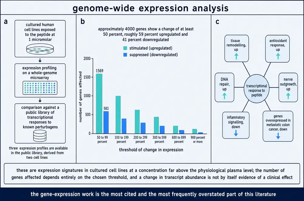
The most widely repeated statement about this molecule is that it changes the expression of about four thousand human genes, resetting them toward a healthier pattern. That claim is real, it is checkable, and it is systematically misunderstood.
Here is what was actually done. The Broad Institute’s Connectivity Map is a public library of transcriptional responses: cultured cells are exposed to a compound and the resulting expression profile is stored, so that any new profile can be matched against the library. It contains three profiles for this tripeptide, all generated by exposing cells to 1 micromolar compound — two in a prostate cancer line and one in a breast cancer line (Pickart et al., 2014). Re-analysing those profiles, the authors calculated that 31.2 per cent of an estimated 13,424 genes changed by at least 50 per cent, split 59 per cent upward and 41 per cent downward (Pickart & Margolina, 2018). Thirty-one per cent of 13,424 is about 4,190, which is where “four thousand genes” comes from.
Every one of those words is defensible. What they add up to is narrower than the headline. This is a threshold-dependent count of transcript changes, in two cancer cell lines, at a concentration a thousand-fold above the plasma level of the compound, from a database designed for compound-matching rather than for mechanism. Move the threshold and the count moves with it. A change in messenger RNA abundance is not by itself a change in protein, in cell behaviour, or in a person.
The same caution applies, with more force, to the study most often cited as showing that the compound reverses emphysema (Campbell et al., 2012). What that study did was profile lung tissue from six people with severe emphysema, derive a 127-gene signature of disease severity, and query it against the Connectivity Map — where this compound’s pre-existing cancer-cell-line profile came back as one of the strongest inverse matches. The authors then ran a single wet-lab follow-up: primary human lung fibroblasts, treated at 0.1 and 10 nanomolar, recovered their ability to contract a collagen gel. That is a real and interesting result. But the compound was never given to a lung, an animal or a patient in that study, and the authors themselves state that whether reversing the signature affects disease progression is unanswered.
The gap between “a database match plus a contraction assay” and “reverses emphysema” is the widest single gap in this molecule’s literature, and it is worth carrying into Part Four, where the human record is examined on its own terms.
16The peptide in human plasma
The claim that anchors the anti-ageing case is a simple one: the tripeptide’s concentration in human plasma falls with age, from about 200 ng/mL at twenty to about 80 ng/mL at sixty, and that decline tracks the loss of regenerative capacity.
Two things should be said about it. The first is that it is traceable, in this corpus, only to the discoverer’s own review articles — and the reviews are not internally consistent about it. One gives the ages as single values, twenty and sixty; another gives ranges, twenty to twenty-five and sixty to eighty, and specifies men (Pickart et al., 2012; Pickart et al., 2015). A third source repeats the figure citing those reviews rather than any measurement (Mavrych et al., 2026). There is also an arithmetical slip: one review equates 200 ng/mL with 10−7 M, but at a molecular mass of 340 Da, 200 ng/mL is closer to 6 × 10⁻⁷ M. None of this makes the figure wrong. It does mean it has never been independently confirmed as stated.
The second thing is that independent measurements now exist, and they tell a subtly different and rather more interesting story. Two Chinese groups, working on chronic lung disease, measured plasma tripeptide in healthy adults as a control condition:
| Source | Population | n | Plasma GHK, ng/mL |
|---|---|---|---|
| Pickart et al., reviews | age 20 (or 20–25) | not stated | ~200 |
| Pickart et al., reviews | age 60 (or 60–80) | not stated | ~80 |
| Bian et al., 2024 | healthy, age-matched controls | 11 | 105.50 ± 31.94 |
| Bian et al., 2024 | silicosis | 15 | 35.67 ± 13.69 |
| Deng et al., 2023 | healthy controls | 11 | 133.0 ± 54.54 |
| Deng et al., 2023 | chronic obstructive pulmonary disease | 9 | 70.27 ± 38.87 |
The two independent healthy-control values, 105 and 133 ng/mL, sit below the quoted young-adult figure and above the quoted sixty-year-old figure — which is unsurprising for middle-aged control groups, and is at least compatible with the decline. What is much better established than the age relationship is the disease relationship. In both cohorts the concentration was roughly a third to a half lower in patients than in controls, and in both it correlated with objective measures of organ function rather than with symptoms. That is a reproducible finding from two independent groups in two different diseases, and it is stronger evidence than the age curve it is usually enlisted to support.
It also does not establish direction. A peptide released by proteolysis of collagen and SPARC might reasonably fall when a tissue is scarred and no longer remodelling, in which case the low concentration is a readout of the disease rather than a contributor to it. Nothing in either study distinguishes those possibilities.
17Does it get through skin?
Everything in Part Three happened in a dish or in an animal, at concentrations the experimenter chose. For the cosmetic use case — which is what almost all actual human exposure to this compound consists of — one question sits in front of all the others. When the complex is applied to skin, does any of it reach the living cells underneath, and at what concentration?
The literature’s answer is not reassuring, and it is not consistent.
The most-cited dataset comes from a pair of companion papers, published in 2010 and 2011, using flow-through diffusion cells and human cadaver skin from three donors (Hostynek et al., 2010; Hostynek et al., 2011). Applied as a 0.68 per cent aqueous solution over 48 hours, copper crossed dermatomed skin to the extent of about 2 per cent of the applied dose, with substantially more retained in the tissue; through isolated stratum corneum alone, nearly 20 per cent crossed; and through isolated epidermis, 0.006 per cent — the epidermis being by far the tighter barrier. Three features of that dataset need stating alongside the numbers. It tracked elemental copper by mass spectrometry, not the intact complex, and the authors themselves attribute much of the retained signal to the complex coming apart and the copper rebinding to endogenous ligands. It used a concentration of roughly 6,800 parts per million, against the 5 to 50 ppm typical of finished cosmetic products — a hundred- to thousand-fold difference. And the two papers disagree with each other about the dermal retention figure.
Against that, a separate experiment reported through a 2025 review found no permeation of either peptide or copper at all through intact human dermatomed skin over nine hours; measurable transport appeared only after the skin was pretreated with microneedles (reviewed in Mortazavi, 2025). For comparison, a conventional cosmetic peptide put through the same kind of assessment left 99.7 per cent of the applied dose in the surface wash, 0.22 per cent in the stratum corneum, 0.01 per cent in the viable epidermis and nothing at all in the dermis or receptor fluid (Bjerke et al., 2026).
Formulation work exists precisely because the base case is poor. Liposomes, ionic-liquid microemulsions, palmitoylation, arginine-rich carrier peptides and covalent conjugates have all been reported to improve delivery, with the better studies claiming roughly two- to five-fold gains over an aqueous control. Those are real improvements on a small number.
18The trials that exist
Set aside the mechanism and ask what has been measured in people. The answer is a short list, and it is best read in order of what each study can actually support.
Randomised, controlled, clinical endpoint
Diabetic neuropathic foot ulcers, 1994. A multicentre, randomised, evaluator-blinded, placebo-controlled trial of the copper complex as Iamin Gel, on top of a standardised protocol of sharp debridement, pressure-relieving footwear and patient education. Median area closure was 98.5 per cent against 60.8 per cent for vehicle (P < 0.05); closure was about three times faster; the effect was larger in ulcers over 100 mm2; and infection occurred in 7 per cent against 34 per cent (P < 0.05) (Mulder et al., 1994). This is the best human result the compound has ever produced.
Venous stasis ulcers, 1992. A prospective, randomised, evaluator-blinded three-arm trial in 86 evaluable patients comparing a 0.4 per cent tripeptide-copper cream, 1 per cent silver sulfadiazine, and an inert vehicle. Silver sulfadiazine significantly reduced ulcer size against both other arms. There was no difference between the copper tripeptide cream and the placebo (Bishop et al., 1992).
Skin after carbon-dioxide laser resurfacing, 2006. Thirteen patients randomised to a post-treatment regimen with or without the complex, assessed by computer analysis and blinded evaluators at twelve weeks. No significant difference in resolution of redness, in wrinkles, or in overall skin quality. The one endpoint that did differ was patient-reported satisfaction (P = 0.04) (Miller et al., 2006).
Randomised, but confounded by co-formulation
Male pattern hair loss, 2016. Forty-five men randomised to two doses of a complex of 5‑aminolevulinic acid and the tripeptide, or placebo, daily for six months. Hair count rose by 52.6 and 71.5 in the two active groups against 9.6 in placebo (P < 0.05); hair length and thickness did not differ (Lee et al., 2016). The active agent is a two-component complex, so the tripeptide’s own contribution cannot be separated.
Not peer-reviewed, and industry-connected
Three studies are cited far more often than any of the above, and they are the source of nearly every cosmetic claim: a facial cream applied for twelve weeks to 71 women with photoageing; an eye cream applied for twelve weeks to 41 women, reported as outperforming placebo and a vitamin K cream; and a thigh-skin application improving collagen production in 70 per cent of women treated against 50 per cent for vitamin C and 40 per cent for retinoic acid.
The first two are conference abstracts from the 60th Annual Meeting of the American Academy of Dermatology, New Orleans, February 2002 (Leyden et al., 2002a; Leyden et al., 2002b). They have never been published as papers, carry no methods section a reader can evaluate, and include an author affiliated with the manufacturer whose product was tested. The third is a ten-participant pilot with a histological rather than clinical endpoint, published in a journal not indexed by PubMed. A later randomised double-blind study reporting a 31.6 per cent reduction in wrinkle volume against an active comparator appeared in a journal published by a house widely regarded as predatory (Badenhorst et al., 2016).
19What happened to the drug, and what is sold now
The most consequential fact about this molecule’s human record is one that appears in no peer-reviewed paper.
Through the early 1990s ProCyte Corporation of Redmond, Washington developed the complex as a wound-healing drug under the name prezatide copper acetate, brand name Iamin, and pursued full approval from the US Food and Drug Administration. The 1994 diabetic-ulcer trial above was part of that programme. According to contemporaneous business reporting, a larger pivotal trial in 511 patients completed in October 1994 and failed to beat its control group, though the compound was shown to be safe. ProCyte cut more than a third of its staff, abandoned the drug pathway, and refiled the same compound under the Class I medical device route, which requires considerably less evidence. Clearance to market Iamin Gel as a wound-care product was granted on 7 February 1996 — with the FDA expressly prohibiting the company from claiming that the product heals wounds (The Seattle Times, 7 February 1996).
That account rests on press reporting, not on a peer-reviewed publication or a primary regulatory document, and this monograph flags it as such. But the absence itself is evidence: a 511-patient pivotal trial in a compound with this much subsequent commercial life has no entry in the peer-reviewed literature. The largest human experiment ever conducted on this molecule is known only because a newspaper reported that it failed.
Registered clinical research is, finally, beginning. A search of ClinicalTrials.gov returns no completed controlled trial of this compound with posted results. It returns one Phase 2 trial that began recruiting in February 2026: a 60-participant, vehicle-controlled study of a 0.1 per cent topical gel in a standardised punch-biopsy wound model, with time to re-epithelialisation as its primary endpoint (NCT07437586). It is the first properly registered, controlled, endpoint-driven trial in the compound’s history.
Meanwhile the compound is sold in a third form that has no relationship to any of the evidence above. Vendors offer it as a lyophilised powder for reconstitution and injection, under research-use-only labelling. There is no approved injectable product, no human pharmacokinetic study of a parenteral route in this corpus, and no controlled trial of injected material at all. In April 2020 the FDA issued a warning letter to a compounding pharmacy that named this compound among some twenty peptides being made into sterile injectable products outside the conditions that exempt compounders from drug-approval requirements. The animal studies in Part Three used intraperitoneal, intra-articular, intranasal and oral routes at doses selected for rodents; none of them establishes anything about a human dose, and this document specifies none.
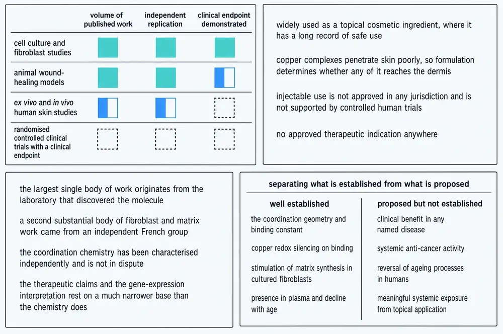
20Established, proposed, contradicted
Fifty-three years of work on a molecule of three residues has produced an evidence base with an unusual shape: extremely solid at the bottom, broad in the middle, and almost absent at the top.
| Claim | Status | What it rests on |
|---|---|---|
| Binds Cu(II) in a 3N square-planar sphere with a labile fourth site | Established | crystallography, EPR, NMR, potentiometry; multiple independent groups over 40 years |
| Affinity comparable to albumin’s N-terminal copper site | Established | equilibrium dialysis and calorimetry — with the caveat that low-molecular-weight carriers hold ~6% of plasma copper |
| Bound copper is redox-attenuated toward mild reductants | Established | inert to ascorbate and dithiothreitol; out-competes amyloid for copper |
| Bound copper is redox-silent | Contradicted | reduced by glutathione within minutes, catalytically; measurable oxidase activity in vitro |
| The sequence occurs in collagen α2(I) and SPARC and is released by proteolysis | Established | sequence verified at defined positions; the release mechanism is inferred, not directly observed in vivo |
| Stimulates matrix synthesis and turnover in cultured fibroblasts at 1–10 nM | Established | independent replication by the Reims group across a decade |
| Accelerates wound repair in rodents | Established | controlled wound-chamber studies with specificity controls |
| Protects lung and muscle in rodent injury models | Established | multiple independent 2016–2024 groups; one identified molecular target |
| Plasma concentration is reduced in chronic lung disease | Established | two independent cohorts, correlating with organ function |
| Plasma concentration falls from ~200 to ~80 ng/mL between 20 and 60 | Single-source | the discoverer’s reviews only; internally inconsistent; never independently confirmed as stated |
| Changes the expression of ~4,000 human genes | Established, and narrower than it sounds | two cancer cell lines at 1 µM; threshold-dependent; transcript abundance only |
| Reverses the gene signature of emphysema | Misstated | a database match plus one fibroblast contraction assay; never given to a lung, animal or patient |
| Topical application delivers effective concentrations to living dermis | Not demonstrated | penetration studies conflict; none uses a realistic dose and reports a tissue concentration |
| Improves the appearance of ageing human skin | Weakly supported | the principal studies are 2002 conference abstracts; the one blinded trial with objective endpoints was null |
| Accelerates healing of diabetic foot ulcers | Contested | one strongly positive randomised trial in 1994; a 511-patient pivotal trial failed and was never published |
| Any systemic or injectable therapeutic effect in humans | Untested | no human pharmacokinetics, no controlled trial, no approved indication |
Read down that middle column and a pattern emerges that is worth naming. Everything in the established rows is a measurement of the molecule itself — where the copper sits, how tightly it is held, what happens when a reductant reaches it, what a fibroblast does when it is added to the medium. Everything in the weak or contradicted rows is a claim about what the molecule does to a person. The dividing line is not between good and bad science. It is between questions that can be answered in a flask or a mouse and questions that require a clinical trial, and it falls exactly where the funding for clinical trials stops.
21Why the evidence has this shape
It is tempting to read the pattern above as a scientific failure, and it is not one. The chemistry was done well, and the animal work of the last decade is better than the animal work of the first three. The gap is not where the science is weak. It is where the science stops, and the reason it stops there is structural rather than scientific.
Three forces point the same way. The molecule is a naturally occurring human peptide of three residues, first described in 1973, and is therefore very difficult to protect with composition-of-matter patents — which removes the mechanism that ordinarily pays for a Phase 3 programme. Its principal commercial market is cosmetic, and the regulatory route for a cosmetic ingredient requires safety data and no efficacy evidence whatsoever. And when the one serious attempt at drug approval was made, in the 1990s, the pivotal trial failed, which converted the compound from a drug candidate into a device and then into an ingredient.
The result is a molecule that has been in the hands of consumers for three decades, in products relying on published effects that were never tested in the form or at the dose in which those products deliver them. That is not fraud, and it is not a mystery. It is what happens when a compound’s route to market does not require the evidence its marketing implies.
22What would settle it
Four studies do not exist, and each of them would resolve something the present literature cannot.
A penetration study at a real dose, reporting a real concentration. Apply a finished product at its actual use concentration to human skin, and measure the concentration of the intact complex — not elemental copper — in viable epidermis and dermis, in molar units. If that number lands in the nanomolar range, the entire cosmetic literature acquires a foundation it has never had. If it does not, that is equally decisive, and equally worth knowing.
The wound trial, run again and reported either way. The 1994 diabetic-ulcer result was strong and the 1994 pivotal trial that followed it was not, and the field has been arguing past that fact for thirty years because the second one was never published. The Phase 2 trial that began recruiting in February 2026 is the first serious attempt to reopen the question.
Human pharmacokinetics for any systemic route. Material is being sold for injection with no published human data of any kind on what happens to it — how quickly it is cleared, whether the complex survives in plasma, where the copper ends up. Those are answerable questions and nobody has answered them.
A copper control, consistently applied. The single most informative control in this entire literature is the one that showed an effect being reproduced by copper ions alone and not by the peptide alone. Running that arm in every experiment would settle, endpoint by endpoint, how much of this molecule’s biology belongs to the peptide and how much belongs to the metal it carries. On the evidence in Part Two, the answer may be uncomfortable for the peptide.
Until then, the honest summary is the one the chemistry supports. This is a real endogenous molecule, cut out of damaged collagen by the injury that damaged it, holding one atom of copper tightly enough to move it safely and loosely enough to give it up. What it does after that, in a person, at a dose anybody actually uses, is not known.
23References
Generated from verified NCBI records rather than from recall. Author lists, journal names, volumes, pages and identifiers are read off the PubMed record for each citation, and the build refuses to run if any identifier fails to resolve. This series has twice shipped reference lists drafted from memory in which identifiers pointed at real but unrelated papers, and once shipped a harvest in which a parsing error assigned each paper the identifiers of the last work it had itself cited.
The eleven entries at the end carry no PubMed record. Their presence is not an oversight: two of them are the conference abstracts that supply most of the cosmetic literature’s human evidence, and one is a newspaper report that is the only located account of the largest clinical trial this compound has ever undergone.
- Adnan SB, Maarof M, Fauzi MB, Fadilah NIM. Exploring the Role of Tripeptides in Wound Healing and Skin Regeneration: A Comprehensive Review. Int J Med Sci. 2025;22(16):4175-4200.
PMID 41209547 · doi:10.7150/ijms.118118 · PMC12595317 - Bian Y, Deng M, Liu J, Li J, Zhang Q, Wang Z, et al.. The glycyl-l-histidyl-l-lysine-Cu2+ tripeptide complex attenuates lung inflammation and fibrosis in silicosis by targeting peroxiredoxin 6. Redox Biol. 2024;75:103237.
PMID 38879894 · doi:10.1016/j.redox.2024.103237 · PMC11228880 - Bishop JB, Phillips LG, Mustoe TA, VanderZee AJ, Wiersema L, Roach DE, et al.. A prospective randomized evaluator-blinded trial of two potential wound healing agents for the treatment of venous stasis ulcers. J Vasc Surg. 1992;16(2):251-7.
PMID 1495150 · doi:10.1067/mva.1992.37086 - Bjerke DL, Li J, Gao Y, Hu P, Lintner K, Hakozaki T. A framework for the safety evaluation of peptides in cosmetics. Curr Res Toxicol. 2026;10:100291.
PMID 41953401 · doi:10.1016/j.crtox.2026.100291 · PMC13054063 - Bossak-Ahmad K, Wiśniewska MD, Bal W, Drew SC, Frączyk T. Ternary Cu(II) Complex with GHK Peptide and Cis-Urocanic Acid as a Potential Physiologically Functional Copper Chelate. Int J Mol Sci. 2020;21(17).
PMID 32867146 · doi:10.3390/ijms21176190 · PMC7503498 - Campbell JD, McDonough JE, Zeskind JE, Hackett TL, Pechkovsky DV, Brandsma CA, et al.. A gene expression signature of emphysema-related lung destruction and its reversal by the tripeptide GHK. Genome Med. 2012;4(8):67.
PMID 22937864 · doi:10.1186/gm367 · PMC4064320 - Chen H, Yang P, Xue P, Li S, Dan X, Li Y, et al.. Food-Derived Tripeptide-Copper Self-Healing Hydrogel for Infected Wound Healing. Biomater Res. 2025;29:0139.
PMID 39902373 · doi:10.34133/bmr.0139 · PMC11788471 - Chen JS, Zhu H, Chai TQ, Yang FQ. The Laccase-like Property of GHK-Cu and Its Applications in Colorimetric Sensing of Phenolic Compounds. Biosensors (Basel). 2026;16(4).
PMID 42041438 · doi:10.3390/bios16040217 · PMC13115362 - Deng M, Zhang Q, Yan L, Bian Y, Li R, Gao J, et al.. Glycyl-l-histidyl-l-lysine-Cu2+ rescues cigarette smoking-induced skeletal muscle dysfunction via a sirtuin 1-dependent pathway. J Cachexia Sarcopenia Muscle. 2023;14(3):1365-1380.
PMID 36905132 · doi:10.1002/jcsm.13213 · PMC10235902 - Dymek M, Olechowska K, Hąc-Wydro K, Sikora E. Liposomes as Carriers of GHK-Cu Tripeptide for Cosmetic Application. Pharmaceutics. 2023;15(10).
PMID 37896245 · doi:10.3390/pharmaceutics15102485 · PMC10610410 - Freedman JH, Pickart L, Weinstein B, Mims WB, Peisach J. Structure of the Glycyl-L-histidyl-L-lysine--copper(II) complex in solution. Biochemistry. 1982;21(19):4540-4.
PMID 6291585 · doi:10.1021/bi00262a004 - Fu SC, Cheuk YC, Chiu WY, Yung SH, Rolf CG, Chan KM. Tripeptide-copper complex GHK-Cu (II) transiently improved healing outcome in a rat model of ACL reconstruction. J Orthop Res. 2015;33(7):1024-33.
PMID 25731775 · doi:10.1002/jor.22831 - Hailu KT, Abriha FN, Duguma YM, Haddad RR, Liyew T, Kasagga A. Unregulated Peptide Use in the Age of Biohacking: Digital Promotion, Gray-Market Access, and Emerging Public Health Risks. Cureus. 2026;18(6):e110657.
PMID 42437212 · doi:10.7759/cureus.110657 · PMC13355462 - Hostynek JJ, Dreher F, Maibach HI. Human skin retention and penetration of a copper tripeptide in vitro as function of skin layer towards anti-inflammatory therapy. Inflamm Res. 2010;59(11):983-8.
PMID 20703511 · doi:10.1007/s00011-010-0214-4 · PMC2945467 - Hostynek JJ, Dreher F, Maibach HI. Human skin penetration of a copper tripeptide in vitro as a function of skin layer. Inflamm Res. 2011;60(1):79-86.
PMID 20721598 · doi:10.1007/s00011-010-0238-9 · PMC3016279 - Kang YA, Choi HR, Na JI, Huh CH, Kim MJ, Youn SW, et al.. Copper-GHK increases integrin expression and p63 positivity by keratinocytes. Arch Dermatol Res. 2009;301(4):301-6.
PMID 19319546 · doi:10.1007/s00403-009-0942-x - Lau SJ, Sarkar B. The interaction of copper(II) and glycyl-L-histidyl-L-lysine, a growth-modulating tripeptide from plasma. Biochem J. 1981;199(3):649-56.
PMID 7340824 · doi:10.1042/bj1990649 · PMC1163421 - Lee WJ, Sim HB, Jang YH, Lee SJ, Kim do W, Yim SH. Efficacy of a Complex of 5-Aminolevulinic Acid and Glycyl-Histidyl-Lysine Peptide on Hair Growth. Ann Dermatol. 2016;28(4):438-43.
PMID 27489425 · doi:10.5021/ad.2016.28.4.438 · PMC4969472 - Li H, Toh PZ, Tan JY, Zin MT, Lee CY, Li B, et al.. Selected Biomarkers Revealed Potential Skin Toxicity Caused by Certain Copper Compounds. Sci Rep. 2016;6:37664.
PMID 27892491 · doi:10.1038/srep37664 · PMC5124859 - Liu T, Liu Y, Zhao X, Zhang L, Wang W, Bai D, et al.. Thermodynamically stable ionic liquid microemulsions pioneer pathways for topical delivery and peptide application. Bioact Mater. 2024;32:502-513.
PMID 38026438 · doi:10.1016/j.bioactmat.2023.10.002 · PMC10643103 - Mao S, Huang J, Li J, Sun F, Zhang Q, Cheng Q, et al.. Exploring the beneficial effects of GHK-Cu on an experimental model of colitis and the underlying mechanisms. Front Pharmacol. 2025;16:1551843.
PMID 40672369 · doi:10.3389/fphar.2025.1551843 · PMC12263609 - Maquart FX, Pickart L, Laurent M, Gillery P, Monboisse JC, Borel JP. Stimulation of collagen synthesis in fibroblast cultures by the tripeptide-copper complex glycyl-L-histidyl-L-lysine-Cu2+. FEBS Lett. 1988;238(2):343-6.
PMID 3169264 · doi:10.1016/0014-5793(88)80509-x - Maquart FX, Bellon G, Chaqour B, Wegrowski J, Patt LM, Trachy RE, et al.. In vivo stimulation of connective tissue accumulation by the tripeptide-copper complex glycyl-L-histidyl-L-lysine-Cu2+ in rat experimental wounds. J Clin Invest. 1993;92(5):2368-76.
PMID 8227353 · doi:10.1172/JCI116842 · PMC288419 - Mavrych V, Shypilova I, Bolgova O. Therapeutic peptides in gerontology: mechanisms and applications for healthy aging. Front Aging. 2026;7:1790247.
PMID 42021992 · doi:10.3389/fragi.2026.1790247 · PMC13095733 - Mazzola J, Rosenfeld M, Tucker M, Wezeman J, Ladiges W, Liao G. Middle-aged mice treated with GHK-Cu peptide administered intraperitoneally or intranasally show behavioral rescue but divergent hippocampal aging programs. Res Sq. 2026.
PMID 42245779 · doi:10.21203/rs.3.rs-9520102/v1 · PMC13232416 - Mehr A, Henneberg F, Chari A, Görlich D, Huyton T. The copper(II)-binding tripeptide GHK, a valuable crystallization and phasing tag for macromolecular crystallography. Acta Crystallogr D Struct Biol. 2020;76(Pt 12):1222-1232.
PMID 33263328 · doi:10.1107/S2059798320013741 · PMC7709198 - Miller TR, Wagner JD, Baack BR, Eisbach KJ. Effects of topical copper tripeptide complex on CO2 laser-resurfaced skin. Arch Facial Plast Surg. 2006;8(4):252-9.
PMID 16847171 · doi:10.1001/archfaci.8.4.252 - Min JH, Sarlus H, Harris RA. Glycyl-l-histidyl-l-lysine prevents copper- and zinc-induced protein aggregation and central nervous system cell death in vitro. Metallomics. 2024;16(5).
PMID 38599632 · doi:10.1093/mtomcs/mfae019 · PMC11135135 - Mortazavi SM, Mohammadi Vadoud SA, Moghimi HR. Topically applied GHK as an anti-wrinkle peptide: Advantages, problems and prospective. Bioimpacts. 2025;15:30071.
PMID 39963574 · doi:10.34172/bi.30071 · PMC11830136 - Mulder GD, Patt LM, Sanders L, Rosenstock J, Altman MI, Hanley ME, et al.. Enhanced healing of ulcers in patients with diabetes by topical treatment with glycyl-l-histidyl-l-lysine copper. Wound Repair Regen. 1994;2(4):259-69.
PMID 17147644 · doi:10.1046/j.1524-475X.1994.20406.x - Ogórek K, Nowak K, Wadych E, Ruzik L, Timerbaev AR, Matczuk M. Are We Ready to Measure Skin Permeation of Modern Antiaging GHK-Cu Tripeptide Encapsulated in Liposomes?. Molecules. 2025;30(1).
PMID 39795193 · doi:10.3390/molecules30010136 · PMC11721469 - Park JR, Lee H, Kim SI, Yang SR. The tri-peptide GHK-Cu complex ameliorates lipopolysaccharide-induced acute lung injury in mice. Oncotarget. 2016;7(36):58405-58417.
PMID 27517151 · doi:10.18632/oncotarget.11168 · PMC5295439 - Pickart L, Thaler MM. Tripeptide in human serum which prolongs survival of normal liver cells and stimulates growth in neoplastic liver. Nat New Biol. 1973;243(124):85-7.
PMID 4349963 - Pickart L, Thayer L, Thaler MM. A synthetic tripeptide which increases survival of normal liver cells, and stimulates growth in hepatoma cells. Biochem Biophys Res Commun. 1973;54(2):562-6.
PMID 4356974 · doi:10.1016/0006-291x(73)91459-9 - Pickart L, Thaler MM, Millard M. Effect of transition metals on recovery from plasma of the growth-modulating tripeptide glycylhistidyllysine. J Chromatogr. 1979;175(1):65-73.
PMID 546910 · doi:10.1016/s0021-9673(00)86403-1 - Pickart L, Thaler MM. Growth-modulating tripeptide (glycylhistidyllysine): association with copper and iron in plasma, and stimulation of adhesiveness and growth of hepatoma cells in culture by tripeptide-metal ion complexes. J Cell Physiol. 1980;102(2):129-39.
PMID 6246126 · doi:10.1002/jcp.1041020205 - Pickart L, Freedman JH, Loker WJ, Peisach J, Perkins CM, Stenkamp RE, et al.. Growth-modulating plasma tripeptide may function by facilitating copper uptake into cells. Nature. 1980;288(5792):715-7.
PMID 7453802 · doi:10.1038/288715a0 - Pickart L. The human tri-peptide GHK and tissue remodeling. J Biomater Sci Polym Ed. 2008;19(8):969-88.
PMID 18644225 · doi:10.1163/156856208784909435 - Pickart L, Vasquez-Soltero JM, Margolina A. The human tripeptide GHK-Cu in prevention of oxidative stress and degenerative conditions of aging: implications for cognitive health. Oxid Med Cell Longev. 2012;2012:324832.
PMID 22666519 · doi:10.1155/2012/324832 · PMC3359723 - Pickart L, Vasquez-Soltero JM, Margolina A. GHK and DNA: resetting the human genome to health. Biomed Res Int. 2014;2014:151479.
PMID 25302294 · doi:10.1155/2014/151479 · PMC4180391 - Pickart L, Vasquez-Soltero JM, Margolina A. GHK Peptide as a Natural Modulator of Multiple Cellular Pathways in Skin Regeneration. Biomed Res Int. 2015;2015:648108.
PMID 26236730 · doi:10.1155/2015/648108 · PMC4508379 - Pickart L, Vasquez-Soltero JM, Margolina A. The Effect of the Human Peptide GHK on Gene Expression Relevant to Nervous System Function and Cognitive Decline. Brain Sci. 2017;7(2).
PMID 28212278 · doi:10.3390/brainsci7020020 · PMC5332963 - Pickart L, Margolina A. Regenerative and Protective Actions of the GHK-Cu Peptide in the Light of the New Gene Data. Int J Mol Sci. 2018;19(7).
PMID 29986520 · doi:10.3390/ijms19071987 · PMC6073405 - Schlesinger DH, Pickart L, Thaler MM. Growth-modulating serum tripeptide is glycyl-histidyl-lysine. Experientia. 1977;33(3):324-5.
PMID 858356 · doi:10.1007/BF02002806 - Siméon A, Monier F, Emonard H, Gillery P, Birembaut P, Hornebeck W, et al.. Expression and activation of matrix metalloproteinases in wounds: modulation by the tripeptide-copper complex glycyl-L-histidyl-L-lysine-Cu2+. J Invest Dermatol. 1999;112(6):957-64.
PMID 10383745 · doi:10.1046/j.1523-1747.1999.00606.x - Siméon A, Emonard H, Hornebeck W, Maquart FX. The tripeptide-copper complex glycyl-L-histidyl-L-lysine-Cu2+ stimulates matrix metalloproteinase-2 expression by fibroblast cultures. Life Sci. 2000;67(18):2257-65.
PMID 11045606 · doi:10.1016/s0024-3205(00)00803-1 - Siméon A, Wegrowski Y, Bontemps Y, Maquart FX. Expression of glycosaminoglycans and small proteoglycans in wounds: modulation by the tripeptide-copper complex glycyl-L-histidyl-L-lysine-Cu(2+). J Invest Dermatol. 2000;115(6):962-8.
PMID 11121126 · doi:10.1046/j.1523-1747.2000.00166.x - Tosto R, Vecchio G, Bellia F. New Biotinylated GHK and Related Copper(II) Complex: Antioxidant and Antiglycant Properties In Vitro against Neurodegenerative Disorders. Molecules. 2023;28(18).
PMID 37764500 · doi:10.3390/molecules28186724 · PMC10538196 - Tucker M, Keely A, Park JY, Rosenfeld M, Wezeman J, Mangalindan R, et al.. Intranasal GHK peptide enhances resilience to cognitive decline in aging mice. bioRxiv. 2023.
PMID 38014118 · doi:10.1101/2023.11.16.567423 · PMC10680828 - Tucker M, Liao GY, Keely A, Park JY, Rosenfeld M, Wezeman J, et al.. Behavioral and neuropathological features of Alzheimer's disease are attenuated in 5xFAD mice treated with intranasal GHK peptide. Aging Pathobiol Ther. 2024;6(3):102-108.
PMID 40766919 · doi:10.31491/apt.2024.09.148 · PMC12323558 - Ufnalska I, Drew SC, Zhukov I, Szutkowski K, Wawrzyniak UE, Wróblewski W, et al.. Intermediate Cu(II)-Thiolate Species in the Reduction of Cu(II)GHK by Glutathione: A Handy Chelate for Biological Cu(II) Reduction. Inorg Chem. 2021;60(23):18048-18057.
PMID 34781677 · doi:10.1021/acs.inorgchem.1c02669 · PMC8653159 - Wegrowski Y, Maquart FX, Borel JP. Stimulation of sulfated glycosaminoglycan synthesis by the tripeptide-copper complex glycyl-L-histidyl-L-lysine-Cu2+. Life Sci. 1992;51(13):1049-56.
PMID 1522753 · doi:10.1016/0024-3205(92)90504-i - Yu X, Yang T, Bei Z, Liu J, Song Y, Wang Q, et al.. Microenvironment-responsive injectable dynamic hydrogel for sequential antioxidant and tissue regeneration therapy of radiation-induced skin injury. Bioact Mater. 2026;63:778-794.
PMID 42058630 · doi:10.1016/j.bioactmat.2026.03.057 · PMC13123401 - Zhang H, Wang Y, He Z. Glycine-Histidine-Lysine (GHK) Alleviates Neuronal Apoptosis Due to Intracerebral Hemorrhage via the miR-339-5p/VEGFA Pathway. Front Neurosci. 2018;12:644.
PMID 30294253 · doi:10.3389/fnins.2018.00644 · PMC6158323 - Zhang H, Wang Y, Lian L, Zhang C, He Z. Glycine-Histidine-Lysine (GHK) Alleviates Astrocytes Injury of Intracerebral Hemorrhage via the Akt/miR-146a-3p/AQP4 Pathway. Front Neurosci. 2020;14:576389.
PMID 33192260 · doi:10.3389/fnins.2020.576389 · PMC7658812 - Zhang Q, Yan L, Lu J, Zhou X. Glycyl-L-histidyl-L-lysine-Cu2+ attenuates cigarette smoke-induced pulmonary emphysema and inflammation by reducing oxidative stress pathway. Front Mol Biosci. 2022;9:925700.
PMID 35936787 · doi:10.3389/fmolb.2022.925700 · PMC9354777 - Zhou XM, Wang GL, Wang XB, Liu L, Zhang Q, Yin Y, et al.. GHK Peptide Inhibits Bleomycin-Induced Pulmonary Fibrosis in Mice by Suppressing TGFβ1/Smad-Mediated Epithelial-to-Mesenchymal Transition. Front Pharmacol. 2017;8:904.
PMID 29311918 · doi:10.3389/fphar.2017.00904 · PMC5733019 - Leyden J, Stephens T, Finkey MB, Appa Y, Barkovic S. Skin care benefits of copper peptide containing facial cream. Proceedings of the American Academy of Dermatology 60th Annual Meeting, New Orleans, 22–27 February 2002; abstract 68. Conference abstract; not peer-reviewed; no methods section published.
- Leyden J, Stephens T, Finkey MB, Barkovic S. Skin care benefits of copper peptide containing eye creams. Proceedings of the American Academy of Dermatology 60th Annual Meeting, New Orleans, 22–27 February 2002; abstract 69. Conference abstract; not peer-reviewed; no methods section published.
- Abdulghani AA, Sherr A, Shirin S, Solodkina G, Tapia E, Wolf B, Gottlieb AB. Effects of topical creams containing vitamin C, a copper-binding peptide cream and melatonin compared with tretinoin on the ultrastructure of normal skin. Disease Management and Clinical Outcomes. 1998;1(4):136–141. Ten participants received the copper-peptide arm; the endpoint was histological, not clinical. Not indexed in PubMed.
https://doi.org/10.1016/S1088-3371(98)00011-4 - Badenhorst T, Svirskis D, Merrilees M, Bolke L, Wu Z. Effects of GHK-Cu on MMP and TIMP expression, collagen and elastin production, and facial wrinkle parameters. Journal of Aging Science. 2016;4:166. Published by a house widely identified as predatory; reported here for completeness rather than as evidence.
https://doi.org/10.4172/2329-8847.1000166 - Pickart L. A Tripeptide in Human Serum That Promotes the Growth of Hepatoma Cells and the Survival of Normal Hepatocytes. PhD thesis, University of California, San Francisco, 1973. The doctoral work from which the 1973 papers derive, and the source of the San Francisco attribution in section 01.
- The Seattle Times. ProCyte wins FDA approval for its Iamin gel. 7 February 1996. Contemporaneous business reporting; the only located account of the 511-patient pivotal trial that completed in October 1994 and failed to beat its control group, of the resulting staff reductions, and of the switch from the drug pathway to Class I medical-device clearance. No peer-reviewed publication of that trial has been located.
https://archive.seattletimes.com/archive/19960207/2313001/procyte-wins-fda-approval-for- - Cosmetic Ingredient Review Expert Panel. Safety Assessment of Tripeptide-1, Hexapeptide-12, their Metal Salts and Fatty Acyl Derivatives, and Palmitoyl Tetrapeptide-7 as Used in Cosmetics. Final report, 30 June 2014. Concludes copper tripeptide-1 (CAS 89030-95-5) safe as used; typical use concentration under 10 ppm; contains no human testing of copper tripeptide-1 itself.
https://www.cir-safety.org/sites/default/files/tripep062014final.pdf - United States National Library of Medicine. A Study of Topical GHK-Cu Gel for Acute Skin Wound Healing. ClinicalTrials.gov identifier NCT07437586. Phase 2; sponsor Hudson Biotech; 60 participants; vehicle-controlled split-arm punch-biopsy model; recruiting from February 2026. The first registered controlled endpoint-driven trial of the compound.
https://clinicaltrials.gov/study/NCT07437586 - United States Food and Drug Administration. Warning letter to Tailor Made Compounding LLC, 1 April 2020. Names this compound among some twenty peptides compounded into sterile injectable drug products outside the conditions of section 503A of the Federal Food, Drug, and Cosmetic Act.
- PubChem, National Center for Biotechnology Information. Compound summaries CID 78384 (glycyl-L-histidyl-L-lysine, C14H24N6O4, 340.38) and CID 71587328 (copper tripeptide-1, C14H23CuN6O4+, 402.92). Source of the formulae and masses in section 04.
https://pubchem.ncbi.nlm.nih.gov/compound/78384 - UniProt Consortium. Entries P08123 (collagen alpha-2(I) chain), P09486 (SPARC), P02452 (collagen alpha-1(I) chain) and P02768 (serum albumin). Source of the verified Gly-His-Lys sequence positions and of the two negatives in section 09.
https://www.uniprot.org/
24How this document was assembled
The corpus was built against project 05, the Therapeutic Peptide Research Library, extended by a PubMed and PubMed Central harvest. Both halves needed an identity gate before anything else, for a reason specific to this compound.
GHK is also the Goldman–Hodgkin–Katz equation. That abbreviation appears in most electrophysiology papers that compute a reversal potential or an ionic flux, and a case-insensitive substring search for “GHK” across a biomedical corpus returns a great many ion-channel papers that have nothing to do with a copper tripeptide. The matcher used here is therefore case-sensitive, boundaried on both sides, and — for a bare GHK token — admits it only when subject-matter corroboration appears within 400 characters and no electrophysiology term appears in the same window. Unambiguous designations such as the systematic name or the residue string confirm on their own.
A local sweep opened every document-extension file in twenty stores and returned 530 matching assets. Classifying those by what they actually are is the step that matters, and it removes most of them: 280 are dated web snapshots of vendor product pages, 189 are vendor and affiliate blog copy collected as writing samples, 28 are the same handful of vendor product PDFs triplicated across three folders, and 8 are this project’s own earlier internal write-ups. Only 25 were peer-reviewed scientific full texts. Reporting 530 as a corpus would have been a claim about coverage this document does not have.
Because the local snapshot held so little primary science, the pipeline queried NCBI directly by two routes. A PubMed harvest returned 256 relevant indexed records spanning 1973 to 2026. PubMed indexes titles, abstracts and MeSH terms only, so a second stage searched PubMed Central’s full text, returning 1,150 matches. Stage 03 fetched the union.
The far-side screen then classified each fetched article by how substantively it uses the compound. 147 report their own experiments and name the compound inside a Methods section; 118 discuss it substantively without one. 143 mention it once or twice and were counted rather than read. 725 never name the tripeptide at all — they were returned by the broad "copper peptide" query term and are copper complexes of unrelated peptides. One was a Goldman–Hodgkin–Katz homograph and 17 had no body text deposited.
Merging the local and fetched sets by PMCID rather than adding them — 21 documents appear in both — gives the reading corpus this monograph is written from: 269 peer-reviewed scientific full texts, 1,781,143 words, roughly 3,562 printed-page equivalents, together with the complete 256-record metadata layer.
| Stage | What it does | Result |
|---|---|---|
| 01b | Targeted scan of the project’s document stores, corroborated matcher | 530 assets |
| 01g | Classification by source kind, de-duplicated by content hash | 25 scientific |
| 02 | PubMed E-utilities harvest | 256 records |
| 02b | PubMed Central full-text search | 1,150 matches |
| 03 | Open-access full-text retrieval of the union | 1,151 fetched |
| 03c | Substantive-use screen and homograph rejection | 265 kept |
| 04 | Keyed merge and inventory | 269 unique |
| 05 | Reference list from verified NCBI records | 68 citations |
| 06 | Assembly of this document | 1 deliverable |
25Evidence handling
Findings are labelled by the kind of study that produced them, in the sentence that reports them. Randomised human trials, uncontrolled human studies, conference abstracts, animal experiments, cell and tissue measurements, cell-free solution chemistry and computational signature matches are different kinds of claim, and the difference is stated rather than left to the reader to infer. Animal and in-vitro findings are never phrased so as to imply a human outcome, and the species is named every time.
Three distinctions were held throughout because this literature routinely collapses them. The free peptide against the copper complex: several influential animal studies used uncomplexed GHK and said so, and at least one set of authors stated explicitly that they could not say whether the complex would have performed better. The concentration at which an effect was seen: matrix effects reported at one nanomolar and gene-expression profiles generated at one micromolar are separated by a factor of a thousand and are not evidence for the same proposition. And what was actually done: the most cited claim in the field — that this compound reverses the gene signature of emphysema — rests on a database match between two lists of genes plus one fibroblast contraction assay, and the compound was never given to a lung, an animal or a patient in that study.
Recency is weighted but not blindly. The 2020–2026 literature is the best in the field methodologically — it produced the first identified molecular target, the first independent plasma measurements, and the chemistry that qualifies the redox-silencing claim — and it is treated as current. Its regulatory and market statements are treated as current. Where a recent review restates an older claim without new data, the claim is traced back to the primary work and reported at the strength that primary work supports.
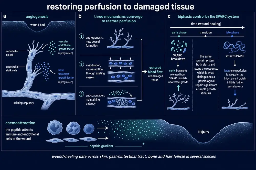
Where a number could be traced only to the laboratory that discovered the molecule, this document says so rather than laundering it through a citation. That applies to the plasma concentration decline, to the anticoagulation mechanism, and to the binding-constant comparison with albumin, whose usual presentation omits the same study’s finding that low-molecular-weight carriers hold only about six per cent of plasma copper.
On the artwork. Eight illustration plates were commissioned for this monograph and every value, label and structure printed on them was checked against the evidence dossier. Nineteen values were confirmed; two were confirmed and improved with residue positions the plates did not carry; five were corrected or qualified in their captions; and one, an absorbance maximum, could not be verified either way and is printed nowhere in this document. Two plates were withheld. Both are chemical structure drawings, and in both the drawn skeleton is not this molecule — one adds a carbon to the glycine residue and prints a molecular formula wrong by two hydrogens, and the other draws a chelate whose donor atoms are five backbone atoms apart where the real complex closes a five-membered ring. A caption cannot repair either, because a reader reads the bonds. Both were replaced by authored figures carrying verified values. The plate-by-plate record is in the delivery bundle.
Continue exploring