
Mitochondrial Dysfunction
Why the cell's power plants work less well with age — what that means for energy, signaling, and disease, and why the interventions sold to 'fix' it are still mostly biomarker-deep.
Abstract
Mitochondria generate most of the cell's ATP through oxidative phosphorylation and also act as signaling organelles that govern metabolism, cell death, and immune activation. 'Mitochondrial dysfunction' is an umbrella term for the age-related decline in that machinery — falling respiratory capacity, loss of membrane potential, and reduced biogenesis — and it is one of the twelve hallmarks of aging in the López-Otín framework. Several mechanisms are proposed: the accumulation of mitochondrial DNA (mtDNA) mutations and deletions with age, documented in human muscle and shown causal in mtDNA-mutator mice; a decline in mitophagy and other quality control; and a fall in cellular NAD+. The free-radical theory of aging (Harman, 1956) once framed reactive oxygen species (ROS) as the central driver, but has been substantially qualified: antioxidant supplement trials did not extend life and some increased mortality, mutator mice age without a rise in oxidative-stress markers, and ROS are now understood as physiological signals whose mild elevation can be beneficial (mitohormesis). Mitochondria also drive senescence, chronic inflammation, and innate immunity through released mtDNA. Exercise is the best-evidenced booster of mitochondrial biogenesis in humans; NAD+ precursors and urolithin A have raised their target biomarkers in small human trials but have not been shown to extend lifespan or prevent disease. Throughout, mechanism and animal data are distinguished from demonstrated human outcomes.
Key findings
- Mitochondria produce most of the cell's ATP through oxidative phosphorylation, and 'mitochondrial dysfunction' — declining respiratory capacity, loss of membrane potential, and reduced biogenesis — is one of the twelve hallmarks of aging (Nunnari & Suomalainen, 2012; López-Otín et al., 2023).
- Mitochondrial DNA mutations and deletions accumulate with age: in human vastus lateralis muscle, the share of fibers with respiratory-chain abnormalities rose from an estimated 6% at age 49 to 31% at age 92, and mtDNA-mutator mice show these mutations can causally accelerate aging — a mouse result, at mutation loads above ordinary aging (Bua et al., 2006; Trifunovic et al., 2004).
- The mitochondrial free-radical theory of aging (Harman, 1956) has been substantially revised, not confirmed: large antioxidant-supplement trials did not extend life and some increased mortality, mtDNA-mutator mice aged without a rise in oxidative-stress markers, and ROS are now recognized as physiological signals (Bjelakovic et al., 2007; Kujoth et al., 2005; Sies & Jones, 2020).
- Some reactive oxygen species act as beneficial signals: in a human trial, antioxidant vitamins C and E blocked the insulin-sensitizing benefit of exercise, a phenomenon called mitohormesis (Ristow et al., 2009; Yun & Finkel, 2014).
- Mitochondria are signaling hubs, not just power plants — they run apoptosis, communicate stress to the nucleus (retrograde signaling), and, when their DNA leaks into the cytoplasm, trigger innate-immune inflammation and can drive a distinct form of cellular senescence (Chandel, 2015; West & Shadel, 2017; Wiley et al., 2016).
- Exercise is the best-evidenced way to boost mitochondrial biogenesis and respiration in humans, including older adults; NAD+ precursors and urolithin A have raised their target biomarkers in small human trials but have not been shown to extend human lifespan or prevent disease (Menshikova et al., 2006; Robinson et al., 2017; Martens et al., 2018; Andreux et al., 2019).
- Methodology
- Narrative evidence review. Every source was retrieved from its primary record via PubMed / PubMed Central and each citation, date, and quantitative figure transcribed from that record, not from memory; the two hallmarks papers and the mtDNA-mutator and antioxidant-trial literature were independently re-verified. Each finding is labelled in-sentence by study type, and animal, worm, and cell-culture results are never phrased as human outcomes. Intervention claims are held to the level of their strongest human data — biomarker, surrogate, or safety — and no dose, supplement, or training protocol is recommended. Evidence cutoff: sources as retrieved 24 August 2026.
Almost everything a cell does costs energy, spent in one currency: a molecule called ATP. Most of it is made inside mitochondria — compartments so central to animal life that a single cell may carry hundreds or thousands of them, burning fuel and oxygen to keep the ATP flowing. When people call mitochondria the cell's power plants, this is what they mean: they are the site of oxidative phosphorylation, the process that captures energy from food and stores it as ATP (Nunnari & Suomalainen, 2012).
"Mitochondrial dysfunction" is the shorthand for what happens to that machinery as a body ages: it runs less efficiently. Respiratory capacity falls, the electrical charge mitochondria maintain across their inner membrane weakens, and the cell makes fewer new mitochondria to replace worn ones. The decline is real and measurable, and it is one of the recognized hallmarks of aging — the short list of recurring processes biologists use to organize how bodies grow old (López-Otín et al., 2013, 2023). Two cautions belong in the same breath. The mechanisms proposed to cause it are drawn largely from worms, mice, and cultured cells, so in humans much of the case is associational rather than proven. And the treatments marketed to "recharge" your mitochondria are, with the honest exception of exercise, supported so far by biomarker changes in small trials, not by any demonstrated effect on how long or how well people live.
What mitochondria do — and what "dysfunction" means
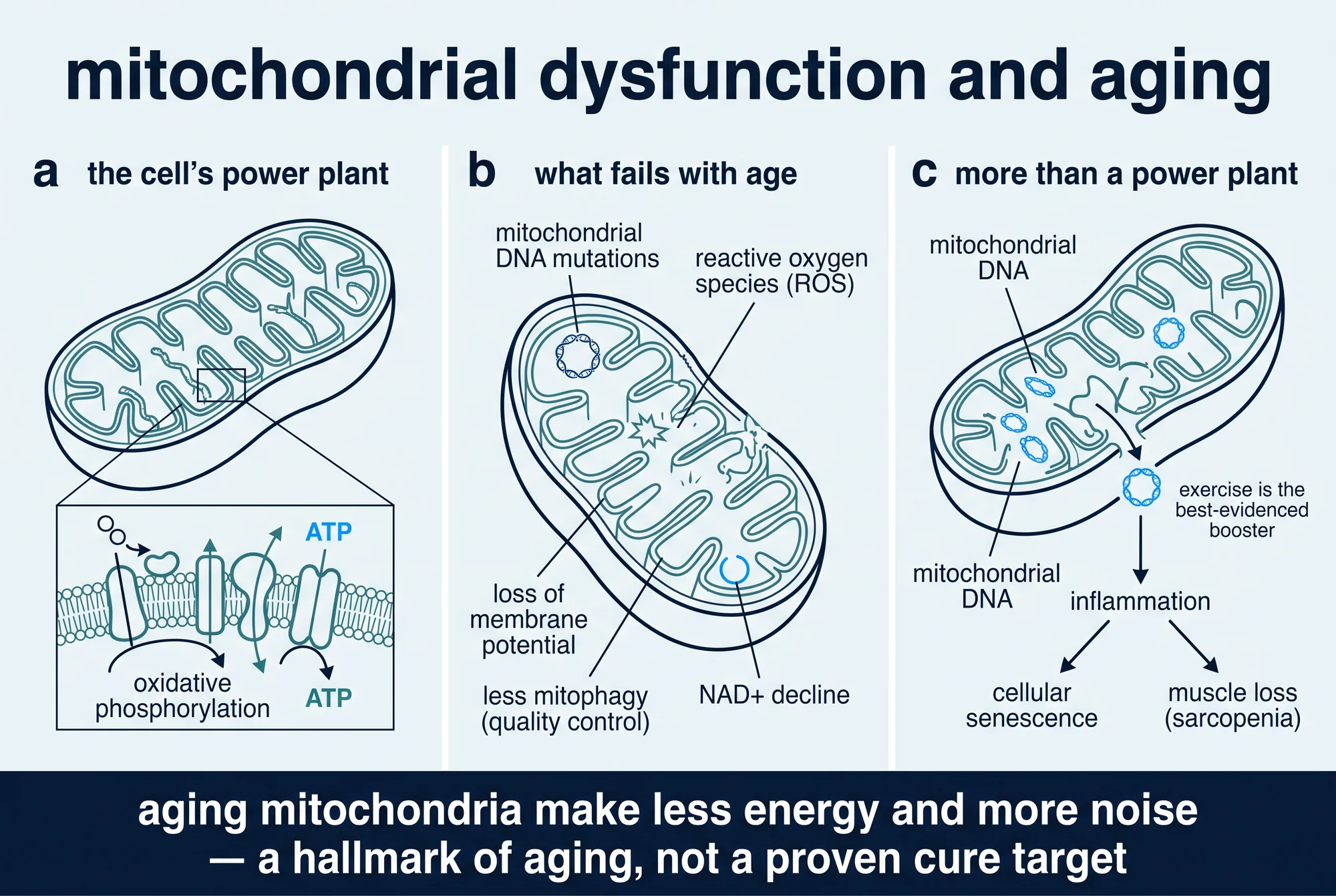
Start with the job. Mitochondria take the products of digested food and, using oxygen, extract the energy in their chemical bonds. Along the folded inner membrane, a series of protein complexes called the electron transport chain passes electrons down a line and pumps protons across the membrane, building up a charge — the mitochondrial membrane potential. A final enzyme, ATP synthase, lets those protons flow back through like water through a turbine and uses the force to stamp out ATP. This sequence is oxidative phosphorylation, and it is why a cell needs oxygen to live. Mitochondria also supply building blocks for other molecules and help run stress responses, but ATP is the headline (Nunnari & Suomalainen, 2012).
One quirk matters for everything below: mitochondria carry their own small loop of DNA, a relic of their descent from an ancestral bacterium that never surrendered its whole genome. That mitochondrial DNA (mtDNA) still encodes a handful of the respiratory chain's essential proteins, so damage to it degrades the power plant directly (West & Shadel, 2017).
Against that backdrop, "mitochondrial dysfunction" is less a single defect than a family of related declines. In an aging cell, mitochondria respire less efficiently, hold a weaker membrane potential, and are renewed — a process called biogenesis — at a reduced rate, so the population skews older and shabbier; this decline in mitochondrial quality is a well-documented correlate of normal aging and of many age-related diseases (Sun et al., 2016). "Dysfunction" names a cluster of measurable changes, not one proven lesion, and separating which changes cause aging from those that merely accompany it is much of the open work.
One of the hallmarks of aging
The reason mitochondrial dysfunction gets so much attention is that it earned a place on a short, influential list. In 2013, Carlos López-Otín and colleagues proposed nine "hallmarks of aging," and mitochondrial dysfunction was one of them; a 2023 update expanded the framework to twelve and kept it on the list (López-Otín et al., 2013, 2023). For the full framework and its limits, see The Hallmarks of Aging.
In the original scheme, mitochondrial dysfunction sits among the "antagonistic" hallmarks — protective at low levels, harmful when they run too hot or too long. A mitochondrion that signals for help is doing its job; the same signaling, sustained over decades, contributes to decline. The framework's authors were candid that the hallmarks are a working map rather than a settled parts-list, and that their relative contributions to human aging remain to be dissected (López-Otín et al., 2013). That caution travels with mitochondrial dysfunction: it is a genuine feature of aging tissue, but its rank among causes is not fixed.
The oldest theory, substantially revised
The most famous idea about mitochondria and aging is also the one that has changed the most. In 1956, Denham Harman proposed that aging is driven by the accidental byproducts of using oxygen — reactive oxygen species (ROS), the unstable molecules popularly called free radicals — which he argued gradually damage the body's own components (Harman, 1956). Because the electron transport chain is a major source of these molecules, the idea later sharpened into a mitochondrial free-radical theory of aging: mitochondria leak ROS, ROS damage mitochondria and their DNA, damaged mitochondria leak still more, and the vicious cycle grinds the cell down. For decades this tidy story dominated the field.
It is also, in its strong form, badly incomplete, which is why careful reviews now describe it as substantially qualified rather than confirmed. Three lines of evidence forced the revision.
The first is the failure of the obvious prediction. If free radicals drive aging, mopping them up with antioxidants should slow it. Tested at scale, they did not. A meta-analysis of 68 randomized trials with more than 232,000 participants found that antioxidant supplements produced no reduction in mortality overall, and that in the higher-quality trials, beta-carotene, vitamin A, and vitamin E were associated with increased death rates (Bjelakovic et al., 2007). Whatever these supplements do, extending life is not it.
The second came from mice built to test the mtDNA side of the theory. When researchers engineered "mutator" mice to accumulate mitochondrial DNA mutations rapidly, the animals did age prematurely — but, revealingly, the acceleration was not accompanied by the rise in oxidative-stress markers the theory would predict; instead it tracked with the induction of apoptosis, or programmed cell death (Kujoth et al., 2005). Mutations that damage mitochondria could speed aging without doing it through a flood of free radicals.
The third is a change in what ROS are understood to be. Far from being only agents of damage, they are now recognized as physiological signaling molecules: at low, controlled levels, species such as hydrogen peroxide act as messengers that adjust metabolism and stress responses, a state one authoritative review calls "oxidative eustress" as distinct from damaging "oxidative distress" (Sies & Jones, 2020). Indiscriminately scavenging ROS can blunt useful signals, not just harmful ones — and the same review notes that broad antioxidant supplementation failed to counter disease in clinical trials.
The clearest demonstration of the upside of ROS is a phenomenon called mitohormesis: a mild, transient burst of mitochondrial stress that leaves the cell better defended afterward (Yun & Finkel, 2014). In a human trial, young men who took vitamin C and vitamin E during four weeks of exercise trained just as hard but lost the metabolic payoff — the antioxidants blocked the exercise-induced improvement in insulin sensitivity, along with the muscle's own adaptive antioxidant response (Ristow et al., 2009). The exercise-generated ROS were part of the benefit. None of this makes free radicals harmless or oxidative damage irrelevant to aging. It means the tidy 1956 picture — radicals as pure poison, antioxidants as pure remedy — does not survive contact with the human evidence.
What has held up better is the narrower claim about mitochondrial DNA. Point mutations and deletions in mtDNA genuinely accumulate with age, and the strongest human evidence comes from muscle. In human vastus lateralis (a thigh muscle), the proportion of fibers showing respiratory-chain abnormalities rose from an estimated 6% at age 49 to about 31% at age 92, and within the affected regions deletion-bearing mtDNA had reached more than 90% of the total — enough to cripple those fibers (Bua et al., 2006). The mutator mice give a causal link in a mammal, with the caveat that their mutation loads are engineered well above those of ordinary aging (Trifunovic et al., 2004; Kujoth et al., 2005). Whether the lower burden of normal human aging drives decline or is one contributor among many is, for humans, still open.
More than power plants: mitochondria as signaling hubs
A shift in the field over the past two decades changes how mitochondrial dysfunction matters. Mitochondria are not only bioenergetic factories; they are signaling organelles that constantly communicate with the rest of the cell to trigger responses under both normal and stressful conditions (Chandel, 2015). Much of the damage attributed to failing mitochondria may run through this signaling role rather than an energy shortfall alone. Three channels stand out. The first is retrograde signaling: a stressed mitochondrion sends messages back to the nucleus that change which genes are switched on, so mitochondrial trouble reshapes the whole cell's behavior (Chandel, 2015). The second is cell death — mitochondria hold the triggers of apoptosis, releasing proteins such as cytochrome c that commit a damaged cell to controlled self-destruction, a normal process that, deregulated, contributes to cell loss in aging tissue (Nunnari & Suomalainen, 2012; Kujoth et al., 2005).
The third channel is the most striking. Because mtDNA resembles bacterial DNA, the immune system reads it as a danger signal: when mitochondria are damaged and their DNA leaks into the cytoplasm or bloodstream, it engages innate-immune pattern-recognition receptors and sets off pro-inflammatory and interferon responses (West & Shadel, 2017). This gives mitochondrial dysfunction a direct line to the chronic, low-grade inflammation that accompanies aging — itself now counted as a separate hallmark. It also links mitochondria to senescence, the state in which cells stop dividing but linger and secrete inflammatory signals: compromising mitochondrial function in dividing human cells drives them into a distinctive senescent state with an altered secretory profile, and mice that accumulate mtDNA mutations build up such cells in their tissues (Wiley et al., 2016). Failing mitochondria, in other words, help produce two of aging's other hallmarks — inflammation and cellular senescence — and also influence the decline of stem cells (Sun et al., 2016).
Quality control: mitophagy and the NAD+ decline
A healthy cell does not just make mitochondria; it culls the bad ones. Worn or damaged mitochondria are supposed to be tagged and digested through mitophagy — the selective form of autophagy, the cell's recycling system, that targets mitochondria. A decline in this quality control is one proposed reason dysfunctional mitochondria accumulate: when the disposal system slows, the population drifts toward the damaged end (Sun et al., 2016). As with much of this biology, the sharpest causal demonstrations of mitophagy's importance come from short-lived model organisms rather than from people.
Running alongside is NAD+ (nicotinamide adenine dinucleotide), a coenzyme essential both to energy production and to enzymes that repair DNA and regulate stress. Tissue and cellular NAD+ levels fall with age across organisms, including rodents and humans, and this decline has been linked to numerous age-related conditions (Covarrubias et al., 2021) — which is why NAD+ is an intervention target. But the strong claim, that restoring NAD+ reverses aging, rests mostly on model organisms; the human evidence, covered below, is so far confined to raising the molecule and moving biomarkers, not to demonstrated health outcomes.
Where it connects to disease
Mitochondrial dysfunction is associated with several conditions of unhealthy aging, and associated is doing deliberate work: these are correlations and mechanistic links, not proof that mitochondrial decline is the root cause of any of them in humans. The tightest tissue link is muscle — the loss of muscle mass and strength with age, called sarcopenia, tracks with the mitochondrial abnormalities documented in aging human muscle fibers, and NAD+ decline has been tied to sarcopenia and frailty (Bua et al., 2006; Covarrubias et al., 2021). For why muscle is more than a motor, see muscle as a metabolic organ. Mitochondrial dysfunction is also a recurring theme in neurodegeneration — neurons are long-lived and energy-hungry — and in metabolic disease, where impaired mitochondrial function accompanies insulin resistance (Nunnari & Suomalainen, 2012; Sun et al., 2016). These associations are strong enough to make mitochondria a serious research target. They are not, on their own, evidence that fixing mitochondria would prevent or cure those diseases in people.
What actually helps, honestly
Does anything reliably improve mitochondrial function? The evidence separates into one well-supported answer and several promising-but-unproven ones.
The well-supported answer is exercise, the best-evidenced booster of mitochondrial biogenesis and function in humans — and, unlike most interventions here, it has been tested in people, including older ones. In older adults, structured exercise increased both mitochondrial content and electron-transport-chain activity in muscle (Menshikova et al., 2006). A more detailed human trial found that high-intensity interval training raised skeletal-muscle mitochondrial respiration and protein synthesis and reversed many of the age-related differences in the muscle proteome, with the largest gains in older participants (Robinson et al., 2017). This is genuine human evidence that mitochondrial capacity is trainable at any age. Two qualifiers keep it in proportion: these studies measure mitochondrial and fitness biomarkers, not lifespan, and exercise does far more than act on mitochondria. But if the goal is better mitochondrial function, the evidence points first at physical activity.
The promising-but-unproven interventions are the supplements built around the mechanisms above, and each deserves a precise label.
NAD+ precursors. Because NAD+ falls with age, compounds that raise it — chiefly nicotinamide riboside (NR) and nicotinamide mononucleotide (NMN) — are widely sold as anti-aging supplements. The best human data show they do raise NAD+: a randomized crossover trial found that chronic NR supplementation was well-tolerated and effectively increased NAD+ in healthy middle-aged and older adults (Martens et al., 2018). What it did not show is a clinical benefit; it offered only "initial insight" and suggested future trials assess possible effects on blood pressure and arterial stiffness. No NAD+ precursor has been shown to extend human lifespan or prevent an age-related disease.
Urolithin A. This compound, produced by gut bacteria from precursors in pomegranates, nuts, and berries, activates mitophagy. In model systems the results are striking: it induced mitophagy, extended lifespan in the worm C. elegans, and improved muscle function in rodents (Ryu et al., 2016). In a first-in-human trial it was safe, and four weeks of supplementation induced a molecular signature of improved mitochondrial gene expression in the muscle of sedentary older adults (Andreux et al., 2019). Read that precisely: the human result is a favorable safety profile plus a biomarker signature — not a demonstrated improvement in strength, endurance, disease risk, or lifespan. The lifespan extension was in a worm.
For NAD+ precursors and urolithin A alike, the mechanism is real and the animal data encouraging, but the human evidence stops at safety and surrogate biomarkers — neither has been shown to make people live longer or prevent disease, and neither is an approved treatment for aging. For how to read longevity-intervention claims, see what longevity science actually shows.
What remains uncertain
Three honest limits sit beside the science. The first is cause versus consequence: mitochondrial dysfunction is firmly associated with aging and age-related disease, but showing that it drives human aging rather than accompanying or resulting from it is hard, and most direct causal evidence comes from mice, worms, and cultured cells (Sun et al., 2016; Kujoth et al., 2005). The mutator mouse proves mitochondrial damage can accelerate aging in a mammal, but at mutation loads above those of normal aging (Trifunovic et al., 2004). The second is that the field's founding theory required major revision, which should induce caution about the current one — simple, single-cause stories about mitochondria have a poor track record, and the newer emphasis on signaling, quality control, and NAD+ has not yet been validated by human outcomes either (Bjelakovic et al., 2007; Sies & Jones, 2020). The third is the gap between biomarker and benefit: the most-marketed interventions, NAD+ precursors and urolithin A, have human data that reach only as far as safety and surrogate measures (Martens et al., 2018; Andreux et al., 2019), and moving a biomarker is a reason to keep studying a compound, not evidence that it changes how a person ages.
The fair summary holds on both sides. Mitochondrial dysfunction is a genuine, measurable hallmark of aging, and mitochondria are far more than passive power plants — they are signaling hubs wired into inflammation, cell death, and senescence. But the mechanisms that connect them to human aging are still mostly shown in animals, the field's oldest theory had to be substantially rewritten, and the promise of a supplement that repairs your mitochondria for a proven health benefit is, for now, unproven in people.
This explainer covers what mitochondria do, what "mitochondrial dysfunction" means, the mtDNA-mutation and free-radical mechanisms and the substantial revision of the free-radical theory, mitochondria as signaling hubs linked to apoptosis, innate immunity, senescence, and inflammation, the disease associations, and the human evidence for exercise, NAD+ precursors, and urolithin A — drawn from primary studies, meta-analyses, and authoritative reviews retrieved from PubMed. Evidence cutoff: sources as retrieved 24 August 2026. It is educational, not medical advice, and names no dose or protocol; every causal aging result is reported with the organism that produced it, and the supplement evidence is labeled as biomarker- or surrogate-level, not demonstrated lifespan or disease-prevention. For the wider framework, see The Hallmarks of Aging and what longevity science actually shows, and browse the Aging biology and Longevity hubs.
References
- 1.Andreux PA, Blanco-Bose W, Ryu D, et al. The mitophagy activator urolithin A is safe and induces a molecular signature of improved mitochondrial and cellular health in humans. Nat Metab. 2019;1(6):595-603. doi:10.1038/s42255-019-0073-4
- 2.Bjelakovic G, Nikolova D, Gluud LL, Simonetti RG, Gluud C. Mortality in randomized trials of antioxidant supplements for primary and secondary prevention: systematic review and meta-analysis. JAMA. 2007;297(8):842-857. doi:10.1001/jama.297.8.842
- 3.Bua E, Johnson J, Herbst A, et al. Mitochondrial DNA-deletion mutations accumulate intracellularly to detrimental levels in aged human skeletal muscle fibers. Am J Hum Genet. 2006;79(3):469-480. doi:10.1086/507132
- 4.Chandel NS. Evolution of mitochondria as signaling organelles. Cell Metab. 2015;22(2):204-206. doi:10.1016/j.cmet.2015.05.013
- 5.Covarrubias AJ, Perrone R, Grozio A, Verdin E. NAD+ metabolism and its roles in cellular processes during ageing. Nat Rev Mol Cell Biol. 2021;22(2):119-141. doi:10.1038/s41580-020-00313-x
- 6.Harman D. Aging: a theory based on free radical and radiation chemistry. J Gerontol. 1956;11(3):298-300. doi:10.1093/geronj/11.3.298
- 7.Kujoth GC, Hiona A, Pugh TD, et al. Mitochondrial DNA mutations, oxidative stress, and apoptosis in mammalian aging. Science. 2005;309(5733):481-484. doi:10.1126/science.1112125
- 8.López-Otín C, Blasco MA, Partridge L, Serrano M, Kroemer G. The hallmarks of aging. Cell. 2013;153(6):1194-1217. doi:10.1016/j.cell.2013.05.039
- 9.López-Otín C, Blasco MA, Partridge L, Serrano M, Kroemer G. Hallmarks of aging: an expanding universe. Cell. 2023;186(2):243-278. doi:10.1016/j.cell.2022.11.001
- 10.Martens CR, Denman BA, Mazzo MR, et al. Chronic nicotinamide riboside supplementation is well-tolerated and elevates NAD+ in healthy middle-aged and older adults. Nat Commun. 2018;9(1):1286. doi:10.1038/s41467-018-03421-7
- 11.Menshikova EV, Ritov VB, Fairfull L, Ferrell RE, Kelley DE, Goodpaster BH. Effects of exercise on mitochondrial content and function in aging human skeletal muscle. J Gerontol A Biol Sci Med Sci. 2006;61(6):534-540. doi:10.1093/gerona/61.6.534
- 12.Nunnari J, Suomalainen A. Mitochondria: in sickness and in health. Cell. 2012;148(6):1145-1159. doi:10.1016/j.cell.2012.02.035
- 13.Ristow M, Zarse K, Oberbach A, et al. Antioxidants prevent health-promoting effects of physical exercise in humans. Proc Natl Acad Sci U S A. 2009;106(21):8665-8670. doi:10.1073/pnas.0903485106
- 14.Robinson MM, Dasari S, Konopka AR, et al. Enhanced protein translation underlies improved metabolic and physical adaptations to different exercise training modes in young and old humans. Cell Metab. 2017;25(3):581-592. doi:10.1016/j.cmet.2017.02.009
- 15.Ryu D, Mouchiroud L, Andreux PA, et al. Urolithin A induces mitophagy and prolongs lifespan in C. elegans and increases muscle function in rodents. Nat Med. 2016;22(8):879-888. doi:10.1038/nm.4132
- 16.Sies H, Jones DP. Reactive oxygen species (ROS) as pleiotropic physiological signalling agents. Nat Rev Mol Cell Biol. 2020;21(7):363-383. doi:10.1038/s41580-020-0230-3
- 17.Sun N, Youle RJ, Finkel T. The mitochondrial basis of aging. Mol Cell. 2016;61(5):654-666. doi:10.1016/j.molcel.2016.01.028
- 18.Trifunovic A, Wredenberg A, Falkenberg M, et al. Premature ageing in mice expressing defective mitochondrial DNA polymerase. Nature. 2004;429(6990):417-423. doi:10.1038/nature02517
- 19.West AP, Shadel GS. Mitochondrial DNA in innate immune responses and inflammatory pathology. Nat Rev Immunol. 2017;17(6):363-375. doi:10.1038/nri.2017.21
- 20.Wiley CD, Velarde MC, Lecot P, et al. Mitochondrial dysfunction induces senescence with a distinct secretory phenotype. Cell Metab. 2016;23(2):303-314. doi:10.1016/j.cmet.2015.11.011
- 21.Yun J, Finkel T. Mitohormesis. Cell Metab. 2014;19(5):757-766. doi:10.1016/j.cmet.2014.01.011
Disclosures
Educational review of published evidence. Not medical advice, diagnosis, or a treatment recommendation, and it names no dose, supplement schedule, or protocol for human use. The core biology of oxidative phosphorylation is well established; the causal aging mechanisms are drawn largely from animal and cell models, reported with the organism that produced them. Exercise, NAD+ precursors (nicotinamide riboside), and urolithin A are discussed only as research; none is an approved treatment for aging or any age-related disease, and their human evidence is biomarker- or surrogate-level.