
ARA-290 Eleven residues from the face of a blood hormone, and the receptor nobody was looking for
Erythropoietin has two jobs. One of them builds red blood cells, and has been a billion-dollar medicine since 1989. The other one protects injured tissue — nerve, brain, kidney, heart — and has never been usable, because the dose that protects tissue also thickens blood, and in large trials thickened blood killed people. ARA-290 is the most aggressive attempt yet made to keep the second job and discard the first: not a modified hormone but eleven amino acids, copied from a single face of a single helix. It is an elegant piece of protein engineering. It has been given to roughly two hundred human beings in twenty years, and the receptor it is named after has never been shown to bind it.
Every finding is labelled by study type in the sentence that reports it. A result in a rat is called a result in a rat; a result in a dish of human cells is not called a result in a person. This matters more than usual here, because the animal literature on this peptide is broad and the human literature is very small, and the two are easy to blur.
Doses appear only as trials and experiments administered them, with the population and duration attached. Nothing in this document is a recommendation, and no dose, route or schedule is proposed for any person. Where the evidence is thin, contested or absent, that is stated in the same breath as the finding rather than deferred to a later section.
01The molecule, in one page
ARA-290 is a chain of eleven amino acids. Written in the one-letter code chemists use, it is QEQLERALNSS, and the glutamine at the front curls around on itself to form a ring — a modification called pyroglutamate that protects the exposed end from enzymes. Its molecular formula is C51H84N16O21 and it weighs 1,257.32 daltons. For comparison, the hormone it was carved out of weighs about thirty thousand.
The compound has four names and they cause real confusion. ARA-290 is the development code assigned by the company that made it. Cibinetide is the international nonproprietary name, adopted in 2015. pHBSP stands for pyroglutamate helix B surface peptide, which describes its origin. HBSP is the same peptide named without reference to the pyroglutamate, and most of the kidney and heart literature uses it as a straight synonym. All four appear throughout this document and all four mean the same eleven residues.
One name that looks similar means something else entirely. CHBP, cyclic helix B peptide, is a chemically distinct derivative in which the chain is closed into a ring through a sulphur bridge. It has a half-life roughly two orders of magnitude longer, a substantial independent literature, and results that are frequently reported in the same journals by overlapping groups. None of its findings appears in this document as evidence about ARA-290.
02The hormone that turned out to do something else
Erythropoietin was named in 1948 for the only thing anyone knew it did. Your kidneys sense how much oxygen is reaching them; when the answer is "not enough," they release erythropoietin into the blood, and the marrow responds by making more red cells. It is the reason people who move to high altitude eventually stop feeling breathless, and the reason people whose kidneys have failed become profoundly anaemic. The protein was isolated in 1977, the gene was cloned in the mid-1980s, and recombinant erythropoietin was approved in 1989. It became one of the most commercially successful biological drugs ever made, and one of the most notorious doping agents in endurance sport.
The second job was found by accident, and it took a while to believe. Across the 1990s, researchers kept finding erythropoietin receptors in places that have nothing to do with blood: on neurons, on the cells lining blood vessels, in heart muscle, in the retina, in the kidney's own tubules. Then came the functional experiments. Erythropoietin given into the abdominal cavity of a rat reduced the damage from an experimental stroke, and biotin-tagged hormone was recovered from inside the brain, meaning it had crossed the blood-brain barrier — something a thirty-kilodalton glycoprotein has no business doing. By 2002 it was protecting the compressed spinal cord in rats; by 2003, the ischaemic heart. (Brines, 2014)
The emerging picture was that erythropoietin is not only a hormone. It is also made locally, in small amounts, by tissues that are being injured, and it acts on the cells around it as a kind of local distress signal — telling them to stop dying, stop recruiting inflammatory cells, and start repairing. The hormonal job travels in the bloodstream and lasts for days. The local job happens at the site of injury and is over in hours.
03Why the second job could not be used
If a drug already on the market protects damaged brain and kidney, the obvious move is to give it to people with damaged brains and kidneys. That was tried, repeatedly, and it is the reason this monograph has a subject at all.
The obstacle is a number. The receptor complex that appears to mediate tissue protection binds erythropoietin somewhere between one hundred and one thousand times less tightly than the receptor that makes red cells. That is not a nuisance to be engineered around with careful dosing — it is the shape of the dose-response curve. Any concentration high enough to engage the protective machinery is, necessarily, far above the concentration that has already told the marrow to go to work. You cannot get the second effect without a very large helping of the first. (Brines et al., 2004; Peng et al., 2020)
Thickened blood is dangerous in a specific way. Erythropoiesis-stimulating agents given to cancer patients were associated in a systematic review with increased on-study mortality (hazard ratio 1.17, 95% confidence interval 1.06 to 1.29) and with a roughly fifty per cent increase in thromboembolism (relative risk 1.51, 1.30 to 1.74). Several oncology trials were halted early. In a German multicentre stroke trial, erythropoietin alone appeared to improve neurological scores, but patients who also received thrombolysis — that is, most patients who present in time to be treated — had more deaths, more intracerebral haemorrhage, more brain oedema and more thromboembolic events than those on placebo. In a randomised trial of kidney transplantation from donors after circulatory death, high-dose erythropoietin improved one-year creatinine clearance and raised thromboembolic events at one month from 4.3 per cent to 17.8 per cent. (Peng et al., 2020)
The kidney literature is not even internally consistent. A single-centre trial of 71 patients undergoing cardiac bypass surgery reported that erythropoietin cut acute kidney injury from 29 per cent to 8 per cent with no thrombotic events. A larger trial, EARLYARF, found no renoprotective effect at all. Reviews within this corpus attribute the discrepancy to the biomarkers used to select patients, to the inclusion of kidney injury that would have resolved on its own, and to treating too late — which may all be true, and which is also what an inconclusive literature looks like. (Peng et al., 2020)
Erythropoietin's tissue-protective effect is real enough to have been reproduced in many laboratories and many organs, and its haematological effect is dangerous enough that no dose exists which delivers the first without the second. Every molecule described in the next section is an attempt to break that link.
04Three ways to cut a hormone down
The first two attempts kept the whole protein and changed it chemically.
AsialoEPO, described in 2003, is erythropoietin with its sialic acid sugars stripped off by enzyme. Those sugars are what keep the hormone in circulation; without them the liver clears it almost instantly, giving a plasma half-life of about 1.4 minutes. The result was startling and important: in mice and rats it was fully neuroprotective in cerebral ischaemia, spinal cord compression and sciatic nerve crush, and it did not raise the haematocrit at doses and schedules where ordinary erythropoietin did. This was the first clear demonstration that the two functions could be separated at all — and, by implication, that tissue protection does not require sustained exposure. A drug present for ninety seconds was enough. (Erbayraktar et al., 2003)
It was also impractical. AsialoEPO has to be made by producing recombinant erythropoietin in cell culture and then enzymatically stripping it, which is an expensive process applied to an expensive starting material.
Carbamylated erythropoietin, characterised across 2004 to 2006, converts the protein's lysine residues to homocitrulline. It, too, protected tissue without stimulating erythropoiesis, and it did something the field found persuasive: in rats it separated cleanly from erythropoietin on exactly the axis that mattered. Erythropoietin raised systemic blood pressure, reduced renal blood flow, and increased platelet counts and procoagulant activity. The carbamylated version increased renal blood flow, promoted sodium excretion, reduced injury-induced procoagulant activity, and left platelet production alone. (Coleman et al., 2006) It was carbamylated erythropoietin, more than anything else, that made the case that the two effects run through two different receptors.
But it remained a chemically modified thirty-kilodalton glycoprotein: costly to manufacture, structurally unstable, and a plausible target for antibody formation. One study of chronic dosing in a muscular dystrophy mouse model reported signs suggestive of anaemia at twelve weeks, worse at the higher dose — which the authors speculated might reflect neutralising antibodies against the modified protein. (Peng et al., 2020)
The third attempt was structural rather than chemical, and it is the subject of this document. Instead of modifying erythropoietin until it stopped making blood, take the part of erythropoietin that does the protecting and throw the rest away. Whether that was possible depended on a claim about the hormone's three-dimensional shape — and on a claim about a receptor that, twenty years later, is still being argued about.
05A face, not a sequence
A protein is a chain that folds. Erythropoietin folds into a bundle of four helices, and when it docks into its receptor, some of that surface is buried against the receptor and some of it is left pointing outward at the surrounding water, touching nothing. The insight behind ARA-290 is that the outward-facing part is exactly where you would look for a second function, because it is the part the first function is not using.
The discovery paper, published in the Proceedings of the National Academy of Sciences in 2008, states the reasoning directly. Helix B is amino acid residues 58 to 82 of erythropoietin, and it "faces the aqueous medium when EPO is bound to the receptor homodimer." The authors showed that helix B on its own — twenty-five residues, no longer part of a protein — was neuroprotective in cell culture and tissue-protective in living animals, in models of ischaemic stroke, diabetes-induced retinal swelling and peripheral nerve trauma. Then they went further, and this is the step that matters: an eleven-residue peptide "composed of adjacent amino acids forming the aqueous face of helix B" did it too. (Brines et al., 2008)
The word doing the work in that sentence is adjacent, and it does not mean what a casual reader assumes. A helix turns once every 3.6 residues, so residues that point the same way are spaced roughly three or four apart along the chain, not next to each other in it. To copy a face of a helix, you must skip residues — take the fourth, then the seventh, then the eleventh — and string the survivors together into a new, straight chain. The residues of ARA-290 are neighbours in space. They are not neighbours in erythropoietin.
This has a consequence that can be checked, and checking it is worthwhile because a great many secondary descriptions of this compound get it wrong. QEQLERALNSS is routinely called "an eleven-amino-acid fragment of erythropoietin." It is not a fragment. Aligned against the mature 166-residue human protein, the best contiguous eleven-residue window matches at four positions out of eleven. The sequence does not occur in erythropoietin at all. It is a synthetic chain that reproduces a surface. Alignment performed for this document against UniProt P01588.
It would be satisfying to give the residue numbers — to say that position four of the peptide is residue 69 of the hormone, and so on. The primary paper does not print that mapping, and more than one in-order assignment of the eleven residues onto erythropoietin's helix B region is arithmetically possible. Any specific list would look authoritative and would be this document's invention rather than the literature's finding, so none is given. The design principle and the verified non-contiguity are stated instead; both come from sources that can be checked.
One further caveat about the discovery paper itself. It is the single most important document in this literature, and its publisher deposits only the abstract for open access. Every quotation above comes from that abstract, which is all there is. No dose, group size or effect size is attributed to it anywhere in this document, because none can be read.
06What the molecule actually is
The eleven residues are pyroglutamate, glutamate, glutamine, leucine, glutamate, arginine, alanine, leucine, asparagine, serine, serine. The first is the interesting one. In the parent sequence that position is a glutamine; a free glutamine at the N-terminus of a peptide will spontaneously and irreversibly cyclise, its side chain closing onto its own backbone nitrogen to form a five-membered ring. The result is pyroglutamate, and it is chemically inert in a useful way: aminopeptidases, the enzymes that chew peptides from the front, cannot get a grip on a ring. This is not a modification anyone had to design. It is what the molecule does on its own, and the name pHBSP simply records that it has happened.
Three independent sources agree on the composition, and they were checked against each other rather than taken on trust. Summing the eleven free amino acids and subtracting ten molecules of water for the ten peptide bonds gives C51H84N16O21, average mass 1,257.32 daltons. The ChEMBL database record gives C51H84N16O21 and 1,257.32. And the European Medicines Agency's orphan designation instrument names the substance in full as L-Pyr-L-Glu-L-Gln-L-Leu-L-Glu-L-Arg-L-Ala-L-Leu-L-Asn-L-Ser-L-Ser — a regulatory document, written by people with no stake in the chemistry, spelling out the same eleven residues in the same order. (Peng et al., 2020)
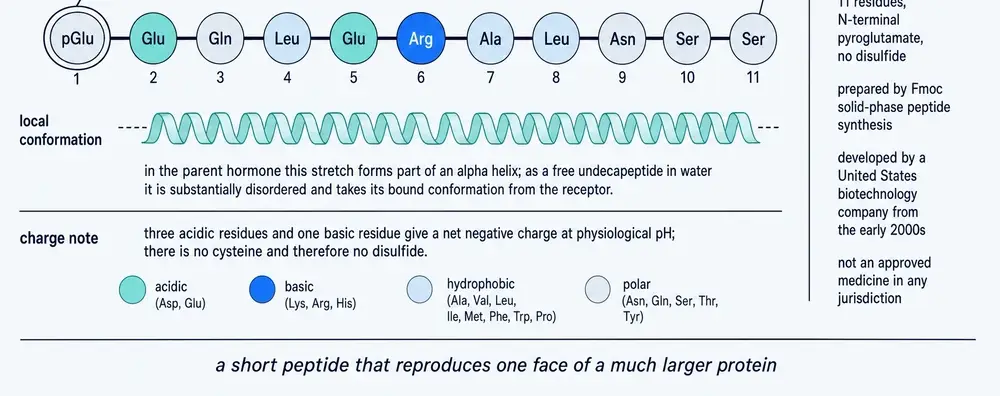
There is nothing exotic here. It is a short, linear, unmodified peptide of common amino acids with one spontaneous cyclisation, which is why it can be made by ordinary solid-phase synthesis for a fraction of what a recombinant glycoprotein costs. That cheapness is part of the point, and it is also the reason the compound has a substantial grey market.
07Two minutes
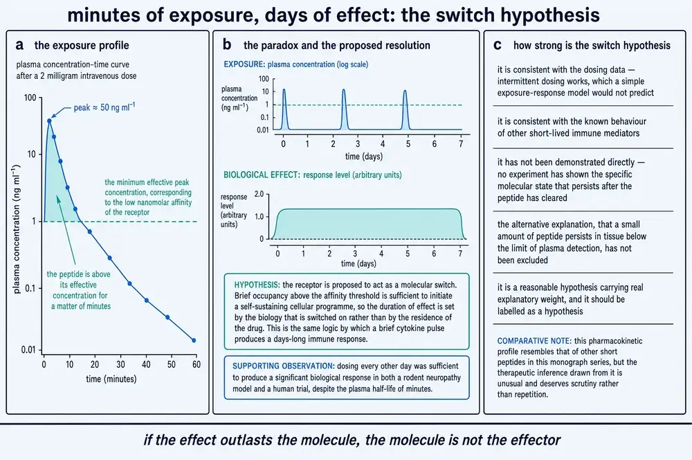
Here is the fact that everything else in this document has to be reconciled with. ARA-290's plasma half-life is approximately two minutes.
Reported figures range from "less than two minutes" to "less than ten minutes," and it is worth being honest about their provenance: no dedicated human pharmacokinetic study of this compound appears to have been published. Every human exposure figure in the literature — a peak concentration around 1.8 nanomolar after a 4 mg subcutaneous dose, an area under the curve of about 245 nanogram-minutes per millilitre after 6 mg — is cited second-hand as unpublished company data. The animal figures are firmer: the renal ischaemia studies in rats and pigs state a plasma half-life of about two minutes as a working parameter of their design. (van Rijt et al., 2013a; van Rijt et al., 2013b)
A two-minute half-life also shapes how the compound can be given at all. Continuous infusion is the obvious way to hold a short-lived drug at a working concentration, and no trial has used it. Every human study instead gave a bolus — intravenously in the earliest work, subcutaneously thereafter — which produces a brief spike and then nothing for a day. That choice only makes sense if the spike is what matters, which is precisely the assumption the next few paragraphs examine. It is worth noticing that the assumption was built into the trial designs before it was tested.
Two minutes is a strange number for a drug. It means that within twenty minutes of an injection, effectively none of the compound remains in circulation. And it produces a genuine puzzle that the literature has not resolved.
On one side, the short half-life clearly constrains dosing. In a mouse model of stroke, 30 micrograms per kilogram given twice daily significantly reduced infarct volume. The same 30 micrograms per kilogram given once daily did not. Neither did 100 micrograms per kilogram once daily — more than three times the dose, given half as often, produced nothing. The authors attributed the failure directly to the two-minute half-life: what matters is how often the receptor is engaged, not how much peptide is present when it is. (Wang et al., 2024)
On the other side, the effects routinely outlast the exposure by an absurd margin. In rats with surgically injured sciatic nerves, five doses given over the first ten days relieved allodynia — pain from a stimulus that should not hurt — for twenty weeks. That is nineteen weeks during which no drug was given and none could possibly have remained. (Swartjes et al., 2014)
The explanation offered in the literature is that the peptide does not act as a conventional receptor agonist that must be present to have an effect, but as a switch: a brief engagement reprograms the local injury response from a destructive mode into a repairing one, and the tissue then stays in the new mode. That is a plausible story and it is consistent with the asialoEPO result from 2003, where a molecule cleared in ninety seconds was fully neuroprotective. (Erbayraktar et al., 2003) It is also, at present, a story rather than a demonstrated mechanism. Nobody has shown what is being switched.
08The relatives, and why they are not the subject
This literature is unusually easy to get wrong, because at least five other molecules are discussed in the same papers, by the same authors, in the same journals, under names that differ by one letter.
CHBP, cyclic helix B peptide, is the important one. It takes the same eleven residues and closes them into a ring through a thioether bridge, stabilising the helical shape and extending the half-life by roughly 2.5-fold in human hepatocytes. (Yang et al., 2014; Peng et al., 2020) It has a large literature of its own, principally in kidney injury and transplantation, and it is a different chemical entity. This document reports its results only as its results. Twenty-six documents in the corpus are dominated by CHBP rather than by the subject, and they are excluded from every claim about ARA-290 rather than quietly absorbed into the total.
Carbamylated erythropoietin and asialoEPO are the earlier whole-protein derivatives described in Section 04. EMP-1, peginesatide and pegmolesatide are erythropoietin-mimetic peptides that engage the classical receptor and are designed to make red cells — the opposite goal. JM-4 is the most interesting relative: a nineteen-residue peptide derived by a different laboratory from a completely different region of erythropoietin, the loop between helices A and B, on the same reasoning that erythropoiesis and tissue protection live in different parts of the molecule. It, too, is reported as tissue-protective and non-erythropoietic. Neither group's work explains the other's, and no paper in this corpus reconciles them. If both are right, erythropoietin has at least two separable tissue-protective surfaces, which is a more interesting claim than either paper makes on its own. (Yuan et al., 2015)
Identity discipline is not pedantry in this literature; it is the difference between a true statement and a false one. The abbreviation HBSP is shared with the hepatitis B spliced protein, whose papers carry the words liver, apoptosis, fibrosis and PI3K/Akt — the same vocabulary as this peptide's own hepatic and renal work. It is also shared with a plasma protease, a hospital school programme, a computer-vision algorithm, an aerosol-optics term and a chromatography medium. The screening used to build this document admitted four protease papers on its first run before the output was read. Section 25 records how, and what was changed.
09The innate repair receptor, as proposed
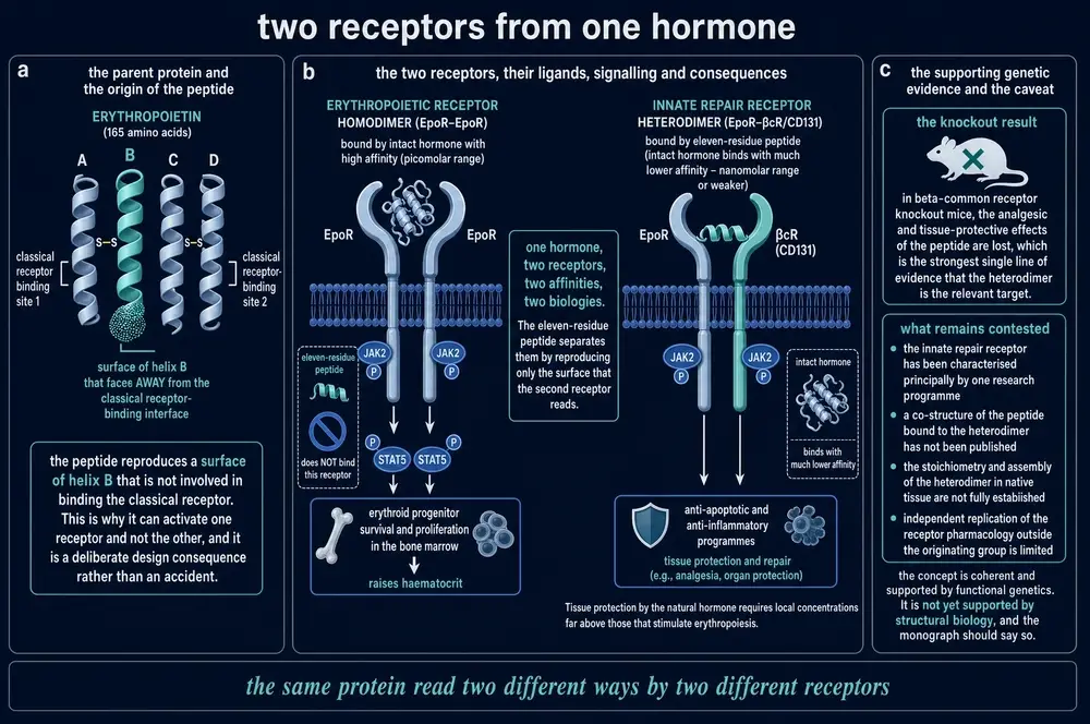
ARA-290's design rests on a claim about receptors, and the claim is more specific than it usually sounds when repeated. It is not merely that erythropoietin has two effects. It is that the two effects run through two physically different receptor assemblies, and that a molecule can be built to engage one and not the other.
The classical erythropoietin receptor works as a homodimer: two identical receptor chains, bound tightly by one molecule of hormone, signalling through JAK2 and STAT5 to tell a marrow progenitor to become a red cell. That machinery has been understood for decades and is not in dispute.
The proposal, made in 2004, is that in injured tissue erythropoietin instead engages a heterocomplex — one erythropoietin receptor chain partnered with CD131, the beta-common receptor. CD131 is not an obscure protein: it is the shared signalling subunit of the receptors for GM-CSF, interleukin-3 and interleukin-5, sitting at the centre of myeloid inflammatory signalling. A receptor that combines the repair machinery of erythropoietin with the inflammatory machinery of the myeloid cytokines is a genuinely attractive idea, and it was later given a name that has stuck: the innate repair receptor. (Brines et al., 2004; Brines, 2014)
The idea explains a great deal. It explains the affinity gap, because a hybrid receptor need not bind the hormone as tightly as the dedicated one. It explains why tissue protection appears only at high concentrations. It explains why the peptide is anti-inflammatory as well as anti-apoptotic. And it predicts exactly the drug ARA-290 is: something shaped to fit the hybrid and too small to fit the homodimer.
10What the supporting evidence can and cannot show
The 2004 paper offered four lines of evidence, and it is worth being precise about what each one establishes, because they are frequently reported as though they were interchangeable.
Affinity chromatography. Membrane preparations from rat brain, heart, liver and kidney were passed over columns bearing erythropoietin or its carbamylated derivative; the erythropoietin receptor came off in a complex with CD131. This shows the two proteins can be recovered together. It does not show that they touch, because anything held in the same membrane patch, or bridged by a third protein, will co-purify.
Co-immunoprecipitation. Pulling on the erythropoietin receptor in a neuronal cell line brought CD131 with it. This is a stronger proximity argument and the same caveat applies: co-immunoprecipitation demonstrates association within a complex, not direct contact between two partners.
Co-localisation. The two proteins were seen in the same places in spinal cord neurons and cardiomyocytes. At the resolution of light microscopy, co-localisation means "within a few hundred nanometres," which on a protein scale is a considerable distance.
Genetic knockout. In mice lacking CD131 — animals with entirely normal erythropoiesis — neither erythropoietin nor its carbamylated derivative protected cardiomyocytes or spinal cord neurons. This is the strongest of the four and it is a different kind of evidence: it shows that CD131 is required for the effect. It does not show that CD131 is part of the receptor. A protein can be required for a response without being in the complex that initiates it — it may be downstream, permissive, or needed to keep the responding cell in a state where it can respond at all. All four lines of evidence are from (Brines et al., 2004).
11The experiment that looked directly
For fourteen years the heterocomplex was inferred and never directly observed. In 2018 a group in Australia set out to observe it, using purified protein and the standard biophysical methods for detecting whether two things bind.
They expressed the extracellular portions of the human erythropoietin receptor and the human beta-common receptor and put them together under six independent conditions. Computational docking suggested the arrangement was sterically implausible: with erythropoietin bound, the membrane-proximal parts of the two receptors sit too far apart. Analytical size-exclusion chromatography gave two separate peaks in multiple buffers and molar ratios, with no complex. Pull-down assays showed no co-elution of either partner, with mass spectrometry confirming the identities. Microscale thermophoresis produced no binding curve. Surface plasmon resonance detected nothing up to 32 micromolar of one partner. Analytical ultracentrifugation found no higher-order species.
And, critically for this document, they ran the experiments with ARA-290 present — at concentrations up to 64 micromolar in the resonance experiments — on the reasoning that if the peptide is the ligand that assembles the complex, it should assemble it. It did not. The peptide also did not co-elute with either receptor. (Cheung Tung Shing et al., 2018)
The same paper added an animal result. If CD131 matters to erythropoietin signalling in life, mice lacking it should behave differently under erythropoietic stress. Mice were made anaemic with phenylhydrazine and recovered indistinguishably from wild-type animals. The authors' conclusion is blunt: the extracellular regions of the two receptors do not specifically associate, in the absence or the presence of erythropoietin or ARA-290, and the beta-common receptor plays no role in erythropoietin signalling in vivo. (Cheung Tung Shing et al., 2018)
No measurement of ARA-290 binding to any receptor — no dissociation constant, no half-maximal effective concentration, no occupancy figure — was located anywhere in this corpus of 419 retrieved full texts. The only study that attempted to measure it directly reported no detectable binding.
This is not a claim that the peptide does nothing. Its biological effects have been reproduced by many independent groups across many models, and Part Four describes them. It is a statement about what is known: after eighteen years, the fundamental pharmacological parameter of this molecule — what it binds, and how tightly — has not been published.
12Living with the contradiction
The obvious reading is that the receptor model is wrong and the compound's effects have another explanation. That reading is too quick, because the functional evidence has not gone away and some of it is very hard to dismiss.
In mice with surgically injured sciatic nerves, ARA-290 attenuated tactile allodynia in wild-type animals and had no effect whatever in mice lacking the beta-common receptor — the treatment effect against vehicle in the knockouts was p = 0.730. In a mouse stroke model, knocking down the beta-common receptor with siRNA injected into the brain's ventricles abolished the neuroprotection of both ARA-290 and erythropoietin. These are not proximity arguments. They are direct demonstrations that removing this protein removes this drug's effect, in two different injuries, by two different techniques. (Swartjes et al., 2013; Wang et al., 2024)
So the evidence says both that CD131 is necessary for the effect and that CD131 does not stick to the erythropoietin receptor. Three ways of reconciling that are on the table.
The complex may form through the membrane. A 2019 study found that anti-CD131 antibodies pulled down the erythropoietin receptor even with no ligand added at all, and that removing CD131 by gene editing abolished the activity of a synthetic ligand designed to act at the transmembrane segments. Since the 2018 negative study used only the extracellular fragments — which is what you must use for purified biophysics — a complex assembled by the parts embedded in the membrane would be invisible to it. This reconciles both bodies of evidence neatly. Its weakness for present purposes is that the ligand tested was not ARA-290. (He et al., 2019)
CD131 may be required without being the receptor. Nothing in the knockout experiments distinguishes a receptor subunit from a downstream requirement. CD131 sits at the hub of myeloid inflammatory signalling, and a drug whose principal effect is to quiet myeloid inflammation might well need that hub intact for reasons that have nothing to do with binding.
The receptor may be something else entirely. One striking result in this corpus points that way. The peptide protects neurons from hypoxia-induced death in the brain of the migratory locust — an animal with no erythropoietin receptor and no CD131 at all — apparently through CRLF3, an evolutionarily ancient cytokine receptor. That is an insect result and must not be read as mammalian mechanism. But it does establish that the peptide can produce a protective effect through a receptor that is not the one it was designed for. (Hahn et al., 2025)
Meanwhile the literature has divided. Some recent papers cite the negative study by name, concede that the definite role of the beta-common receptor remains to be addressed, and substitute knockdown experiments for the missing binding data. Some reviews list the heterocomplex as one of three candidate receptors alongside EphB4 and CRLF3. And a substantial number of therapeutic papers continue to describe the innate repair receptor as settled mechanism without citing the contrary evidence at all.
One further contradiction belongs here, because it cuts against the universality of the model rather than its existence. Darbepoetin — a long-acting engineered erythropoietin — remained fully cardioprotective after myocardial infarction in mice lacking the beta-common chain. (Kanellakis et al., 2010) If the heterocomplex were the universal route for tissue protection, that experiment should have failed. The most economical reading of the whole body of evidence is that erythropoietin protects tissue by more than one mechanism, that CD131 is genuinely required for some of them, and that ARA-290 depends on whichever of them CD131 serves.
13Nerve injury, and an effect that outlasts the drug
The strongest preclinical result for this compound is in neuropathic pain, and it is strong for an unusual reason: not the size of the effect but its duration.
The model is spared nerve injury. Two of the three branches of a rat's sciatic nerve are cut and tied; the third is left intact. The animal develops persistent hypersensitivity in the territory of the spared branch — light touch and cold become painful. It is a standard model of the chronic neuropathic pain that follows nerve damage in people, and it does not spontaneously resolve.
Female Sprague-Dawley rats received ARA-290 intraperitoneally at 3, 10, 30 or 60 micrograms per kilogram on days 1, 3, 6, 8 and 10 after the injury — five doses, then nothing. Mechanical allodynia improved dose-dependently, reaching significance at 30 micrograms per kilogram (p = 0.049) and 60 (p < 0.001), with a linear dose-response of R² = 0.56. Cold allodynia improved significantly at every dose (p < 0.0001 each, R² = 0.78). The animals were then followed for twenty weeks, and the higher doses kept them allodynia-free for longer, by log-rank analysis, p < 0.001. (Swartjes et al., 2014)
A mechanism was offered in the same work. Nerve injury activates microglia in the spinal cord, and that activation spreads over months: in untreated rats, microglial reactivity was confined to one spinal segment at two weeks and had spread across five segments by twenty weeks. ARA-290 at 30 micrograms per kilogram prevented that spread at both time points. Astrocytes, measured by GFAP, did not react to the injury at all in this rat model, so there was nothing for the drug to suppress — a null result the authors report plainly. (Swartjes et al., 2014)
A companion study in mice added the control that matters. ARA-290 attenuated allodynia in wild-type animals and did nothing at all in mice lacking the beta-common receptor. The same study found that the peptide produced no acute pain relief of any kind — tail-withdrawal latency was unchanged — which distinguishes it sharply from an analgesic. It is not blocking pain signalling. Whatever it is doing takes days and then persists. (Swartjes et al., 2013)
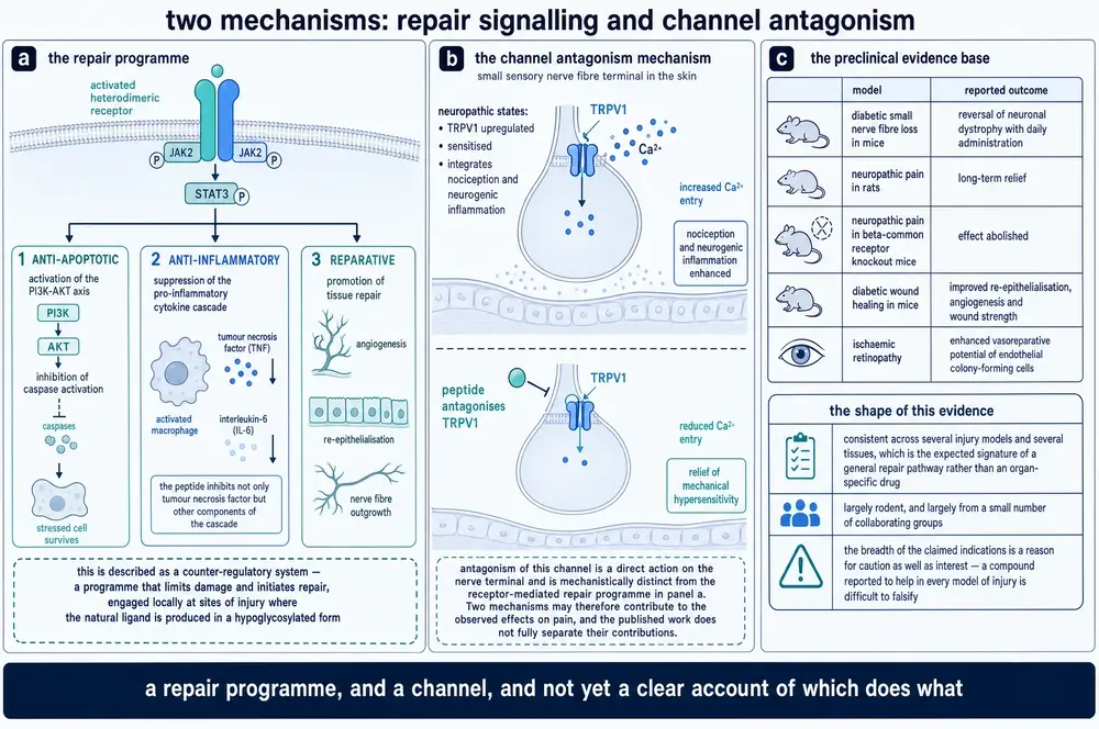
A second mechanism has been proposed for the pain effect, and it is mechanistically unrelated to the receptor. TRPV1 is the ion channel that opens in response to capsaicin, noxious heat and acid; in damaged nerves it is upregulated and sensitised, and it sits at the junction between feeling pain and generating the local inflammation that keeps a nerve irritable. ARA-290 has been reported to antagonise it directly at the nerve terminal, reducing calcium entry and relieving mechanical hypersensitivity.
If that is right, the compound has two ways of acting on pain: a slow one that reprograms the injury response through a receptor, and a fast one that blunts a channel. The published work does not separate their contributions, and the primary evidence for the channel action is thinner than for the receptor action — it rests largely on a single report, and is more often encountered as a citation inside reviews than as a result being replicated. It belongs in any honest account of the mechanism, with that caveat attached.
14The rest of the nervous system
Stroke. In male C57BL/6J mice with middle cerebral artery occlusion, 30 micrograms per kilogram twice daily reduced infarct volume and brain tissue loss, cut neuronal apoptosis, and improved neurological scores, with efficacy comparable to erythropoietin at 5,000 units per kilogram. Brain cytokines were unchanged at one day and significantly reduced at seven — the anti-inflammatory effect is not immediate. Unlike erythropoietin, the peptide changed no haematological parameter and no spleen measurement. (Wang et al., 2024)
Traumatic brain injury. In rats given a mild controlled cortical impact combined with haemorrhagic hypotension, the peptide at 30 micrograms per kilogram every twelve hours for three days reduced contusion volume from 20.8 cubic millimetres to 5.9 — statistically indistinguishable from full erythropoietin at 5,000 units per kilogram, which gave 7.7. In a separate mild injury model it improved Morris water maze performance (latency 22.3 seconds against 26.3 in controls, p = 0.022) while producing no effect on motor performance, which the mild model barely impaired in the first place. (Robertson et al., 2012; Robertson et al., 2013)
Autoimmune neuritis. In Lewis rats with experimental autoimmune neuritis, ARA-290 at 30 milligrams per kilogram per day — a dose roughly a thousand times the one used in the pain studies, which is worth noticing — reduced peak neurological severity, shortened disease duration, reduced inflammatory infiltration of the sciatic nerve, promoted remyelination, and shifted helper T cells away from the Th1 and Th17 phenotypes toward Th2 and regulatory. Red cell count, haemoglobin and haematocrit were unchanged. (Liu et al., 2014)
Diabetic autonomic neuropathy. In Akita diabetic mice with four months of established disease, the peptide at 36.1 micrograms per kilogram per day reduced neuritic dystrophy in sympathetic ganglia to 38 per cent of untreated levels. In the same ganglia of the same animals it did not reduce neuronopathy and did not prevent neuron loss, and it changed no measure of glycaemic control. Insulin, run in parallel, normalised all of them. This is a clean and instructive dissociation: the drug rescued the nerve fibres and not the cell bodies they belong to. (Schmidt et al., 2011)
One feature of this literature deserves more attention than it usually receives: the doses do not agree with each other. The neuropathic-pain work in rats used 3 to 60 micrograms per kilogram. The stroke and brain-injury work used 30. The kidney work used 10 nanomoles per kilogram, which is about 12.6 micrograms per kilogram. The autoimmune neuritis study used 30 milligrams per kilogram — a thousandfold more than the pain studies, in the same species, by the same route. The islet and ageing studies sit in between at 100 to 120 micrograms per kilogram.
A thousandfold spread across a literature that reports consistent effects is not by itself a contradiction; different injuries in different tissues may genuinely need different exposures, and a peptide with a two-minute half-life has a wide margin before anything accumulates. But it does mean that no dose-response relationship has been established across this body of work as a whole, and that a reader cannot infer from one model what exposure another would need. It also means the compound has never been shown to have a ceiling. Where a dose-response was measured within a single study, it was sometimes linear and sometimes not: rats with nerve injury showed a clean gradient across a twentyfold range, while the pivotal human trial's middle dose outperformed its highest.
Read together, the nervous-system evidence has a consistent shape. The compound does not block pain; it changes how injured tissue behaves, over days, and the change persists. It acts on the supporting cells — microglia, Schwann cells, the immune cells that arrive after an injury — more clearly than on neurons themselves. And where an experiment has separated the fibre from the cell body, or the phenotype of a glial population from its number, the compound has moved the first and not the second. That is a coherent picture of a repair modifier rather than an analgesic or a neuroprotectant, and it is the picture the clinical programme then tried to test.
It also explains why the animal work reads as broadly as it does. If a compound acts on the shared machinery of injury rather than on any one tissue, then almost any acute injury model will show something, and the list of successes will grow without the evidence for any single indication getting deeper. That is a strength of the hypothesis and a weakness of the literature at the same time.
15The kidney, and a retraction
Kidney ischaemia-reperfusion injury is the workhorse model of this literature, and the clearest finding in it is about timing rather than dose.
In Lewis rats given thirty minutes of unilateral warm ischaemia, ARA-290 at 10 nanomoles per kilogram given one hour after reperfusion significantly reduced serum creatinine, interleukin-6 messenger RNA, the injury marker Kim-1, and tubular damage on histology. The same dose given at four hours produced non-significant trends only. Giving it at both one and four hours produced non-significant trends only. (van Rijt et al., 2013a) In pigs given forty-five minutes of ischaemia, repeated dosing at 0, 2, 4 and 6 hours raised glomerular filtration rate across seven days and reduced interstitial fibrosis. (van Rijt et al., 2013b)
The consistent reading across the non-retracted literature is that the treatment window is narrow and opens at the moment of injury. One paper in this corpus states the reasoning explicitly: the receptor components are swiftly upregulated on injury, indicating a critical window at onset, and administration at a later point will be much less effective. (Wu et al., 2024)
Against that stood a 2012 paper in Molecular Medicine reporting that the peptide attenuated rat acute kidney injury when given six hours into reperfusion — a far more clinically useful claim, because six hours is long enough to get a patient to a hospital. (Patel et al., 2012)
That paper was retracted in 2026.
The retraction notice states that concerns were raised about western blots in that paper's third figure — that in one panel three lanes appeared highly similar, and that in another a lane appeared highly similar to a lane in a different panel. The authors supplied original autoradiograms. The publisher's further checks found that "a number of bands selected for publication did not appear to be representative of the observed experimental results," and the Editor-in-Chief recorded that he no longer had confidence in the presented data. Three of the authors do not agree with the retraction; the notice records that the others did not respond to correspondence. Michael Brines, the compound's co-inventor, is among the listed authors. (Patel et al., 2026)
Two things follow, and they should be kept separate. The narrow one is that the six-hour window claim is withdrawn, and the remaining evidence says the window is about an hour. The broader one is a caution about the whole literature: this was not an obscure paper, and the images were not detected by peer review, by the fourteen years of citation that followed, or by any automated screen — they were detected by someone looking at the figures. The rest of this literature has not had that examination.
Where the peptide has been tested in other kidney models the results are broadly positive and unremarkable: reduced creatinine, urea and tubular injury in mouse sepsis-associated injury, with macrophages shifted from the inflammatory M1 phenotype toward M2; reduced urea and proteinuria in rats given ciclosporin; reduced proteinuria and preserved podocyte architecture in puromycin nephropathy, in which haemoglobin, haematocrit, urea and creatinine were all unaffected. A biodistribution experiment using an iridium-tagged version found the peptide concentrated on the apical surface of renal tubular epithelium, with weak signal in lung and essentially none in heart or liver. (Huang et al., 2024; Wu et al., 2013; Chen et al., 2024; Wu et al., 2023)
16The heart
The cardiac literature repeats the timing lesson and then complicates it.
In rats with permanent coronary ligation, a single bolus given immediately after ligation reduced apoptosis in the area at risk by about 80 per cent and inflammation by 34 per cent at 24 hours, and cut infarct size by half at 24 hours and by a quarter at six weeks. It worked at doses as low as 1 microgram per kilogram. The same 60 micrograms per kilogram given 24 hours after the infarction did nothing to infarct size. (Ahmet et al., 2011; Ueba et al., 2010)
Yet in animals with established disease the picture reverses. Rats whose treatment began two weeks after infarction — in an already remodelling heart — received 60 micrograms per kilogram twice weekly for ten months, and the results were substantial: mortality halved; end-diastolic volume rose 41 per cent against 86 per cent in controls; ejection fraction fell 4 per cent against 63 per cent; infarct expansion 3 per cent against 38 per cent. No immune response and no haematocrit change over ten months. (Ahmet et al., 2013)
"Delayed" therefore means two different things. Missing the acute window by a day forfeits the acute protection. Beginning months later, in a chronic remodelling process, addresses a different problem and appears to work.
The ageing study is the most interesting and the most sobering. Rats aged 18 months received 100 micrograms per kilogram three times weekly for fifteen months. The peptide blunted the age-related rise in end-systolic volume by about three-quarters and the fall in ejection fraction by about half, blunted the rise in blood pressure, reduced cardiac immune-cell infiltration, enhanced autophagy, raised the mitochondrial permeability-transition threshold, and reduced the frailty index. It did not extend life. Survival between groups did not differ (p = 0.182). (Winicki et al., 2022)
17Metabolism, islets, bone and immunity
In Goto-Kakizaki rats, a spontaneous non-obese model of type 2 diabetes, ARA-290 reduced HbA1c by about 20 per cent without changing whole-body or hepatic insulin sensitivity; the effect came from improved glucose-stimulated insulin secretion in the islets themselves, through better calcium handling and ATP production, and was abolished by inhibiting protein kinase A. (Muller et al., 2016) In diet-induced obese mice the peptide reduced weight and fat mass dose-dependently, normalised glucose and insulin tolerance, reduced adipose tissue TNF-alpha, interleukin-6 and MCP-1, and did not stimulate haematopoiesis. (Liu et al., 2015; Collino et al., 2014)
Isolated human pancreatic islets from deceased donors, stressed with a cocktail of inflammatory cytokines, retained ATP, showed reduced caspase 3/7 activity and secreted more insulin in response to glucose when cibinetide was present at 100 nanomolar. In a loop model using human blood, it reduced platelet consumption. When those human islets were transplanted into mice, the treated recipients had more human insulin in the liver and more human C-peptide in serum. This is a promising result and it is not a human outcome: the recipients were mice, and it is described here as what it is. (Yao et al., 2021)
Cibinetide inhibited osteoclast formation in vitro and raised bone mineral density in mice — cortical by 5.8 per cent, trabecular by 5.2 per cent — without affecting haemoglobin, and it counteracted erythropoietin's own bone-resorbing effect while leaving erythropoietin (Awida et al., 2021) 's red-cell effect intact. In mouse colitis it reduced myeloid chemokine and cytokine output through CD131, JAK2 and PI3K, with the effect confirmed absent in CD131-null and JAK2-null macrophages. (Nairz et al., 2017) In mouse lupus models it reduced antinuclear antibodies, anti-double-stranded-DNA antibodies, creatinine and renal immunoglobulin deposition without changing erythropoiesis. (Huang et al., 2018)
18Where it failed
A drug that works in every model is usually a drug whose failures have not been published. This one has published failures, and they are more informative than most of the successes.
That is worth saying plainly, because a compound whose literature contains no failures is not a compound that never failed — it is a compound whose failures were not written up. Negative results are harder to publish, and a small field working on a single molecule has every incentive to leave them in a drawer. The presence of a real negative ledger here counts in the literature's favour, and it is also what makes the positive results usable: an effect that appears in some models and not others has a shape, and a shape can be tested. An effect that appears everywhere cannot.
Two of these deserve emphasis because they point the same way, and it is not the way one might expect.
In mouse heterotopic heart transplantation, full erythropoietin combined with CTLA4-Ig prolonged graft survival substantially — median 34 days against 17 — and the mechanism was traced to the classical erythropoietin receptor homodimer on myeloid cells. ARA-290, added to the same regimen at 0.5 milligrams per kilogram three times weekly, did not prolong graft survival at all. In human alloreactive T cells in vitro, erythropoietin suppressed CD4-positive T-cell proliferation dose-dependently through the classical homodimer; adding ARA-290 did not alter T-cell proliferation or cytokine production. (Horwitz et al., 2022; Cravedi et al., 2014)
Both are negative results for the compound, and both are evidence for its selectivity. If ARA-290 were simply a weak erythropoietin, it should have produced weak versions of these effects. It produced none, in exactly the two settings where the effect was shown to run through the receptor ARA-290 is designed not to engage. That is the behaviour of a genuinely selective molecule, and it is among the better arguments in the whole corpus that the peptide is doing something specific.
The rest are boundary conditions. In a mouse model of oxygen-induced retinopathy the peptide had no effect on preretinal neovascularisation at 1, 10 or 30 micrograms per kilogram — a complete dose-ranging null, which the authors frame as reassuring on safety grounds, since promoting new vessel growth is one of erythropoietin's liabilities, and which is also simply a failure to work. In a chronic stress model of depression it reversed anhedonia and reduced microglial activation but did not significantly reduce forced-swim immobility and did not change prefrontal cortex TNF-alpha. (McVicar et al., 2011) In diabetic rat retina it shifted microglial morphology without changing microglial numbers. And in the aged-rat study it improved almost every measure of cardiac function and frailty without extending survival by a single day.
19Four trials
Everything in Part Four happened in animals. This part is about the other literature, and the first thing to say about it is how small it is.
Searching ClinicalTrials.gov by intervention returns four registered studies of this compound, in total, across its entire history.
It is worth pausing on what that number means, because "phase 2" is a phrase that sounds further along than it is. A drug that reaches the market has normally been through a dose-finding programme, at least one adequately powered trial designed to detect a difference patients would notice, and then a confirmatory trial in a different population run by different investigators. The purpose of the last of these is specifically to catch the ways a promising mid-size result can be wrong: chance, a surrogate endpoint that does not track the outcome, a population that was easier to treat than the general one, or analytical choices made after the data were seen. None of that has happened here.
The largest enrolled 64 people. The smallest enrolled nine and was terminated. One study's registry record has never been updated and still reads "unknown status," although its results were published in 2015. Another was registered on ClinicalTrials.gov only in October 2024 — seven years after the study ended and four years after it was published — and is recorded as terminated.
Beyond the registered four there is earlier work that was never registered and never published: a single-ascending-dose study in 36 people, a multiple-ascending-dose study in 14, and a single-dose study in people with impaired kidney function. These are described only in the protocol text of one of the registered trials and in a review, always attributed to unpublished company data. Adding everything together gives roughly two hundred people ever exposed to this molecule — a figure the diabetic macular oedema paper independently corroborates when it refers to "over 200 treated." (Lois et al., 2020)
For comparison, a single phase 3 trial of a modern metabolic drug enrolls more people than that in a fortnight.
20Sarcoidosis: the first signal
Sarcoidosis is an inflammatory disease that forms granulomas, most often in the lungs. A substantial minority of patients also develop small fibre neuropathy: the thinnest sensory nerves die back, producing burning feet, pain from bedsheets, disturbed temperature sensation and autonomic symptoms. It is common, disabling, poorly treated, and — importantly for a small company — rare enough to qualify for orphan designation.
The first trial, reported in 2012, gave 2 milligrams intravenously three times weekly for four weeks to 12 patients, against 10 on placebo. On the small fibre neuropathy screening list the treated group improved by 11.5 points against 2.9 on placebo (p < 0.05), and two quality-of-life dimensions improved from baseline. Two other instruments — the Brief Pain Inventory and the Fatigue Assessment Scale — improved significantly and equivalently in both arms, and a depression scale did not move in either. The authors reported no safety concerns. (Heij et al., 2012b; Heij et al., 2012a)
That is a reasonable early signal, and it has the shape that recurs throughout this programme: the disease-specific instrument separates from placebo, and the general symptom instruments do not.
21The pivotal trial, read against its own registry record
The programme's central study is NCT02039687: a double-blind, placebo-controlled phase 2b trial run by Araim Pharmaceuticals at the Cleveland Clinic and Leiden University Medical Center, randomising 64 patients with sarcoidosis-associated small fibre neuropathy and neuropathic pain to 1, 4 or 8 milligrams of cibinetide daily by subcutaneous injection, or placebo, for 28 days, with twelve weeks of follow-up.
The pre-specified primary endpoint was the change at 28 days in corneal nerve fibre area, measured by corneal confocal microscopy — a non-invasive way of imaging the dense nerve plexus beneath the corneal surface, which thins in small fibre neuropathy and can be re-imaged in the same patient.
The published result: the placebo-corrected mean change in corneal nerve fibre area was 109 square micrometres at 1 milligram (95% confidence interval −429 to 647), 697 at 4 milligrams (159 to 1236, p = 0.012), and 431 at 8 milligrams (−130 to 992). The trial met its primary endpoint at one of three doses. (Culver et al., 2017)
The registry's own posted results give the raw arm means, and they agree. Placebo fell by 170.0; the 1 milligram arm fell by 64.3; the 4 milligram arm rose by 533.8; the 8 milligram arm rose by 203.8. Subtracting placebo gives 105.7, 703.8 and 373.8 — close to the paper's covariance-adjusted 109, 697 and 431. There is no discrepancy between the publication and the registry on this endpoint, which is worth stating plainly, because there is one further down. Arm means are the registry-posted results for NCT02039687.
Before reading the numbers it is worth knowing what the instrument does. The cornea is the most densely innervated tissue in the body, and its nerves sit in a thin plexus just beneath the surface, where a confocal microscope can image them through the intact eye in a few minutes without touching the patient. That makes it possible to photograph the same square millimetre of nerve before and after treatment — something no other small-fibre measure allows, since the alternative is a skin biopsy that cannot be repeated in the same spot. It is a genuinely elegant choice of endpoint for a four-week trial.
It is also a surrogate. Nobody's symptoms are caused by their corneal nerve density, and no trial has established that changing it changes how a patient with sarcoidosis feels or functions. The endpoint was chosen because it is sensitive and repeatable, which are the right properties for detecting whether a drug does anything at all — and the wrong properties for concluding that what it does matters.
Three observations follow from the figure, and none of them makes the result wrong.
The dose-response is not monotonic. Eight milligrams did less than four. For a receptor agonist this is not fatal — bell-shaped curves are common where a ligand can drive receptor internalisation or engage a second target — but it is the pattern that a false positive also produces, and with sixteen patients per arm the trial cannot distinguish the two.
The variance is enormous. The 4 milligram arm's standard deviation was 1,110 against a mean of 534. That is what the corneal measurement is like in this population, and it is why the confidence interval on the significant result runs from 159 to 1236 — an eight-fold range.
Withdrawals concentrated at the top dose. Sixteen of sixteen completed at 1 milligram and on placebo; fifteen of sixteen at 4 milligrams; thirteen of sixteen at 8 milligrams. Serious adverse events were two in the 1 milligram arm, none at 4, one at 8 and none on placebo. The single event at 8 milligrams was suicidal ideation, in a participant with a pre-existing history of depression; the protocol was subsequently amended to add formal suicidality screening to all later assessments. Across the whole programme this is the one adverse event that changed how the trials were run. (Lois et al., 2020)
22Four endpoints named "nerve fibre"
This is the part of the record that a reader most needs, and it is the part least visible from any abstract.
Small nerve fibres can be counted in two tissues by several methods, and the resulting numbers are not interchangeable. In the cornea, confocal microscopy yields at least four distinct variables: fibre density, a count of nerve bundles per square millimetre; branch density, a count of branch points; fibre length, total length per unit area; and fibre area, a two-dimensional measure that responds to the thickness of fibres as well as to how many there are. In skin, a punch biopsy yields intraepidermal nerve fibre density — the histological standard for diagnosing small fibre neuropathy — and, with a different stain, a count of GAP-43-positive fibres, which marks the subset actively regrowing.
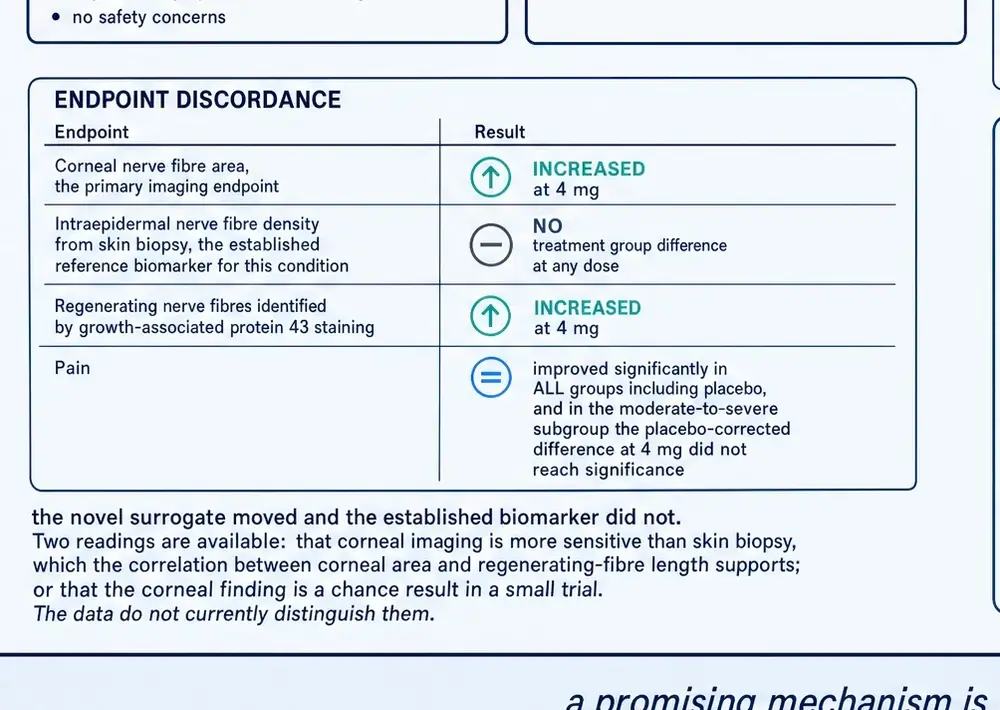
In this one trial, in this one group of patients, over these same 28 days, those measures did the following.
The distinction is not academic. A patient with small fibre neuropathy comes to a clinic because their feet burn and they cannot sleep. The intraepidermal fibre count in a skin biopsy is the measure the diagnostic literature has spent two decades tying to that experience; the corneal measures are newer, easier to repeat, and not yet anchored to how anyone feels. When the two disagree, which one is believed decides whether the trial reads as a success or as a null — and nothing in the data settles that question.
Corneal nerve fibre area rose — the primary endpoint, and the trial's headline. Corneal nerve branch density rose in a later reanalysis. Corneal nerve fibre density, the conventional diagnostic count, did not move. Corneal nerve fibre length did not reach significance by analysis of covariance. In skin, GAP-43-positive regenerating fibres increased in the 4 milligram group (p = 0.035). And total intraepidermal nerve fibre density — the registered secondary endpoint, and the diagnostic gold standard — did this, in fibres per millimetre: placebo +0.8, 1 milligram +0.5, 4 milligrams +0.4, 8 milligrams −0.3. Registry-posted results, NCT02039687.
Placebo numerically beat every active dose on the registered skin endpoint. The publications do not report that comparison. What they report instead, from the same biopsies, is the GAP-43-positive count — a differently defined measure that was not one of the trial's registered outcomes.
None of this establishes that the trial was misreported. The GAP-43 measure is scientifically defensible and arguably more sensitive: a regenerating fibre is exactly what one would expect to see first, before total density has time to recover in a four-week study, and the investigators' own review states plainly that the treatment increased corneal fibres "without affecting proximal and distal limb intraepidermal nerve fiber densities." They did not hide it. (Dahan et al., 2016)
But the reader is entitled to the shape of the whole result. Two tissues were measured by six methods. The measures that improved were the two-dimensional corneal one and the regeneration-specific skin one. The measures that did not improve were the conventional diagnostic counts in both tissues. A reader who saw only the phrase "significantly increased small nerve fiber abundance in the cornea and skin" would not know that.
The 2018 reanalysis compounds the difficulty in a way its authors are candid about. It compares only the 4 milligram arm against placebo, dropping the other two doses, and it introduces a new pixel-counting implementation of fibre area that differs from the registry's primary measure. On that new metric the difference is significant. On the conventional length measure, in the same two arms, analysis of covariance showed no significant difference. The paper describes itself as methods development, and it is. It is also the source most often cited for the claim that the compound regrows nerves. (Brines et al., 2018)
The patient-reported outcomes complete the picture, and they need to be read with one fact in mind: placebo responses in neuropathic pain trials are large. Patients who enrol are usually in a bad phase of a fluctuating condition, they are being examined attentively for the first time in months, and pain is a report rather than a measurement. Improvement on placebo is therefore expected, and the question is never whether the treated group improved but whether it improved more.
Five instruments were used. The Small Fibre Neuropathy Screening List asks twenty-one questions about the specific symptoms of this condition and is the most disease-specific of the set. The Brief Pain Inventory scores pain severity and, separately, how much pain interferes with daily activities. The Neuropathic Pain Symptom Inventory scores ten qualities of pain — burning, electric shocks, pins and needles — that distinguish nerve pain from other kinds. The Fatigue Assessment Scale measures a symptom that dominates sarcoidosis and is largely independent of the neuropathy. On every one of them a lower score is better.
The publication states that pain improved significantly in all groups, including placebo, and that patients with moderate-to-severe baseline pain showed a clinically meaningful placebo-corrected decrease in pain intensity in the 4 milligram group, at p = 0.157. A p-value of 0.157 is not statistically significant, and describing the difference it accompanies as clinically meaningful is a characterisation the number does not support. (Culver et al., 2017)
23Diabetes, the eye, and where the programme stopped
A phase 2 study in Stockholm gave 4 milligrams daily for 28 days to patients with prediabetes or drug-naive type 2 diabetes, with a further 28 days of follow-up. Neuropathic symptoms improved on the PainDetect instrument, and HbA1c and lipid profile improved across the 56 days. Corneal nerve fibre density increased significantly in the subgroup whose baseline density was more than one standard deviation below normal, with no change on placebo — which is a direct disagreement with the sarcoidosis trial, where that same named endpoint did not move. The two populations differ, and one plausible explanation offered in the corpus is that sarcoidosis and diabetes damage corneal fibres differently, changing which measure is sensitive. It remains that the same drug, at the same dose and route, moved a given endpoint in one disease and not in another. Its registry record still reads "unknown status" and its results were never posted. (Brines et al., 2015)
The last trial was in the eye. Nine patients with diabetic macular oedema in Belfast self-administered 4 milligrams daily for twelve weeks; eight completed. Best-corrected visual acuity, the primary endpoint, did not improve — the mean change was a loss of 2.9 letters. Central retinal thickness did not improve (+10 micrometres). Central retinal sensitivity did not improve. Tear production did not improve. No patient gained ten or more letters. The one whole-group improvement was a vision-related quality-of-life questionnaire, by 2.7 points. Three of nine eyes with the thickest baseline oedema improved beyond test-retest variability, and all four patients with abnormal baseline albumin-to-creatinine ratios improved. No serious adverse events occurred and no anti-drug antibodies were detected. The trial is recorded as terminated. Its authors are transparent that the headline result is null and that the individual-level signals warrant further investigation. (Lois et al., 2020)
The molecule's defining property is that it does not stimulate red-cell production. That is the entire reason it exists, and it is well documented in animals: haematocrit, haemoglobin, red cell counts and reticulocytes were unchanged in every animal study in this corpus that measured them.
No published human haematology table for this compound was located anywhere — not in the trial publications, not in the registry's posted results, not in any review. The human non-erythropoietic claim rests on general statements that laboratory assessments raised no safety issues, plus extrapolation from the animal data. That is likely to be correct. It has not been shown.
Where the programme stands is a matter of record rather than inference. Both European orphan designations — EU/3/13/1191 for sarcoidosis, granted 7 October 2013, and EU/3/16/1721 for prevention of graft loss in pancreatic islet transplantation, granted 29 August 2016 — remain formally active, the sponsorship having transferred from a United Kingdom entity to an Irish one in April 2019. A company press release from May 2017 also claims United States orphan and Fast Track designations; the orphan designation is corroborated by the European agency's own record, and the Fast Track claim rests on the press release alone.
There is no marketing authorisation anywhere. ChEMBL records the compound's maximum clinical phase as 2. No trial has recruited since 2017, and no new study is registered. Widely repeated statements that the sponsor has ceased operations could not be traced to any primary source and are not asserted here.
Weighing all of this requires separating two questions that are easy to run together. The first is whether the molecule does anything — whether it has real pharmacological activity in living tissue. On that the evidence is substantial: independent laboratories on three continents, working across five organ systems and at least eight species, report effects in the same direction, with a genetic control that behaves as the hypothesis predicts and two clean negatives in exactly the settings where the mechanism says it should fail. That is a stronger preclinical package than most compounds ever assemble.
The second question is whether any of it helps a patient, and there almost nothing has been established. One trial of sixty-four people, one dose out of three, one surrogate endpoint, twenty-eight days — and the instruments that measure how people actually feel moved about as much on placebo. Distinguishing these two questions is most of the work of reading a file like this one, and running them together is how a compound acquires a reputation its evidence does not support.
It is worth being clear about what this record does and does not mean. ARA-290 is not a failed drug in the ordinary sense — it was never taken to a trial capable of failing. Its one adequately-sized study met its primary endpoint at one of three doses, in 64 people, over 28 days, on a surrogate measure of nerve structure. Whether that surrogate predicts how patients feel or function over years is exactly the question a phase 3 trial exists to answer, and no such trial has been run. The molecule sits in the large category of plausible compounds with a positive small trial and no confirmation, and the correct description of it is not "promising" or "disproven" but unresolved.
What makes it genuinely interesting is not the clinical file, which is thin. It is that a hormone's tissue-protective function was traced to one face of one helix, reproduced by eleven synthetic residues, and shown to work across five organ systems and at least eight species by laboratories with no connection to each other — while the receptor that was supposed to explain all of it failed the only direct test anyone has published. Whatever ARA-290 is doing, the field does not yet know what it binds.
This document describes published research. It does not recommend human use of ARA-290 or of any other compound, and specifies no dose, route or schedule for any person. Doses appear only as investigators administered them in the studies described, with population and duration attached, and their appearance here is not an endorsement of them.
ARA-290 holds no marketing authorisation in any jurisdiction. Orphan designation is an incentive granted to encourage development in a rare disease; it is not a finding of safety or efficacy and it is not an approval. The compound is sold for research use only, and this document found roughly eighty-eight times as many vendor product pages naming it in the project's own archives as peer-reviewed full texts.
24References
Generated from verified NCBI records rather than from recall. Author lists, journal names, volumes, pages and identifiers are taken from the PubMed record for each citation, and the build refuses to run if any identifier fails to resolve. This is not a formality: earlier monographs in this series shipped reference lists drafted from memory in which identifiers pointed at real but unrelated papers, and the failure is invisible to every word count and byte total in a build.
- Ahmet I, Tae HJ, Juhaszova M, Riordon DR, Boheler KR, Sollott SJ, et al.. A small nonerythropoietic helix B surface peptide based upon erythropoietin structure is cardioprotective against ischemic myocardial damage. Mol Med. 2011;17(3-4):194-200.
PMID 21170473 · doi:10.2119/molmed.2010.00235 · PMC3060982 - Ahmet I, Tae HJ, Brines M, Cerami A, Lakatta EG, Talan MI. Chronic administration of small nonerythropoietic peptide sequence of erythropoietin effectively ameliorates the progression of postmyocardial infarction-dilated cardiomyopathy. J Pharmacol Exp Ther. 2013;345(3):446-56.
PMID 23584743 · doi:10.1124/jpet.113.202945 · PMC3657107 - Awida Z, Bachar A, Saed H, Gorodov A, Ben-Califa N, Ibrahim M, et al.. The Non-Erythropoietic EPO Analogue Cibinetide Inhibits Osteoclastogenesis In Vitro and Increases Bone Mineral Density in Mice. Int J Mol Sci. 2021;23(1).
PMID 35008482 · doi:10.3390/ijms23010055 · PMC8744753 - Brines M. Discovery of a master regulator of injury and healing: tipping the outcome from damage toward repair. Mol Med. 2014;20 Suppl 1(Suppl 1):S10-6.
PMID 25549226 · doi:10.2119/molmed.2014.00167 · PMC4374522 - Brines M, Patel NS, Villa P, Brines C, Mennini T, De Paola M, et al.. Nonerythropoietic, tissue-protective peptides derived from the tertiary structure of erythropoietin. Proc Natl Acad Sci U S A. 2008;105(31):10925-30.
PMID 18676614 · doi:10.1073/pnas.0805594105 · PMC2504825 - Brines M, Grasso G, Fiordaliso F, Sfacteria A, Ghezzi P, Fratelli M, et al.. Erythropoietin mediates tissue protection through an erythropoietin and common beta-subunit heteroreceptor. Proc Natl Acad Sci U S A. 2004;101(41):14907-12.
PMID 15456912 · doi:10.1073/pnas.0406491101 · PMC522054 - Brines M, Culver DA, Ferdousi M, Tannemaat MR, van Velzen M, Dahan A, et al.. Corneal nerve fiber size adds utility to the diagnosis and assessment of therapeutic response in patients with small fiber neuropathy. Sci Rep. 2018;8(1):4734.
PMID 29549285 · doi:10.1038/s41598-018-23107-w · PMC5856845 - Brines M, Dunne AN, van Velzen M, Proto PL, Ostenson CG, Kirk RI, et al.. ARA 290, a nonerythropoietic peptide engineered from erythropoietin, improves metabolic control and neuropathic symptoms in patients with type 2 diabetes. Mol Med. 2015;20(1):658-66.
PMID 25387363 · doi:10.2119/molmed.2014.00215 · PMC4365069 - Cerit H, Veer IM, Dahan A, Niesters M, Harmer CJ, Miskowiak KW, et al.. Testing the antidepressant properties of the peptide ARA290 in a human neuropsychological model of drug action. Eur Neuropsychopharmacol. 2015;25(12):2289-99.
PMID 26431906 · doi:10.1016/j.euroneuro.2015.09.005 - Chen T, Shen XY, Liang HM, Shi H, Yuan L. Renal protective effects of helix B surface polypeptide in rats with puromycin aminonucleoside nephropathy. Ren Fail. 2024;46(2):2394637.
PMID 39189638 · doi:10.1080/0886022X.2024.2394637 · PMC11351356 - Cheung Tung Shing KS, Broughton SE, Nero TL, Gillinder K, Ilsley MD, Ramshaw H, et al.. EPO does not promote interaction between the erythropoietin and beta-common receptors. Sci Rep. 2018;8(1):12457.
PMID 30127368 · doi:10.1038/s41598-018-29865-x · PMC6102255 - Coleman TR, Westenfelder C, Tögel FE, Yang Y, Hu Z, Swenson L, et al.. Cytoprotective doses of erythropoietin or carbamylated erythropoietin have markedly different procoagulant and vasoactive activities. Proc Natl Acad Sci U S A. 2006;103(15):5965-70.
PMID 16585502 · doi:10.1073/pnas.0601377103 · PMC1458681 - Collino M, Benetti E, Rogazzo M, Chiazza F, Mastrocola R, Nigro D, et al.. A non-erythropoietic peptide derivative of erythropoietin decreases susceptibility to diet-induced insulin resistance in mice. Br J Pharmacol. 2014;171(24):5802-15.
PMID 25164531 · doi:10.1111/bph.12888 · PMC4290718 - Cravedi P, Manrique J, Hanlon KE, Reid-Adam J, Brody J, Prathuangsuk P, et al.. Immunosuppressive effects of erythropoietin on human alloreactive T cells. J Am Soc Nephrol. 2014;25(9):2003-15.
PMID 24676641 · doi:10.1681/ASN.2013090945 · PMC4147979 - Culver DA, Dahan A, Bajorunas D, Jeziorska M, van Velzen M, Aarts LPHJ, et al.. Cibinetide Improves Corneal Nerve Fiber Abundance in Patients With Sarcoidosis-Associated Small Nerve Fiber Loss and Neuropathic Pain. Invest Ophthalmol Vis Sci. 2017;58(6):BIO52-BIO60.
PMID 28475703 · doi:10.1167/iovs.16-21291 - Dahan A, Dunne A, Swartjes M, Proto PL, Heij L, Vogels O, et al.. ARA 290 improves symptoms in patients with sarcoidosis-associated small nerve fiber loss and increases corneal nerve fiber density. Mol Med. 2013;19(1):334-45.
PMID 24136731 · doi:10.2119/molmed.2013.00122 · PMC3883966 - Dahan A, Brines M, Niesters M, Cerami A, van Velzen M. Targeting the innate repair receptor to treat neuropathy. Pain Rep. 2016;1(1):e566.
PMID 29392190 · doi:10.1097/PR9.0000000000000566 · PMC5741312 - Erbayraktar S, Grasso G, Sfacteria A, Xie QW, Coleman T, Kreilgaard M, et al.. Asialoerythropoietin is a nonerythropoietic cytokine with broad neuroprotective activity in vivo. Proc Natl Acad Sci U S A. 2003;100(11):6741-6.
PMID 12746497 · doi:10.1073/pnas.1031753100 · PMC164517 - Etscheid M, Hunfeld A, König H, Seitz R, Dodt J. Activation of proPHBSP, the zymogen of a plasma hyaluronan binding serine protease, by an intermolecular autocatalytic mechanism. Biol Chem. 2000;381(12):1223-31.
PMID 11209757 · doi:10.1515/BC.2000.150 - Gil JM, Leist M, Popovic N, Brundin P, Petersén A. Asialoerythropoietin is not effective in the R6/2 line of Huntington's disease mice. BMC Neurosci. 2004;5:17.
PMID 15134587 · doi:10.1186/1471-2202-5-17 · PMC434499 - Hahn N, Knorr DY, Twellsieck B, Huang R, Trebilcock A, Schwedhelm-Domeyer N, et al.. Multiple Mammalian Cytokines and Erythropoietin-Mimetic Peptides Protect Insect Neurons via Phylogenetically Conserved Cytokine Receptor-Like Factor 3 (CRLF3). J Neurochem. 2025;169(9):e70207.
PMID 40903881 · doi:10.1111/jnc.70207 · PMC12409108 - He L, Cohen EB, Edwards APB, Xavier-Ferrucio J, Bugge K, Federman RS, et al.. Transmembrane Protein Aptamer Induces Cooperative Signaling by the EPO Receptor and the Cytokine Receptor β-Common Subunit. iScience. 2019;17:167-181.
PMID 31279934 · doi:10.1016/j.isci.2019.06.027 · PMC6614117 - Heij L, Dahan A, Hoitsma E. Sarcoidosis and pain caused by small-fiber neuropathy. Pain Res Treat. 2012a;2012:256024.
PMID 23304492 · doi:10.1155/2012/256024 · PMC3523152 - Heij L, Niesters M, Swartjes M, Hoitsma E, Drent M, Dunne A, et al.. Safety and efficacy of ARA 290 in sarcoidosis patients with symptoms of small fiber neuropathy: a randomized, double-blind pilot study. Mol Med. 2012b;18(1):1430-6.
PMID 23168581 · doi:10.2119/molmed.2012.00332 · PMC3563705 - Horwitz JK, Bin S, Fairchild RL, Keslar KS, Yi Z, Zhang W, et al.. Linking erythropoietin to Treg-dependent allograft survival through myeloid cells. JCI Insight. 2022;7(10).
PMID 35389892 · doi:10.1172/jci.insight.158856 · PMC9220923 - Huang B, Jiang J, Luo B, Zhu W, Liu Y, Wang Z, et al.. Non-erythropoietic erythropoietin-derived peptide protects mice from systemic lupus erythematosus. J Cell Mol Med. 2018;22(7):3330-3339.
PMID 29570934 · doi:10.1111/jcmm.13608 · PMC6010693 - Huang L, Wu Y, Sai W, Wang Y, Feng G, Lu Y, et al.. HBSP inhibits tubular cell pyroptosis and apoptosis, promotes macrophage M2 polarization, and protects LPS-induced acute kidney injury. J Cell Mol Med. 2024;28(22):e70202.
PMID 39584501 · doi:10.1111/jcmm.70202 · PMC11586777 - Kanellakis P, Pomilio G, Agrotis A, Gao X, Du XJ, Curtis D, et al.. Darbepoetin-mediated cardioprotection after myocardial infarction involves multiple mechanisms independent of erythropoietin receptor-common beta-chain heteroreceptor. Br J Pharmacol. 2010;160(8):2085-96.
PMID 20649603 · doi:10.1111/j.1476-5381.2010.00876.x · PMC2958651 - Liu Y, Luo B, Han F, Li X, Xiong J, Jiang M, et al.. Erythropoietin-derived nonerythropoietic peptide ameliorates experimental autoimmune neuritis by inflammation suppression and tissue protection. PLoS One. 2014;9(3):e90942.
PMID 24603865 · doi:10.1371/journal.pone.0090942 · PMC3946253 - Liu Y, Luo B, Shi R, Wang J, Liu Z, Liu W, et al.. Nonerythropoietic Erythropoietin-Derived Peptide Suppresses Adipogenesis, Inflammation, Obesity and Insulin Resistance. Sci Rep. 2015;5:15134.
PMID 26459940 · doi:10.1038/srep15134 · PMC4602313 - Lois N, Gardner E, McFarland M, Armstrong D, McNally C, Lavery NJ, et al.. A Phase 2 Clinical Trial on the Use of Cibinetide for the Treatment of Diabetic Macular Edema. J Clin Med. 2020;9(7).
PMID 32674280 · doi:10.3390/jcm9072225 · PMC7408632 - Lou J, Zhang H, Qi J, Xu Y, Wang X, Jiang J, et al.. Cyclic helix B peptide promotes random-pattern skin flap survival via TFE3-mediated enhancement of autophagy and reduction of ROS levels. Br J Pharmacol. 2022;179(2):301-321.
PMID 34622942 · doi:10.1111/bph.15702 - McVicar CM, Hamilton R, Colhoun LM, Gardiner TA, Brines M, Cerami A, et al.. Intervention with an erythropoietin-derived peptide protects against neuroglial and vascular degeneration during diabetic retinopathy. Diabetes. 2011;60(11):2995-3005.
PMID 21911748 · doi:10.2337/db11-0026 · PMC3198080 - Miller JL, Church TJ, Leonoudakis D, Lariosa-Willingham K, Frigon NL, Tettenborn CS, et al.. Discovery and Characterization of Nonpeptidyl Agonists of the Tissue-Protective Erythropoietin Receptor. Mol Pharmacol. 2015;88(2):357-67.
PMID 26018904 · doi:10.1124/mol.115.098400 · PMC4518087 - Muller C, Yassin K, Li LS, Palmblad M, Efendic S, Berggren PO, et al.. ARA290 Improves Insulin Release and Glucose Tolerance in Type 2 Diabetic Goto-Kakizaki Rats. Mol Med. 2016;21(1):969-978.
PMID 26736179 · doi:10.2119/molmed.2015.00267 · PMC4818260 - Nairz M, Haschka D, Dichtl S, Sonnweber T, Schroll A, Aßhoff M, et al.. Cibinetide dampens innate immune cell functions thus ameliorating the course of experimental colitis. Sci Rep. 2017;7(1):13012.
PMID 29026145 · doi:10.1038/s41598-017-13046-3 · PMC5638901 - Patel NS, Kerr-Peterson HL, Brines M, Collino M, Rogazzo M, Fantozzi R, et al.. Delayed administration of pyroglutamate helix B surface peptide (pHBSP), a novel nonerythropoietic analog of erythropoietin, attenuates acute kidney injury. Mol Med. 2012;18(1):719-27.
PMID 22415011 · doi:10.2119/molmed.2012.00093 · PMC3388125 - Patel NS, Nandra KK, Brines M, Collino M, Wong WF, Kapoor A, et al.. A nonerythropoietic peptide that mimics the 3D structure of erythropoietin reduces organ injury/dysfunction and inflammation in experimental hemorrhagic shock. Mol Med. 2011;17(9-10):883-92.
PMID 21607291 · doi:10.2119/molmed.2011.00053 · PMC3188881 - Patel NSA, Kerr-Peterson HL, Brines M, Collino M, Rogazzo M, Fantozzi R, et al.. Retraction Note: Delayed administration of pyroglutamate helix B surface peptide (pHBSP), a novel nonerythropoietic analog of erythropoietin, attenuates acute kidney injury. Mol Med. 2026;32(1).
PMID 41749083 · doi:10.1186/s10020-026-01439-y · PMC12937552 - Peng B, Kong G, Yang C, Ming Y. Erythropoietin and its derivatives: from tissue protection to immune regulation. Cell Death Dis. 2020;11(2):79.
PMID 32015330 · doi:10.1038/s41419-020-2276-8 · PMC6997384 - Robertson CS, Garcia R, Gaddam SS, Grill RJ, Cerami Hand C, Tian TS, et al.. Treatment of mild traumatic brain injury with an erythropoietin-mimetic peptide. J Neurotrauma. 2013;30(9):765-74.
PMID 22827443 · doi:10.1089/neu.2012.2431 · PMC3653382 - Robertson CS, Cherian L, Shah M, Garcia R, Navarro JC, Grill RJ, et al.. Neuroprotection with an erythropoietin mimetic peptide (pHBSP) in a model of mild traumatic brain injury complicated by hemorrhagic shock. J Neurotrauma. 2012;29(6):1156-66.
PMID 21545288 · doi:10.1089/neu.2011.1827 · PMC4955612 - Schmidt RE, Feng D, Wang Q, Green KG, Snipes LL, Yamin M, et al.. Effect of insulin and an erythropoietin-derived peptide (ARA290) on established neuritic dystrophy and neuronopathy in Akita (Ins2 Akita) diabetic mouse sympathetic ganglia. Exp Neurol. 2011;232(2):126-35.
PMID 21872588 · doi:10.1016/j.expneurol.2011.05.025 · PMC3202026 - Swartjes M, van Velzen M, Niesters M, Aarts L, Brines M, Dunne A, et al.. ARA 290, a peptide derived from the tertiary structure of erythropoietin, produces long-term relief of neuropathic pain coupled with suppression of the spinal microglia response. Mol Pain. 2014;10:13.
PMID 24529189 · doi:10.1186/1744-8069-10-13 · PMC3928087 - Swartjes M, Niesters M, Heij L, Dunne A, Aarts L, Hand CC, et al.. Ketamine does not produce relief of neuropathic pain in mice lacking the β-common receptor (CD131). PLoS One. 2013;8(8):e71326.
PMID 23936499 · doi:10.1371/journal.pone.0071326 · PMC3731332 - Tan R, Tian H, Yang B, Zhang B, Dai C, Han Z, et al.. Autophagy and Akt in the protective effect of erythropoietin helix B surface peptide against hepatic ischaemia/reperfusion injury in mice. Sci Rep. 2018;8(1):14703.
PMID 30279567 · doi:10.1038/s41598-018-33028-3 · PMC6168561 - Ueba H, Brines M, Yamin M, Umemoto T, Ako J, Momomura S, et al.. Cardioprotection by a nonerythropoietic, tissue-protective peptide mimicking the 3D structure of erythropoietin. Proc Natl Acad Sci U S A. 2010;107(32):14357-62.
PMID 20660739 · doi:10.1073/pnas.1003019107 · PMC2922582 - Ueba H, Shiomi M, Brines M, Yamin M, Kobayashi T, Ako J, et al.. Suppression of coronary atherosclerosis by helix B surface Peptide, a nonerythropoietic, tissue-protective compound derived from erythropoietin. Mol Med. 2013;19(1):195-202.
PMID 23648638 · doi:10.2119/molmed.2013.00037 · PMC3745597 - van Rijt WG, Nieuwenhuijs-Moeke GJ, van Goor H, Ottens PJ, Ploeg RJ, Leuvenink HG. Renoprotective capacities of non-erythropoietic EPO derivative, ARA290, following renal ischemia/reperfusion injury. J Transl Med. 2013a;11:286.
PMID 24225194 · doi:10.1186/1479-5876-11-286 · PMC3842642 - van Rijt WG, Nieuwenhuijs-Moeke GJ, van Goor H, Jespersen B, Ottens PJ, Ploeg RJ, et al.. ARA290, a non-erythropoietic EPO derivative, attenuates renal ischemia/reperfusion injury. J Transl Med. 2013b;11:9.
PMID 23302512 · doi:10.1186/1479-5876-11-9 · PMC3567997 - Wang RL, Yang ZH, Huang YY, Hu Y, Wang YL, Yan F, et al.. Erythropoietin-derived peptide ARA290 mediates brain tissue protection through the β-common receptor in mice with cerebral ischemic stroke. CNS Neurosci Ther. 2024;30(3):e14676.
PMID 38488446 · doi:10.1111/cns.14676 · PMC10941562 - Winicki NM, Nanavati AP, Morrell CH, Moen JM, Axsom JE, Krawczyk M, et al.. A small erythropoietin derived non-hematopoietic peptide reduces cardiac inflammation, attenuates age associated declines in heart function and prolongs healthspan. Front Cardiovasc Med. 2022;9:1096887.
PMID 36741836 · doi:10.3389/fcvm.2022.1096887 · PMC9889362 - Wu Y, Huang L, Sai W, Chen F, Liu Y, Han C, et al.. HBSP improves kidney ischemia-reperfusion injury and promotes repair in properdin deficient mice via enhancing phagocytosis of tubular epithelial cells. Front Immunol. 2023;14:1183768.
PMID 37207230 · doi:10.3389/fimmu.2023.1183768 · PMC10188997 - Wu Y, Wang Y, Chen F, Han C, Huang L, Sai W, et al.. Co-treatment with erythropoietin derived HBSP and caspase-3 siRNA: A promising approach to prevent fibrosis after acute kidney injury. J Cell Mol Med. 2024;28(23):e70082.
PMID 39628378 · doi:10.1111/jcmm.70082 · PMC11615408 - Wu Y, Zhang J, Liu F, Yang C, Zhang Y, Liu A, et al.. Protective effects of HBSP on ischemia reperfusion and cyclosporine a induced renal injury. Clin Dev Immunol. 2013;2013:758159.
PMID 24282430 · doi:10.1155/2013/758159 · PMC3824821 - Yang C, Xu Z, Zhao Z, Li L, Zhao T, Peng D, et al.. A novel proteolysis-resistant cyclic helix B peptide ameliorates kidney ischemia reperfusion injury. Biochim Biophys Acta. 2014;1842(11):2306-17.
PMID 25220479 · doi:10.1016/j.bbadis.2014.09.001 - Yao M, Domogatskaya A, Ågren N, Watanabe M, Tokodai K, Brines M, et al.. Cibinetide Protects Isolated Human Islets in a Stressful Environment and Improves Engraftment in the Perspective of Intra Portal Islet Transplantation. Cell Transplant. 2021;30:9636897211039739.
PMID 34498509 · doi:10.1177/09636897211039739 · PMC8436319 - Yuan R, Wang B, Lu W, Maeda Y, Dowling P. A Distinct Region in Erythropoietin that Induces Immuno/Inflammatory Modulation and Tissue Protection. Neurotherapeutics. 2015;12(4):850-61.
PMID 26271954 · doi:10.1007/s13311-015-0379-1 · PMC4604189
Sources without a PubMed record
Registry records, regulatory instruments and database entries have no PubMed record and are listed separately, so the generated list above remains wholly machine-verified. Each was read directly rather than through a summary of it.
- ClinicalTrials.gov. NCT02039687 — A Double Blind, Placebo Controlled Phase 2 Dose Ranging Study of the Effects of ARA 290 on Corneal Nerve Fiber Density and Neuropathic Symptoms of Subjects With Sarcoidosis. Sponsor: Araim Pharmaceuticals. Posted results read from the registry record.
https://clinicaltrials.gov/study/NCT02039687 - ClinicalTrials.gov. NCT02070783 — The Effects of ARA290 on the Cognitive and Neural Processing of Emotions in Healthy Volunteers. Sponsor: Leiden University Medical Center.
https://clinicaltrials.gov/study/NCT02070783 - ClinicalTrials.gov. NCT01933529 — Effects of ARA 290 on Glucose Tolerance, Insulin Secretion, Insulin Sensitivity and Long-term Glucose Control in Individuals With Prediabetes and/or Drug-naive Type 2 Diabetes. Record status: unknown; no results posted.
https://clinicaltrials.gov/study/NCT01933529 - ClinicalTrials.gov. NCT06626971 — A Phase II Clinical Trial on the Use of ARA 290 for the Treatment of Diabetic Macular Oedema. Sponsor: Belfast Health and Social Care Trust. Registered October 2024; status terminated.
https://clinicaltrials.gov/study/NCT06626971 - European Medicines Agency. EU/3/13/1191 — orphan designation for the treatment of sarcoidosis. Active substance L-Pyr-L-Glu-L-Gln-L-Leu-L-Glu-L-Arg-L-Ala-L-Leu-L-Asn-L-Ser-L-Ser (cibinetide). Granted 7 October 2013 to Araim Pharma Europe Ltd; transferred April 2019.
https://www.ema.europa.eu/en/medicines/human/orphan-designations/eu-3-13-1191 - European Medicines Agency. EU/3/16/1721 — orphan designation for prevention of graft loss in pancreatic islet transplantation. Granted 29 August 2016.
https://www.ema.europa.eu/en/medicines/human/orphan-designations/eu-3-16-1721 - ChEMBL database, EMBL-EBI. CHEMBL3545305 — CIBINETIDE. Molecular formula C51H84N16O21; average mass 1257.32; HELM notation PEPTIDE1{[Glp].E.Q.L.E.R.A.L.N.S.S}; maximum clinical phase 2; USAN adopted 2015.
https://www.ebi.ac.uk/chembl/compound_report_card/CHEMBL3545305/ - UniProt Knowledgebase. P01588 — EPO_HUMAN, erythropoietin, Homo sapiens. 193-residue precursor, 166-residue mature chain. Used for the sequence alignment reported in Section 05.
https://www.uniprot.org/uniprotkb/P01588/entry
25How this document was assembled
The corpus was built against project 05, the Therapeutic Peptide Research Library, and extended by a targeted harvest from PubMed and PubMed Central. Three features of this compound shaped the method: its literature is small, its abbreviations collide with a remarkable number of unrelated things, and it has a chemically distinct sibling whose results are easy to inherit by accident.
Four named harvest arms
A compound whose own literature runs to a few hundred records cannot support a monograph on that literature alone — there would be nothing to say about the twenty-five years of biology behind it. The harvest therefore ran four arms, counted and reported separately rather than pooled into one impressive figure:
| Arm | What it retrieves | Records |
|---|---|---|
| Core | the compound under any of its four designations | 131 |
| Receptor | the erythropoietin receptor with CD131; the innate repair receptor | 56 |
| Lineage | non-erythropoietic erythropoietin derivatives, scoped to tissue protection | 517 |
| Sibling | cyclic helix B peptide — harvested so it can be excluded knowingly | 81 |
| Union, de-duplicated | 689 | |
The parent hormone's own MeSH surface — 25,424 records for "Erythropoietin"[MeSH Terms] — was counted and deliberately not harvested. Quoting it as corpus would be a claim about coverage this document does not have.
Retrieval was reconciled against an independent total rather than trusted: the partitioned search recovered 689 unique records against an unpartitioned esearch total of 689, a shortfall of 0. A count that has not been reconciled is not a count. Of those, 585 survived the relevance screen and 104 did not.
Because PubMed indexes only titles, abstracts and subject headings, a paper that reports results for this peptide in its Results section without naming it in the abstract is invisible to that route. A full-text search of PubMed Central found 304 such articles, of which 211 were new. Stage 03 fetched the union: 419 documents, 2,235,309 words, about 4,471 printed-page equivalents.
What was read, and what was only counted
Of the 419 documents retrieved, 330 carry substantive full text. 80 are abstract-only, because their publishers deposit no open full text — and that group includes the 2008 discovery paper itself, which is the single most important document in this literature. Its design rationale is quoted here from its abstract, which is all that is available, and no dose, group size or effect size is attributed to it. A corpus figure that counted those eighty documents as "read" would be a claim this document cannot support.
227 of the retrieved documents name the compound at least once and 60 name it five times or more. 26 are dominated by the cyclic sibling rather than by the subject, and none of their results appears anywhere in this document as evidence about ARA-290.
The identity gate, and the one it missed
The abbreviation HBSP is shared by, at least: the hepatitis B spliced protein; the plasma hyaluronan-binding serine protease; a hospital-based school programme; a business-school publisher's affiliation string; a hybrid bilinear and semidefinite programming method in computer vision; an aerosol-optics abbreviation; and a mixed-mode chromatography stationary phase. Every one of those is a correctly-capitalised, boundaried match on a real string in a real paper.
The matcher used here therefore never admits a bare code alone. It requires corroboration by the parent molecule, the receptor components, or the compound's own designations — and explicitly not by the biology the compound is studied in, because the hepatitis-B literature carries apoptosis, liver, fibrosis and PI3K/Akt too. Rejection causes are counted separately rather than pooled, because they mean opposite things:
| Outcome of the identity test | Records | What it means |
|---|---|---|
| Admitted on an unambiguous designation | 105 | the compound names itself |
| Admitted on a corroborated bare code | 14 | the gate did work |
| Refused — parent molecule only | 264 | context, harvested on purpose |
| Refused — other EPO derivative only | 151 | lineage, harvested on purpose |
| Refused — cyclic sibling only | 33 | a different molecule |
| Refused — unrelated mimetic peptide | 14 | a different molecule |
| Refused — known homograph | 3 | the query was too wide |
| Refused — uncorroborated bare code | 1 | string present, subject absent |
On the first run the matcher treated pHBSP as an unambiguous designation without regard to case. Written that way it also matches PHBSP, the plasma hyaluronan-binding serine protease — better known as factor VII activating protease — and four papers from a 1999–2002 haemostasis literature were admitted as evidence about this peptide.
They were caught by reading the output rather than by any gate, which is the general lesson: a screen that has never been shown to fail has not been shown to work. The lower-case form is now matched case-sensitively, the upper-case form is routed through corroboration, and the protease is named explicitly in the refusal list. The corrected core count is 119 rather than the 123 the first run reported.
The local library, and what it actually holds
The project's own stores returned 16 assets that survived the identity gate. Classified by what they are rather than counted as a total, 3 are peer-reviewed scientific full texts and 13 are bibliographic metadata caches — search results, not readable papers.
In the same tree, 471 files name this compound in a commercial context: archived retailer product pages, vendor catalogue snapshots and web-archive index records, reducing to 266 unique documents by content hash. The ratio — 266 unique vendor pages to 3 peer-reviewed full texts — is not an embarrassment to be hidden in a methods note. For a compound with a large research-chemical market and a small clinical literature it is one of the more informative numbers in this document, and it is why the standing constraint below is written as firmly as it is.
Registry and regulatory records
Every trial-registry and regulatory claim in this document was verified against the registry or the instrument itself, never against a summary of it. The four ClinicalTrials.gov records were retrieved through the registry's own interface and the posted results for NCT02039687 were read from that record rather than from the publication. Both European orphan designations were read from the agency's register, which is also where the peptide's full chemical name was independently confirmed. Where a claim could be found only in a company press release — the United States Fast Track designation — it is reported as a company claim and labelled as one.
26Evidence handling
Study type is named in the sentence that reports the finding. A rat is a rat, a locust is a locust, and a human pancreatic islet in a dish is not a patient. This document contains a large amount of animal evidence and a small amount of human evidence, and the two are never allowed to blur.
The sibling is kept out. Cyclic helix B peptide is a different molecule with a half-life two orders of magnitude longer and a substantial literature of its own. No CHBP result is attributed to ARA-290 anywhere here, and the 26 sibling-dominated documents in the corpus are counted and excluded rather than quietly absorbed.
Conflicts are presented as conflicts. Three are live in this literature and none is resolved here: whether the erythropoietin receptor and CD131 physically associate at all; whether the corneal and cutaneous nerve measurements in the pivotal trial are measuring the same thing; and whether the molecule's durable effects can be reconciled with a two-minute half-life. Each is presented with the evidence on both sides rather than adjudicated.
The retraction is reported where the claim was made, not in a footnote. A 2012 paper reporting that this peptide protected rat kidneys when given six hours after reperfusion was retracted in 2026 for image integrity. That claim is struck where it appears, the notice's own wording is quoted, the dissent of three co-authors is recorded, and the fact that the compound's co-inventor is among the listed authors is stated rather than omitted.
The supplied artwork was checked, not merely placed. Five commissioned plates were delivered for this compound with a caption list, and the caption list was identity-checked against the artwork before anything was used. Three plates are reproduced whole. Two are reproduced as crops, because each contained one defective panel: on the structure plate both chemical drawings are wrong — the pyroglutamate carries a nitroso group on its ring nitrogen and no lactam anywhere, the serine has an extra backbone carbon — and both are refuted by the formula the same plate prints beside them; on the clinical plate the forest panel's sample-size column reads 109 for all three arms, which is a mean-change value copied into it and would have implied 327 participants against the sixty-four the same plate states. Where a plate's figure is merely questionable rather than wrong it is reproduced and the caption says so, which is why several captions in this document are long. The value-by-value record, including the values that were verified and the ones that could not be, travels with the delivery bundle.
What is deliberately not printed. The eleven residues of this peptide correspond to positions on the aqueous face of helix B of erythropoietin, but the primary paper prints no residue-by-residue assignment and more than one in-order mapping onto the protein is arithmetically possible. A specific residue-number mapping would look authoritative and would be this document's invention, so none is given. The design principle and the verified non-contiguity are stated instead.
Recency is weighted, but not blindly. A newer finding takes precedence over an older one unless a preponderance of evidence contradicts it. Applied here that rule cuts against the compound twice: the direct biophysical test of its receptor is newer than the co-immunoprecipitation evidence it contradicts, and the retraction is newer than the paper it withdraws. It also cuts the other way once — the knockout experiments that support the receptor requirement are recent, replicated and were not addressed by the negative study, which tested purified protein fragments rather than living animals.

Continue exploring