
KPV The three-residue tail of a pigment hormone, and what happened when it was cut off
A hormone discovered because it darkened frog skin turned out to also break fevers and quiet inflammation. Those jobs live in different parts of the molecule. Snip off the last three amino acids — lysine, proline, valine — and you are left with a fragment that keeps the anti-inflammatory half and abandons the pigment half. That fragment has been studied for forty-two years. It gets into cells through a door the disease itself opens. It shuts down two of the central signalling pathways of inflammatory bowel disease at concentrations measured in billionths of a mole. It has never been given to a human being in a published study. In July 2026, an advisory committee to the United States Food and Drug Administration voted to let pharmacies compound it anyway — over the written objection of the agency's own reviewers, whose stated reason was that no human data of any kind exists.
Findings are labelled by the kind of study that produced them, in the sentence that reports them. A result in a mouse is called a result in a mouse. A result in a dish of cultured cells is called that. Where a number is reported, the species, the route and the duration travel with it.
Four molecules appear in these pages and they are not interchangeable. KPV is the subject. α-MSH is the hormone it was cut from, and it is a different, larger molecule with its own literature. KdPT is a designed analogue that is not a melanocortin sequence at all — and it, not KPV, is the one that reached human trials. (CKPV)2 is a dimer of KPV that reached a clinic and then vanished. Confusing any of these with KPV would make the evidence base look several times stronger than it is, and the temptation to confuse them is real, because they share authors, laboratories, journals and disease models. Every finding below names the molecule that was actually studied.
Doses appear only as reported experimental parameters. Nothing in this document recommends human use of any compound, and it specifies no dose, route or schedule for any person.
01Three letters, four traps
KPV is three amino acids: lysine, proline, valine, joined in that order. It weighs 342.43 daltons, which makes it roughly one two-hundredth the mass of an insulin molecule and small enough that it could be written out in full on a grain of rice. It contains no sulphur, forms no disulphide bridge, and binds no metal. By the standards of the peptides in this series it is almost absurdly simple.
Its name is not simple at all, and the difficulty is worth stating at the outset because it shaped how this document was assembled. “KPV” is a three-letter code, and three-letter codes collide. In the course of building the corpus for this monograph, the string turned up as a mouse genotype in cancer genetics — animals carrying mutant Kras, deleted p53 and no vimentin are KPV mice, and one such paper used the string 344 times without once mentioning a peptide. It turned up as a bacterial gene locus, as a proportional gain in a control-systems paper, and, in the closely related form CKPV, as the standard abbreviation for chicken parvovirus. Worst of all, because it is unfixable by any amount of cleverness about capitalisation, KPV is simply what lysine–proline–valine looks like in the one-letter code that biologists use to write protein sequences — so it occurs by chance, meaninglessly, inside a great many unrelated proteins.
The practical consequence is that a document about this compound has to earn its evidence rather than collect it. Of 400 files on disk that contained the string, 264 were refused. Of 1,223 scientific articles retrieved from the literature, 934 never mentioned the compound at all and a further 108 mentioned something else by the same name. What remains is the subject of this monograph, and the arithmetic is set out in the Apparatus.
02The hormone that changed colour
The story starts with skin, and with a man who is remembered for something else entirely.
Aaron Bunsen Lerner was the founding chair of dermatology at Yale. He is best known for isolating melatonin in 1958, and it is a fair guess that most people who know his name know it for that. But through the 1950s his laboratory was occupied with a different pituitary product: a substance that made pigment cells darken. In 1954, with Kinji Shizume and Thomas Fitzpatrick, he published a bioassay that could measure it — a frog-skin preparation that turned the question of “how much” from an impression into a number (Shizume et al., 1954). That is usually the moment a substance becomes tractable, and so it proved.
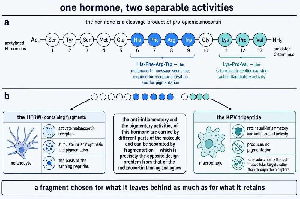
Three years later, in a one-page note in Nature, Lerner and J. Ignatius Harris gave the sequence: thirteen amino acids, beginning with serine and ending with valine (Harris & Lerner, 1957). The last three of those thirteen are the subject of this document, though nobody would have any reason to care about them for another twenty-seven years.
Then in 1961 Lerner and John McGuire did the experiment that closes the loop. They gave the hormone to people and watched their skin darken (Lerner & McGuire, 1961). α-melanocyte-stimulating hormone — α-MSH — was, as far as anyone was concerned, a pigment hormone. It had a name that said so, an assay built on pigment cells, and a demonstrated effect on human skin colour. That framing held for two decades.
03The peptide that lowered a fever
In 1980, in a laboratory 1,500 miles away and in a discipline with no obvious connection to dermatology, James M. Lipton was injecting peptides into the brains of rabbits to see what happened to their body temperature.
Lipton was a physiologist at the University of Texas Southwestern Medical Center in Dallas, and his interest was thermoregulation. His first published screen was frankly exploratory: sixteen peptides administered centrally, with body temperature as the read-out (Lipton & Glyn, 1980). Two of them did something. One was ACTH. The other was α-MSH.
The following year, Glyn and Lipton established the important qualifier: the peptide reduced a fever driven by leukocytic pyrogen at doses that did not lower normal body temperature (Glyn & Lipton, 1981). That distinction is the difference between an antipyretic and a refrigerant, and it is what made the finding interesting rather than merely thermodynamic.
In 1983 the work reached Science. Murphy, Richards and Lipton reported that centrally administered α-MSH was, milligram for milligram, far more potent at reducing fever in rabbits than acetaminophen (Murphy et al., 1983). A pigment hormone was outperforming the most widely used antipyretic on Earth. Something about the received framing of the molecule was incomplete.
It is tempting, and common, to tell this story as though the anti-inflammatory melanocortin field is Lipton's alone. It is not. Two independent groups opened the anti-cytokine line on the whole molecule, and they did it from immunology rather than from thermoregulation. In 1986 Cannon, Tatro, Reichlin and Charles Dinarello — the discoverer of interleukin-1 — showed that α-MSH blocked interleukin-1's actions on thymocytes and fibroblasts, with a minimum effective concentration of 10−11 M, and noted that a highly potent melanotropic analogue did not reproduce the effect, which already suggested the classical pigment receptor was not responsible (Cannon et al., 1986). Two years later Robertson, Dostal and Daynes at Utah showed the same hormone suppressing fever, acute-phase proteins and neutrophilia driven by interleukin-1 and tumour necrosis factor in live mice (Robertson et al., 1988).
04Cutting the message off
If a thirteen-residue hormone does two unrelated things, the obvious experiment is to cut it up and find out which piece does which. Lipton's laboratory ran that experiment, and the first paper to test lysine–proline–valine on its own is not about inflammation at all. It is about fever.
In 1984 Richards and Lipton gave the free tripeptide to rabbits made febrile with leukocytic pyrogen, both directly into the brain and peripherally. It reduced fever by both routes — 0.5 to 2.0 mg centrally, 2 to 200 mg peripherally (Richards & Lipton, 1984). The hundred-fold gap between those two dose ranges is the first appearance of a problem that runs through this entire monograph, and Richards and Lipton were candid about the other half of it: the tripeptide was substantially less potent than the intact hormone, so whatever the last three residues carried, they did not carry everything.
Deeter, Martin and Lipton pinned that down in 1988 by testing a ladder of fragments in the same model. Residues 8–13 were markedly more effective than either 10–13 or 11–13; none matched the full molecule; and, curiously, the four-residue 10–13 fragment was less effective than the three-residue 11–13 (Deeter et al., 1988). Adding amino acids to the tripeptide could help or hurt. The message was not a simple gradient.
051989, and the word “antiinflammatory”
The founding paper of this compound is two and a half pages long, appeared in The FASEB Journal in September 1989, and has two authors: Michael Hiltz and James Lipton (Hiltz & Lipton, 1989).
The design is plain. Mice were painted with picryl chloride, a contact irritant that makes the ear swell. Graded doses of α-MSH(11–13) were compared against saline and against a large dose of corticosteroid. The tripeptide inhibited the swelling, and it did so in proportion to dose.
Two details in that paper deserve to be preserved rather than smoothed over. The first is that its own abstract contains a typographical error — it calls the fragment a “trieptide” — which is a small reminder that the founding document of a research programme is a physical object made by people in a hurry. The second matters more: the abstract states that “in recent preliminary research this tripeptide inhibited increases in vasopermeability.” That prior work is not indexed anywhere, and it could not be located for this monograph. The claim that KPV is anti-inflammatory therefore rests, at its very origin, partly on a result that was never formally published.
The claim did not stay lonely. Within a year the same laboratory extended it to paw oedema and to contact sensitivity, two additional models with different mechanics (Hiltz & Lipton, 1990). Anna Catania, an internist in Milan, joined the collaboration in 1991 and would go on to become the field's most prolific author, eventually co-writing its standard review (Catania et al., 2004).
06Which residues matter
The 1991 paper by Hiltz, Catania and Lipton is the one to read if you want to know what the molecule is actually doing, because it is the one that takes the molecule apart stereochemically (Hiltz et al., 1991). Amino acids come in left- and right-handed forms; nature uses the left. Swapping one residue for its mirror image is the cleanest available way to ask whether that residue's shape is doing the work.
Three results, in mice, on picryl-chloride ear swelling. Replacing lysine with its mirror image changed nothing — L-Lys11 is not essential. Replacing proline with its mirror image abolished activity entirely — L-Pro12 is essential. And replacing valine with its mirror image made the peptide roughly four times more potent than the parent tripeptide.
That is a coherent picture: the proline kink is the shape that matters, the lysine is along for the ride, and the terminal residue can be improved on. It also produced the field's first hint of a complication that recurs throughout this document — the dose–response curves were bell-shaped. More was not monotonically better. A peptide whose dose–response inverts at the top end is a peptide whose optimal exposure is a narrow target rather than a floor.
The proline finding is not unanimous. A 2018 synthetic study reviewing the same question reports that the literature conflicts on whether the L-configuration of proline is essential, citing three papers on different sides (Songok et al., 2018). Where a structure–activity conclusion from 1991 has not been settled by 2018, this document says so rather than choosing.
07The door that opens when you are ill
Here is the problem with giving anyone a tripeptide by mouth. The gastrointestinal tract is a machine for destroying peptides. That is its function. Acid, pepsin, trypsin, and a brush border studded with peptidases exist precisely to reduce dietary protein to fragments small enough to absorb. A three-residue peptide swallowed into that environment ought to last minutes.
Except that the gut does not absorb protein as free amino acids. It absorbs a large fraction of it as di- and tripeptides, through a dedicated transporter, and a three-residue peptide is exactly the right size to be mistaken for food.
The transporter is PepT1, the product of the gene SLC15A1. It sits in the apical brush-border membrane of the small intestine, and it is not powered by ATP directly. It is powered by protons: the lumen of the small intestine is more acidic than the inside of the cell, and PepT1 lets a proton fall down that gradient while carrying a peptide with it. It is famously indiscriminate, handling on the order of four hundred dipeptides and eight thousand tripeptides (Viennois et al., 2018).
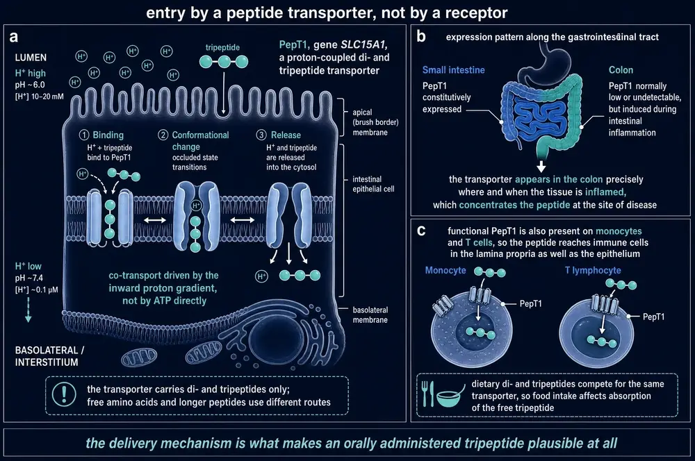
The elegant part is where it appears. PepT1 is abundant in duodenum, jejunum and ileum, and in a healthy colon it is minimal or absent. But in inflammatory bowel disease it is induced in colonic tissue, and both tumour necrosis factor and interferon-γ can drive that induction in cultured intestinal epithelium (Viennois et al., 2018). The transporter appears in the colon precisely where and when the tissue is inflamed. A drug that rides it is concentrated at the site of the disease by the disease.
Guillaume Dalmasso, working with Didier Merlin at Emory, showed in 2008 that KPV is a PepT1 substrate. The experiment was done twice over: unlabelled KPV competed against a radiolabelled PepT1 substrate, and tritiated KPV was used directly to obtain the transport kinetics (Dalmasso et al., 2008). The affinity is genuinely high — a Michaelis constant of roughly 160 µmol/L in Caco2-BBE cells, which is among the lowest reported for this transporter; the workhorse substrate glycyl-sarcosine sits at a millimole per litre or worse (Viennois et al., 2016).
The induction claim is not unanimous either, and the dissent sits inside the same review that makes the claim: alongside the up-regulation data, Viennois and colleagues record an observation of decreased PepT1 expression in the descending colon of patients with inflammatory bowel disease during acute inflammation (Viennois et al., 2018). The direction of a transporter's response to inflammation is the load-bearing assumption behind every colon-targeted KPV formulation in Part Five, and it is not fully settled.
08Inside the cell
Once through the membrane, KPV is in the cytosol, and this is where it stops resembling a hormone at all.
Dalmasso's group stimulated three human cell lines with pro-inflammatory cytokines — two intestinal epithelial lines, Caco2-BBE and HT29-Cl.19A, and the T-cell line Jurkat — and measured what KPV did to the signalling that followed. Their finding, in the paper's own words, is that nanomolar concentrations of KPV inhibit activation of NF-κB and of MAP kinase inflammatory signalling, and reduce pro-inflammatory cytokine secretion (Dalmasso et al., 2008). Nanomolar is a serious number for a tripeptide with no receptor.
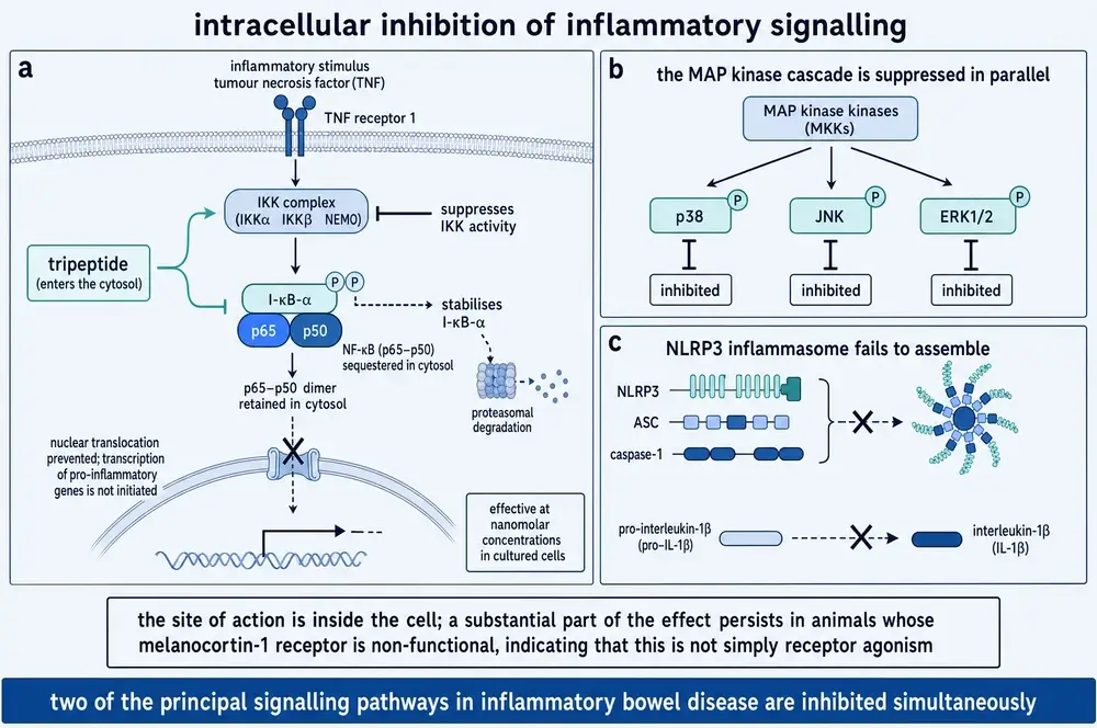
NF-κB is worth a sentence for readers who have not met it. It is a transcription factor that sits in the cytosol bound to an inhibitor, IκB. When an inflammatory signal arrives, a kinase complex tags IκB for destruction, NF-κB is released, moves into the nucleus, and switches on the genes for the inflammatory cytokines. Nearly every anti-inflammatory drug in existence interferes with this cascade somewhere.
The most specific mechanistic proposal for where KPV interferes comes from Stephen Land, working in cultured human bronchial epithelium rather than gut (Land, 2012). His account is that KPV is itself imported into the nucleus, that it stabilises IκBα, and that it suppresses nuclear translocation of the p65 subunit of NF-κB — and, most concretely, that it interacts with the importin-α3 binding site on p65 itself, apparently blocking the armadillo domains that the import machinery uses to grip its cargo. If that is right, KPV is not inhibiting an enzyme in the cascade. It is standing in the doorway.
09The receptor that turns out not to be necessary
α-MSH works through melanocortin receptors. There are five of them. The natural assumption about a fragment of α-MSH is that it works through them too, more weakly. For KPV that assumption appears to be wrong, and the experiment that shows it is a good one.
In 2008, a group at Münster led by Klaus Kannengiesser tested KPV in three mouse models: chemically induced colitis, an immune-mediated transfer colitis, and — the decisive arm — chemically induced colitis in MC1Re/e mice, which carry a frameshift that renders the melanocortin-1 receptor non-functional (Kannengiesser et al., 2008). If KPV needs that receptor, it should do nothing in those animals.
It did something considerable. In the authors' words, KPV treatment “rescued all animals in the treatment group from death” during colitis in the receptor-null mice. Their conclusion is measured — the effects “seem to be at least partially independent of MC1R signalling” — and that measured phrasing is the right one, since survival was rescued while the weight and histology endpoints were not significantly changed in that arm. But the direction is unambiguous: a molecule that works in an animal lacking the receptor is not working only through the receptor.
This finding is worth flagging as a small object lesson. A recent review in this corpus characterises the receptor-null result as a “slightly decreased lethality” with no significant changes — a fair reading of two of the three endpoints and a considerable understatement of the third. The primary report says all treated animals were rescued from death. The difference between “slightly decreased lethality” and “rescued all animals” is the difference between a footnote and a mechanism. Every registry, regulatory and structural claim in this document series is checked against the instrument rather than against a summary of it, for exactly this reason; here the practice rescued a commissioned figure that a secondary source would have condemned.
There is a second, quieter piece of evidence pointing the same way, and it comes from a study of the dimer rather than the monomer. When Ji and colleagues measured cyclic AMP — the second messenger that melanocortin receptors produce — they found that (CKPV)2 raises it and KPV does not. Their sentence is explicit: KPV inhibited inflammation with no cyclic AMP accumulation, suggesting its effects may not be solely dependent on melanocortin receptors (Ji et al., 2013). Two closely related molecules, one signalling like a hormone and one not.
What the corpus does not offer is a settled alternative. Four different receptor stories appear across the literature reviewed here: no receptor at all, with a transporter doing the work; MC1R-dependent calcium signalling in keratinocytes; MC3R-dependence in bronchial epithelium; and, from the traumatic-brain-injury study in Section 13, a suggestion of MC4R. These cannot all be right, and no paper in this corpus reconciles them. The honest position is that KPV's target is not established — and that the strongest single piece of evidence, the receptor-null survival result, argues that at least part of the effect needs no melanocortin receptor at all.
10What the cytokine data do and do not say
It would be satisfying to close this Part with a chart of cytokine suppression: tumour necrosis factor down by so much, interleukin-6 by so much, the anti-inflammatory interleukin-10 untouched, demonstrating that the compound turns down the inflammatory arm without recruiting a counter-regulatory one. That is a meaningful pharmacological distinction, and it is the distinction that would separate this compound from a corticosteroid.
The evidence does not support that chart. The primary study reports “a decrease in pro-inflammatory cytokine expression” and names no individual cytokine (Dalmasso et al., 2008). No paper in this corpus reports an interleukin-10 measurement under KPV in colitis at all — so the proposition that it is unchanged has not been tested, which is a different and weaker statement than the proposition that it was tested and did not move. What can be said, from the delivery studies in Part Five where individual cytokines were measured, is that formulated KPV reduces tumour necrosis factor, interleukin-1β and interleukin-6 in inflamed mouse colon (Zhang et al., 2024; Xu et al., 2026). In one of those studies interleukin-10 rose (Xu et al., 2026), which is the opposite of the tidy story.
11Two colitis models
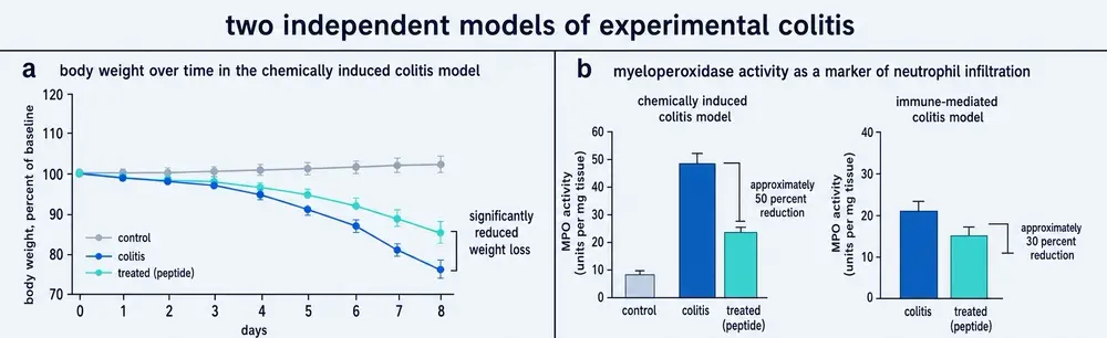
The animal case for KPV rests on colitis, and it rests on two independent laboratories reaching the same conclusion in the same year through different models.
Dalmasso and colleagues put KPV in the drinking water of mice with colitis induced two different ways — chemically, with dextran sulphate sodium, and immunologically, with trinitrobenzene sulphonic acid. Both were reduced, assessed histologically and by pro-inflammatory cytokine messenger RNA (Dalmasso et al., 2008). The exposure reported downstream for that study is 205 µg per day per mouse (Zhang et al., 2021).
Kannengiesser's group at Münster used chemically induced colitis and a quite different second model: transfer colitis, in which naive T cells are moved into an immunodeficient host and attack the bowel over weeks. This is a slow, lymphocyte-driven disease rather than an acute chemical injury, and it engages a different arm of the immune system. KPV produced earlier recovery and significantly stronger regain of body weight; inflammatory infiltrates were significantly reduced on histology; and myeloperoxidase activity — an enzyme released by neutrophils, and therefore a proxy for how many of them have arrived — was significantly reduced in colonic tissue (Kannengiesser et al., 2008).
Two models, two laboratories, one direction. For a compound of this kind that is a respectable evidentiary base, and it is the reason KPV is taken seriously at all.
12Cancer, and a transporter that cuts both ways
Long-standing bowel inflammation causes cancer. That is the clinical justification for treating inflammatory bowel disease aggressively, and it makes inflammation-driven carcinogenesis the natural place to ask whether an anti-inflammatory peptide does anything that matters.
Viennois and colleagues asked it in 2016, with a design that answers two questions at once (Viennois et al., 2016). Mice were given a carcinogen followed by cycles of chemical colitis, which reliably produces colon tumours. Some received KPV at 100 µmol/L in their drinking water throughout.
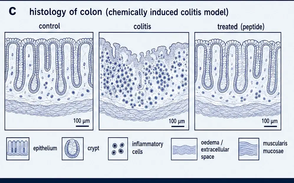
In wild-type mice, KPV produced what the authors describe as a drastic decrease in colon tumorigenesis — fewer tumours, smaller tumours, lower overall tumour burden, fewer aberrant crypt foci, less cellular infiltration and less epithelial proliferation. It did this without changing how much weight the animals lost during the colitis phase, which is a useful control: the benefit was not simply a milder illness.
The second question was mechanistic, and it is the more interesting one. In mice lacking PepT1 entirely, KPV did not protect. Tumour numbers, sizes, burdens and body weights were not significantly different between treated and untreated knockouts. The authors are careful to note a non-significant trend toward lower tumour burden even so, and to allow that another peptide transporter might carry some of the load — but the headline is that removing the transporter removes the drug's effect. It is unusual to have that clean a demonstration that a compound reaches its target the way you think it does.
A third arm sharpens the interpretation. In APCMin/+ mice — which develop intestinal tumours through a purely genetic route, without inflammation driving them — thirteen weeks of KPV in the drinking water reduced intestinal inflammation, measured by lipocalin-2, and did not reduce tumour burden in either small intestine or colon. The anti-inflammatory effect was real and was not sufficient. KPV is not an anti-cancer agent; it is an anti-inflammatory agent that, where inflammation is what is driving the cancer, reduces the cancer.
The same paper shows that PepT1 itself is pro-tumorigenic. Mice engineered to overexpress it developed larger tumours with greater burden and more proliferation; mice lacking it developed fewer and smaller ones; and in a tissue microarray of human samples, the majority of colon tumour specimens showed increased staining relative to normal and benign tissue.
So the door that lets KPV into the diseased colon is a door whose over-activity is associated with worse disease. The authors' resolution is to treat the transporter as a delivery route to be exploited rather than a target to be stimulated, which is reasonable — but it should be stated plainly rather than passed over.
13Outside the gut
Almost everything above concerns the bowel, for the good reason that the transporter story makes the bowel the obvious place to look. Four studies elsewhere are worth reporting, and their unevenness is itself informative.
Brain. Schaible and colleagues gave a single intraperitoneal dose of α-MSH(11–13) at 1 mg/kg to mice thirty minutes after a controlled cortical impact — a mechanical traumatic brain injury — and assessed them at 24 hours (Schaible et al., 2013). Contusion volume was 21.0 ± 3.4 mm3 against 27.7 ± 4.8 in vehicle-treated animals (p = 0.016), and apoptotic neurons were roughly halved, 26 ± 10.3 against 54.8 ± 19.4 cells per region of interest (p = 0.002). Microglia showed a less activated morphology. Two things temper this. Tumour necrosis factor and interleukin-1β messenger RNA did not change significantly, so the mechanism was not the expected one. And the neurological severity score did not differ (2 ± 1.1 against 3.6 ± 2.3, p = 0.139) — the tissue looked better and the animals did not measurably behave better. The authors describe their study as explorative and say their p-values are given for descriptive reasons only, which is an unusually honest caveat and should be respected rather than quietly dropped.
Eye. In rabbits with corneal abrasions, KPV instilled topically four times daily at 1, 5 or 10 mg/mL produced complete re-epithelialisation of 100 per cent of treated corneas by 60 hours, against none of the untreated controls (reported in Dinparastisaleh & Mirsaeidi, 2021). Taken at face value that is the most dramatic single result in this monograph. It rests on one study, reported at second hand, and the corpus copy of the citing review renders the instilled volume as “30 mL”, which for a rabbit eye is not possible and is presumably 30 µL.
Airway. In an immortalised human bronchial epithelial line challenged with tumour necrosis factor and respiratory syncytial virus, KPV produced dose-dependent inhibition of NF-κB activation, matrix metalloproteinase-9 activity, interleukin-8 and eotaxin secretion (Land, 2012). This is the study that supplied the importin mechanism in Section 08. It is cultured cells, not an animal, and not a person.
Liver. The newest finding in this monograph, published in 2026, is also the thinnest. In HepG2 human hepatic cells loaded with oleic acid to model fatty-liver disease, KPV at 100 µg/mL reduced lipid accumulation and suppressed fatty acid synthase without cytotoxicity, apparently by lowering reactive oxygen species and thereby modulating the PPARγ pathway (Lee et al., 2026). One cell line, one concentration, no magnitudes reported, no animal. It is a hypothesis, and it is recorded here as one.
14The antimicrobial arm, and the paper that could not reproduce it
In 2000, Cutuli, Cristiani, Lipton and Catania reported that α-MSH and its C-terminal tripeptide inhibited two very different pathogens: the Gram-positive bacterium Staphylococcus aureus and the yeast Candida albicans (Cutuli et al., 2000). Three features of that paper made it influential.
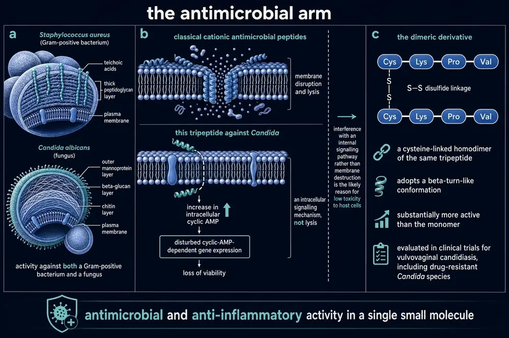
The activity extended across a broad concentration range including the picomolar — that is, the concentrations at which the hormone actually circulates, which raises the possibility that this is a physiological function rather than a pharmacological curiosity. The mechanism appeared not to be membrane destruction: cyclic AMP rose in peptide-treated yeast, and an inhibitor of the enzyme that makes cyclic AMP partly reversed the killing. And — the point the authors clearly cared about most — the peptides did not impair human neutrophils' ability to kill either organism. They enhanced it. Conventional anti-inflammatory drugs suppress host defence; this one did not.
It is not settled. In 2018 a synthetic-chemistry group set out to make improved analogues, and needed the parent compound to work first. They synthesised acetylated, amidated KPV, tested it by agar diffusion following the published protocols, and saw nothing. Suspecting their own synthesis, they then bought the peptide from the same commercial supplier used in the earlier work and tested that. Again nothing (Songok et al., 2018). Their own description of the result is “surprising and disappointing.”
They were not alone. The same paper records that a head-to-head comparison of thirty ultra-short antimicrobial peptides against a panel of skin pathogens found no tripeptide active below 100 µM, and quotes Grieco's assessment that these molecules have weak activity under standard microbiological conditions in a way that hampers realistic clinical use. A separate strand of work, in which the tripeptide's activity is compared against longer α-MSH fragments, finds α-MSH(11–13) achieving only a 0.7 log reduction against stationary-phase S. aureus at 150 µM, where a six-to-eight-residue fragment achieved roughly two logs at 20–50 µM (Mumtaz et al., 2020).
There is also a recurring caveat that cuts across the positive results: the α-MSH peptides retain activity in physiological saline but are reported not to be active in the culture media routinely used in laboratory microbiology. A compound whose antimicrobial activity depends that sharply on assay conditions is a compound about which two laboratories can honestly disagree.
This document takes no side. What can be said is that the antimicrobial claim for KPV specifically has been reported, has been contested by direct attempted replication using material from the same source, and has not been resolved in the literature reviewed here. Where the compound's mechanism is described elsewhere as combining anti-inflammatory and antimicrobial action in one small molecule, the second half of that description should be read as disputed. Note also that the great majority of the mechanistic antimicrobial work — the membrane depolarisation studies, the DNA and protein synthesis inhibition — was done on α-MSH or on longer fragments, not on the tripeptide.
15Three molecules that are not KPV
Most compounds in this series have a name that belongs to them alone. KPV does not. It sits in a small family whose members were made by the same people, tested in the same disease models, published in the same journals, and are routinely discussed in the same paragraph — and whose evidence bases are of very different strengths.
This is a harder problem than a name that merely collides with something unrelated. When a compound's name accidentally matches a physics variable, the subject matter separates them instantly. Here the subject matter is shared by construction: every term that would confirm a paper is about KPV — melanocortin, anti-inflammatory, colitis, Candida, NF-κB — confirms just as strongly that it is about α-MSH, or KdPT, or the dimer. The only thing that separates them is the designator itself.
The reason to be strict about it is arithmetic. KPV has no human data. Its analogue KdPT has two completed European clinical trials. Its dimer has an unpublished company trial in twenty patients. Pool the three and the compound appears to have a clinical record. It does not.
16KdPT — the analogue that reached people
KdPT is lysine, D-proline, threonine. Two changes from KPV: proline is switched to its mirror image, and valine is replaced by threonine. It is sometimes described as an α-MSH-derived tripeptide, and one review in this corpus states flatly that it is not — that its sequence occurs in no member of the melanocortin family (Gravina et al., 2023). The proposed rationale for the molecule is entirely different from KPV's: the L-enantiomer of KdPT is homologous to residues 193–195 of interleukin-1β, and it is thought to act as an antagonist at the interleukin-1 receptor rather than anywhere in the melanocortin system.
Bettenworth and colleagues at Münster characterised it in 2011 (Bettenworth et al., 2011). In two mouse colitis models it markedly reduced severity. In colonic epithelial cells it increased proliferation, accelerated wound closure, and improved transepithelial electrical resistance after cytokine challenge; it preserved tight-junction protein expression and improved barrier function in living animals. It acted independently of the interleukin-1 receptor type I in vivo, complicating its own mechanistic story. And — the selling point — melanin assays confirmed it does not induce pigmentation.
Then KdPT did the thing KPV has never done. It went into people.
Two trials were run in Europe by Dr August Wolff GmbH of Bielefeld, both in ulcerative colitis, both completed, and the efficacy trial was published (Kucharzik et al., 2017). It was a multicentre, randomised, double-blind phase IIa study of oral KdPT twice daily at 20, 50 or 100 mg, added to existing medication, in patients with mild-to-moderate active disease. The primary objective was time to a sustained 50 per cent improvement in the colitis activity index at week 8.
The result deserves to be read carefully, because it is the only human efficacy data anywhere near this compound family. The pooled KdPT group showed significantly higher remission rates at two and four weeks (p = 0.0349 and 0.0278) and a significantly higher proportion with a response at week 8 (p = 0.0434). The primary endpoint, in the authors' own words, was met “after additional analyses.” The reason given is a very high placebo response rate after week 4, which prompted subgroup analyses restricted to patients with unquestionably active or more severe disease; those subgroups responded earlier and significantly. All doses were well tolerated and the safety profile was described as excellent.
That is a preliminary positive signal in a small phase IIa study whose primary endpoint required post-hoc work to reach, in a disease notorious for placebo response. It is not proof of efficacy, the authors do not claim it is, and development does not appear to have continued. It is also, emphatically, not evidence about KPV. Different molecule, different proposed target, different stereochemistry.
17(CKPV)2 — the dimer that reached a clinic and then stopped
The other relative is closer. Take two copies of KPV, put a cysteine on the front of each, and join the two cysteines through a disulphide bond. The result is an eight-residue molecule, (CKPV)2, also called CZEN-002. Nuclear magnetic resonance shows it adopts an extended backbone with a β-turn-like element, and it was designed deliberately: Catania and colleagues report inserting the Cys-Cys linker between two KPV units and finding excellent candidacidal effects in pilot tests, including against azole-resistant Candida species (Catania et al., 2005).
It is more active than the monomer, and unlike the monomer it behaves like a melanocortin. Gatti and colleagues found it inhibited tumour necrosis factor production by endotoxin-stimulated human mononuclear cells as effectively as a superpotent α-MSH analogue and more potently than KPV; in rats it reduced circulating tumour necrosis factor after endotoxin and restored ultrafiltration in dialysis peritonitis (Gatti et al., 2006). Ji and colleagues later showed it raises cyclic AMP in macrophages, that knocking down MC1R by interfering RNA largely abolishes that rise, and that it produces no cyclic AMP at all in cells lacking the receptor unless the receptor is put back (Ji et al., 2013). In a rat model of Candida vaginitis, a gel at 2 mg/kg daily reduced fungal survival to 12.0 per cent by day 11 against 44.7 per cent for miconazole, and to essentially zero by day 18.
So: a dimer of KPV that is more potent than KPV, works through the receptor KPV apparently does not use, and beat a standard antifungal in an animal model. It went into the clinic.
The peer-reviewed literature records that it happened. A 2006 paper states the dimer “is currently under clinical investigation for antimicrobial use” (Gatti et al., 2006); a 2012 review states it “is being currently evaluated in clinical trials for treatment of vulvovaginal candidiasis” (Seo et al., 2012); a 2014 review states it is “already in clinical trial” (Singh & Mukhopadhyay, 2014). None of them reports a result.
Searches of ClinicalTrials.gov for CZEN-002, CKPV, KPV and for the sponsor Zengen return nothing. The EU Clinical Trials Register returns nothing. The trial predates the 2005 registration requirement, so its absence from the registries is expected rather than sinister — but the consequence is that the only account of what happened is a company press release from May 2004, describing an open-label phase I/II in vulvovaginal candidiasis with twenty patients enrolled and seventeen completing. This document does not report those results as trial findings, because they have never been through peer review, never appeared on a registry, and cannot be checked. A 2020 review that went looking concluded that a larger dose-ranging phase IIb was planned for 2005 and that the compound's development status is not currently known (Mercer & O'Neil, 2020).
The patents — filed by Anna Catania and James Lipton, assigned first to Zengen, then to MSH Pharma, then to MSH Biopharma — are listed as expired for non-payment of fees. The programme did not fail in public. It stopped.
There is a detail in the patent record worth keeping. The antimicrobial patent claims a priority date of 24 March 1999 — nearly a year before the Journal of Leukocyte Biology paper that is universally cited as the founding antimicrobial report appeared in February 2000. The commercial claim was staked before the scientific one was published. That sequence is unremarkable in drug development and it is worth noticing anyway, because the antimicrobial finding is the one that later failed to replicate.
18Why the free peptide is hard to use
Everything attractive about KPV as a molecule — that it is tiny, polar, uncharged apart from one amine, and made entirely of ordinary amino acids — is also what makes it difficult to deliver. It looks exactly like food.
The most direct measurement in this corpus was made in 2026 as a control experiment. Free KPV was incubated in simulated gastric fluid (pH 1.2 with pepsin) and in simulated intestinal fluid (pH 7.4 with trypsin) at body temperature. After two hours, chromatography showed nearly complete hydrolysis in both. More tellingly, free KPV recovered from those conditions had completely lost its ability to suppress cytokine production in stimulated macrophages (Cheng et al., 2026). It is not merely degraded; it is inactivated.
The protected form is not much better in the presence of a broad protease. Acetylated, amidated KPV incubated with pronase was degraded to its three constituent amino acids within 24 hours — a result the authors of that study established in passing while demonstrating that a sugar-modified analogue was completely stable under the same conditions (Songok et al., 2018).
What happens after absorption is not known. The field says so itself: a 2019 review states plainly that the pharmacokinetics of KPV in circulation are still not clear, while noting that intravenously administered α-MSH and its tripeptides can last only a few minutes because of serum proteases (Wang et al., 2019). There is no published half-life for KPV, no published oral bioavailability figure, no Cmax, no volume of distribution and no clearance. The nearest approach is a fluorescent-label study in mice showing plasma signal falling to baseline within four hours after intravenous dosing — and that tracks the dye, not an assay of the peptide (Cheng et al., 2026).
One further absence is worth stating early because it bears directly on Section 22. There is no measurement anywhere in this corpus of KPV penetrating skin. The one relevant datum comes from outside the peer-reviewed literature: an in vitro study cited by the United States Food and Drug Administration reports that KPV does not permeate cadaver human skin (FDA, 2026).
19Nine ways to carry three residues
The response to all this has been formulation, and there is now a substantial literature devoted to getting KPV somewhere useful. Nearly all of it points at the inflamed colon, for the reason given in Part Two: the transporter that carries the peptide is upregulated exactly there.
The best-known approach is Bo Xiao and Merlin's hyaluronic-acid-functionalised nanoparticle: a protein core carrying KPV, coated in chitosan, surfaced with hyaluronic acid to engage CD44 on inflamed epithelium and macrophages, and embedded in a hydrogel that survives the stomach and collapses in the colon. Particles run about 272 nm. In mice with chemically induced colitis, oral administration accelerated mucosal healing while reducing inflammation, and did both better than the same nanoparticles without the hyaluronic acid (Xiao et al., 2017).
Others have used KPV as a ligand rather than as the drug — decorating a nanoparticle with it so that PepT1 pulls the whole particle in, with cyclosporine A or a small interfering RNA as the actual payload (Liu et al., 2021; Zeng et al., 2026). Those studies are evidence about targeting, not about KPV's own pharmacology, and are labelled as such throughout.
Two of the more inventive recent designs are worth describing because they show where the field has gone. Zhang and colleagues built a carrier-free co-assembly — KPV and the immunosuppressant tacrolimus simply assembled together into 200-nanometre particles with a little pegylated lipid, no carrier polymer at all. Given intravenously at 1 mg/kg daily to mice with chronic colitis, the combination took survival to 100 per cent against 70 per cent for untreated animals, and outperformed either agent alone on colon length, cytokine levels, myeloperoxidase and tight-junction protein restoration (Zhang et al., 2024). And Xu and colleagues engineered Escherichia coli Nissle 1917 — a probiotic strain — to secrete a protein containing twenty-two copies of KPV, each preceded by a linker that neutrophil elastase cuts. The bacteria are encapsulated in a silk-fibroin shell that scavenges reactive oxygen species. KPV is therefore released only where neutrophils have arrived, which is to say only where there is inflammation; colonic KPV peaked four hours after oral administration and the construct improved disease in two different colitis models (Xu et al., 2026).
The dose-sparing claim. Several reviews report that nanoparticle encapsulation allowed a 12,000-fold lower delivered concentration of KPV to achieve the same effect as the free peptide. One of those same reviews states the figure as 1,200-fold in a different paragraph of the same paper, and two reviews attribute it to two different formulations. The primary source is not in this corpus. A tenfold internal contradiction inside a single document is sufficient reason to leave the number out of the running text, and it is left out.
The negative results. A lipid nanocapsule programme first tried attaching KPV to the particle surface through a lipid tail and reported no effect on pro-inflammatory cytokines after seven daily doses; that failure is why the design was changed to a hyaluronic-acid conjugate. Even the revised version showed no significant difference from vehicle on weight loss, colon weight-to-length ratio or histology in the acute model, and in the chronic model its mucin-2 result lost significance under the appropriate multiple-comparison correction, with no differences on histology or colonoscopy (Marotti et al., 2024). Formulation is not a solved problem, and papers that solved it are easier to find than papers that did not.
202026: the conjugate that made the free peptide look inert
The most technically impressive work on this compound was published this year, and it contains the most uncomfortable finding in this monograph.
Cheng and colleagues built what they call a self-immolative peptide prodrug conjugate (Cheng et al., 2026). A hydrolysable phosphazene scaffold carries a polyethylene glycol chain, four peroxide-sensitive boronate ester units, and one molecule of KPV. In water it assembles spontaneously into 81-nanometre micelles. When it meets reactive oxygen species — which are elevated at sites of inflammation — the boronate units are oxidised, and the scaffold then unzips itself, falling apart into glycine, ammonium and phosphate and releasing intact KPV. It is a molecule designed to disassemble only where it is needed.
It works. Only about 9 per cent of the peptide is released after two hours in simulated gastric fluid, and release accelerates sharply on reaching intestinal conditions with peroxide present. Colonic exposure in mice with colitis was 3.8-fold higher than for free peptide by area under the fluorescence curve, and 2.7-fold higher in inflamed than in healthy animals. Given orally at 2.5 mg/kg it protected against weight loss, disease activity and colon shortening, restored tight-junction proteins and the mucus layer, reduced tumour necrosis factor, interleukin-1β, interleukin-6, myeloperoxidase and oxidative markers, and shifted macrophages toward the regulatory phenotype. In a head-to-head comparison it outperformed mesalazine at 50 mg/kg — roughly a twenty-fold higher dose of the standard of care.
And the control arm: free KPV given orally at 1 mg/kg showed no beneficial effects. Not a smaller effect. None — on any endpoint, including every mechanistic read-out. That 1 mg/kg is four times the peptide content of the 2.5 mg/kg conjugate dose that worked.
This does not overturn the earlier work, and it should not be read as doing so. The older studies gave KPV continuously in drinking water, at 205 µg per mouse per day or 100 µmol/L, over days to weeks; Cheng's control was a single daily gavage. Continuous low-level exposure and once-daily bolus are not the same experiment, and for a compound with a half-life the field admits it cannot state, the difference could be decisive. But the two results sit in the corpus together, and a reader is entitled to know that the most recent and most carefully controlled study to test free oral KPV head-to-head found it did nothing.
21What is established and what is not
It is worth setting this out plainly, because the gap between what the laboratory literature supports and what the compound is sold for is unusually wide.
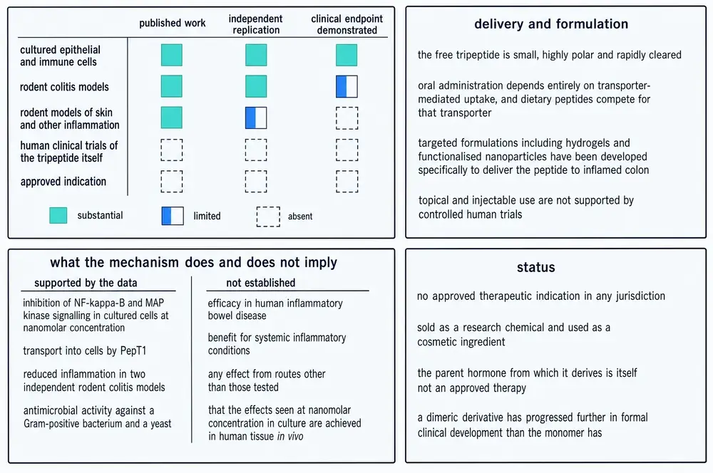
| Supported by the evidence reviewed here | Not established |
|---|---|
| Inhibition of NF-κB and MAP kinase signalling in cultured human epithelial and T cells at nanomolar concentration | Efficacy in human inflammatory bowel disease, or in any human condition |
| Transport into cells by PepT1, with the transporter shown to be necessary for the anticancer effect in a knockout | Any effect by any route in a human being — there are no human data |
| Reduced inflammation in two independent rodent colitis models, from two laboratories | Benefit in systemic inflammatory conditions |
| Reduced tumour burden in inflammation-driven colon carcinogenesis in mice, absent in PepT1-null animals | Benefit in genetically driven tumorigenesis — explicitly tested and negative |
| Reduced contusion volume and neuronal apoptosis after traumatic brain injury in mice | Improved neurological function in that model — explicitly measured and not significant |
| Activity at least partly independent of the melanocortin-1 receptor | Which target it does act on; four incompatible accounts exist |
| No pigmentation, which is the reason the fragment exists | Antimicrobial activity, which has been contested by direct attempted replication |
| Rapid destruction in simulated gastrointestinal fluid, with loss of activity | Half-life, oral bioavailability, clearance, volume of distribution — none published |
| Delivery to inflamed colon by several engineered formulations, in mice | That nanomolar culture concentrations are reached in human tissue in vivo |
| Failure to permeate cadaver human skin, in the one study located | Any topical, subcutaneous, intramuscular, intranasal or transdermal use — no data exist for any of these routes |
22July 2026
On 23 July 2026, the Pharmacy Compounding Advisory Committee of the United States Food and Drug Administration met at White Oak to consider whether several peptides should be added to the 503A Bulks List — the register of substances that compounding pharmacies may lawfully make into medicines without an approved application. KPV was the second item on the agenda. The use under consideration was “wound healing and inflammatory conditions”, and the proposed product was a topical cream or gel at 0.1 per cent (FDA, 2026).
The agency's own briefing document, published before the meeting, is the most comprehensive assessment of this compound in existence and its findings are almost entirely absences. Reading it in sequence:
- FDA “did not identify clinical studies in humans assessing pharmacokinetics or pharmacodynamics of KPV (free base) or KPV acetate via any route of administration.”
- It did not identify pharmacokinetic or toxicokinetic studies, acute toxicity studies, repeat-dose toxicity studies, genotoxicity studies, developmental and reproductive studies, or carcinogenicity studies.
- It did not identify nonclinical toxicity studies relevant to topical application to skin — the route being proposed.
- It noted that an in vitro study reports KPV does not permeate cadaver human skin.
- It found the substance “not well-characterized” physically and chemically, citing naming conventions that do not follow established chemical nomenclature, the absence of a Unique Ingredient Identifier, and the absence of microbiological testing data required for a topical product.
- There is no United States Pharmacopeia or National Formulary monograph for either form, and neither is a component of any approved drug.
- A search of the adverse-event reporting system through December 2025 retrieved no reports, and no outsourcing facility reported compounding any KPV product between January 2017 and June 2025.
The agency's conclusion is one sentence: “we propose not adding KPV (free base) or KPV acetate to the 503A Bulks List.”
The committee voted the other way. Press accounts of the meeting report the vote on KPV as 8 to 6 with one abstention in favour of adding it, alongside similar votes on BPC-157, TB-500, MOTS-c, Semax and Epitalon — five of which also have monographs in this series — with emideltide rejected. FDA has posted the agenda, the questions put to the committee and the final roster, but had not published minutes or a transcript at the time of writing, so the tally here is reported from press coverage rather than from the agency's own record. The votes are advisory and non-binding. Adding a substance to the list requires a proposed rule, a comment period and a final rule, none of which has occurred. KPV is not on the 503A Bulks List.
FDA also surveyed how the compound is marketed. The claims it found promoted for KPV include inflammatory conditions, wound healing, skin health, immune strengthening, protection against nerve damage and stroke, gut health, antimicrobial action, reducing tumour growth, psoriasis, inflammatory bowel disease, mast cell activation syndrome, histamine intolerance, recovery from COVID-19, Lyme disease, mould toxicity and pain syndromes.
The number of published human studies supporting any of them is zero.
The absence of adverse-event reports should not be read as reassurance. FDA notes in the same document that compounders operating under section 503A generally do not report to the adverse-event system, and the outsourcing-facility database shows no KPV production at all — so the zero reflects an absence of surveillance as much as an absence of harm.
23What this compound is, at the end of it
KPV is a genuinely interesting molecule and a genuinely thin therapy, and the two facts are not in tension.
The interesting part is the design logic, which is unusual and clean. A hormone does two things; the two things live in different parts of the chain; remove the part you do not want and you are left with a fragment that does the job you do want and nothing else. That fragment then turns out to enter cells through a transporter that the target disease itself upregulates, so the drug is concentrated where the pathology is by the pathology. Very few compounds have that shape. It is why the molecule has held attention for four decades on the strength of a fairly small literature.
The thin part is everything downstream of the mouse. Forty-two years after the first paper, this compound has no human pharmacokinetics, no human safety data, no human efficacy data, no regulatory approval anywhere, no pharmacopoeial monograph, and no identifier in the standard chemical registries. Its most recent and best-controlled test found the free peptide, given orally, did nothing at all — while the same peptide inside an engineered carrier beat the standard of care at a twentieth of the dose. Its closest analogue has been into humans and produced a preliminary signal that needed post-hoc analysis to reach its endpoint. Its dimer went into a clinic in 2004 and the result was never published.
The most defensible reading of the evidence is that KPV is a promising payload whose value depends almost entirely on the vehicle carrying it, and that the interesting scientific question about it in 2026 is not whether the peptide works but whether anything can get enough of it to the right place for long enough to matter. That question has been answered convincingly in mice, and not at all in people.
This document describes published research on KPV. It does not recommend human use of the compound and specifies no dose, route or schedule for any person. Every dose, concentration and route reported above is a parameter of a published experiment, given with the species and the duration in which it was used, and the great majority were used in mice, rats or rabbits.
No published study has administered KPV to a human being. There is therefore no human safety data, no human dose-response, no characterised adverse-effect profile and no established route of administration. The compound has no approved therapeutic indication in any jurisdiction. It is sold for research use only.
24References
Generated from verified NCBI records rather than from recall. Author lists, journal names, volumes, pages and identifiers are taken from the PubMed record for each citation, and the build refuses to run if any identifier fails to resolve. This series has twice shipped reference lists drafted from memory in which identifiers pointed at real but unrelated papers.
- Adnan SB, Maarof M, Fauzi MB, Fadilah NIM. Exploring the Role of Tripeptides in Wound Healing and Skin Regeneration: A Comprehensive Review. Int J Med Sci. 2025;22(16):4175-4200.
PMID 41209547 · doi:10.7150/ijms.118118 · PMC12595317 - Beloqui A. Gut hormone stimulation as a therapeutic approach in oral peptide delivery. J Control Release. 2024;373:31-37.
PMID 38971429 · doi:10.1016/j.jconrel.2024.07.007 · PMC11413617 - Bettenworth D, Buyse M, Böhm M, Mennigen R, Czorniak I, Kannengiesser K, et al.. The tripeptide KdPT protects from intestinal inflammation and maintains intestinal barrier function. Am J Pathol. 2011;179(3):1230-42.
PMID 21741932 · doi:10.1016/j.ajpath.2011.05.013 · PMC3157275 - Cannon JG, Tatro JB, Reichlin S, Dinarello CA. Alpha melanocyte stimulating hormone inhibits immunostimulatory and inflammatory actions of interleukin 1. J Immunol. 1986;137(7):2232-6.
PMID 3489761 - Capsoni F, Ongari AM, Reali E, Catania A. Melanocortin peptides inhibit urate crystal-induced activation of phagocytic cells. Arthritis Res Ther. 2009;11(5):R151.
PMID 19814819 · doi:10.1186/ar2827 · PMC2787256 - Catania A, Gatti S, Colombo G, Lipton JM. Targeting melanocortin receptors as a novel strategy to control inflammation. Pharmacol Rev. 2004;56(1):1-29.
PMID 15001661 · doi:10.1124/pr.56.1.1 - Catania A, Grieco P, Randazzo A, Novellino E, Gatti S, Rossi C, et al.. Three-dimensional structure of the alpha-MSH-derived candidacidal peptide [Ac-CKPV]2. J Pept Res. 2005;66(1):19-26.
PMID 15946192 · doi:10.1111/j.1399-3011.2005.00265.x - Cheng J, Wu P, Li C, Han Y, Sun M, Dou Y, et al.. Inflammation-triggered self-immolative conjugates enable oral peptide delivery by overcoming gastrointestinal barriers. Sci Adv. 2026;12(3):eaea2989.
PMID 41533788 · doi:10.1126/sciadv.aea2989 · PMC12802832 - Cutuli M, Cristiani S, Lipton JM, Catania A. Antimicrobial effects of alpha-MSH peptides. J Leukoc Biol. 2000;67(2):233-9.
PMID 10670585 · doi:10.1002/jlb.67.2.233 - Dalmasso G, Charrier-Hisamuddin L, Nguyen HT, Yan Y, Sitaraman S, Merlin D. PepT1-mediated tripeptide KPV uptake reduces intestinal inflammation. Gastroenterology. 2008;134(1):166-78.
PMID 18061177 · doi:10.1053/j.gastro.2007.10.026 · PMC2431115 - Deeter LB, Martin LW, Lipton JM. Antipyretic properties of centrally administered alpha-MSH fragments in the rabbit. Peptides. 1988;9(6):1285-8.
PMID 2854626 · doi:10.1016/0196-9781(88)90193-3 - Dinparastisaleh R, Mirsaeidi M. Antifibrotic and Anti-Inflammatory Actions of α-Melanocytic Hormone: New Roles for an Old Player. Pharmaceuticals (Basel). 2021;14(1).
PMID 33430064 · doi:10.3390/ph14010045 · PMC7827684 - Gatti S, Carlin A, Sordi A, Leonardi P, Colombo G, Fassati LR, et al.. Inhibitory effects of the peptide (CKPV)2 on endotoxin-induced host reactions. J Surg Res. 2006;131(2):209-14.
PMID 16413580 · doi:10.1016/j.jss.2005.08.009 - Glyn JR, Lipton JM. Hypothermic and antipyretic effects of centrally administered ACTH (1--24) and alpha-melanotropin. Peptides. 1981;2(2):177-87.
PMID 6270634 · doi:10.1016/s0196-9781(81)80032-0 - Gravina AG, Pellegrino R, Durante T, Palladino G, Imperio G, D'Amico G, et al.. The Melanocortin System in Inflammatory Bowel Diseases: Insights into Its Mechanisms and Therapeutic Potentials. Cells. 2023;12(14).
PMID 37508552 · doi:10.3390/cells12141889 · PMC10378568 - HARRIS JI, LERNER AB. Amino-acid sequence of the alpha-melanocyte-stimulating hormone. Nature. 1957;179(4574):1346-7.
PMID 13451616 · doi:10.1038/1791346a0 - Hiltz ME, Lipton JM. Antiinflammatory activity of a COOH-terminal fragment of the neuropeptide alpha-MSH. FASEB J. 1989;3(11):2282-4.
PMID 2550304 - Hiltz ME, Lipton JM. Alpha-MSH peptides inhibit acute inflammation and contact sensitivity. Peptides. 1990;11(5):979-82.
PMID 2284205 · doi:10.1016/0196-9781(90)90020-6 - Hiltz ME, Catania A, Lipton JM. Anti-inflammatory activity of alpha-MSH(11-13) analogs: influences of alteration in stereochemistry. Peptides. 1991;12(4):767-71.
PMID 1788140 · doi:10.1016/0196-9781(91)90131-8 - Ji HX, Zou YL, Duan JJ, Jia ZR, Li XJ, Wang Z, et al.. The synthetic melanocortin (CKPV)2 exerts anti-fungal and anti-inflammatory effects against Candida albicans vaginitis via inducing macrophage M2 polarization. PLoS One. 2013;8(2):e56004.
PMID 23457491 · doi:10.1371/journal.pone.0056004 · PMC3573073 - Kannengiesser K, Maaser C, Heidemann J, Luegering A, Ross M, Brzoska T, et al.. Melanocortin-derived tripeptide KPV has anti-inflammatory potential in murine models of inflammatory bowel disease. Inflamm Bowel Dis. 2008;14(3):324-31.
PMID 18092346 · doi:10.1002/ibd.20334 - Kucharzik T, Lemmnitz G, Abels C, Maaser C. Tripeptide K(D)PT Is Well Tolerated in Mild-to-moderate Ulcerative Colitis: Results from a Randomized Multicenter Study. Inflamm Bowel Dis. 2017;23(2):261-271.
PMID 28092306 · doi:10.1097/MIB.0000000000001000 - Land SC. Inhibition of cellular and systemic inflammation cues in human bronchial epithelial cells by melanocortin-related peptides: mechanism of KPV action and a role for MC3R agonists. Int J Physiol Pathophysiol Pharmacol. 2012;4(2):59-73.
PMID 22837805 · PMC3403564 - Lee JY, Lee J, Jung WK, Je JY, Lee SJ. Lysine-proline-valine peptide attenuates hepatic lipid accumulation through ROS-dependent regulation of the PPARγ pathway in HepG2 cells. Cytotechnology. 2026;78(3):98.
PMID 42064835 · doi:10.1007/s10616-026-00967-z · PMC13125694 - LERNER AB, MCGUIRE JS. Effect of alpha- and betamelanocyte stimulating hormones on the skin colour of man. Nature. 1961;189:176-9.
PMID 13761067 · doi:10.1038/189176a0 - Lin X, Wang L, Lin Z, Yang Z, Zhao Z, Wen Y, et al.. Recent advances in albumin-based nanoparticle drug delivery systems for intestinal disease treatment. Int J Pharm X. 2025;10:100387.
PMID 40978009 · doi:10.1016/j.ijpx.2025.100387 · PMC12446670 - Lipton JM, Glyn JR. Central administration of peptides alters thermoregulation in the rabbit. Peptides. 1980;1(1):15-8.
PMID 7243607 · doi:10.1016/0196-9781(80)90029-7 - Liu P, Gao C, Chen H, Vong CT, Wu X, Tang X, et al.. Receptor-mediated targeted drug delivery systems for treatment of inflammatory bowel disease: Opportunities and emerging strategies. Acta Pharm Sin B. 2021;11(9):2798-2818.
PMID 34589398 · doi:10.1016/j.apsb.2020.11.003 · PMC8463263 - Marinho A, Nunes C, Reis S. Hyaluronic Acid: A Key Ingredient in the Therapy of Inflammation. Biomolecules. 2021;11(10).
PMID 34680150 · doi:10.3390/biom11101518 · PMC8533685 - Marotti V, Xu Y, Bohns Michalowski C, Zhang W, Domingues I, Ameraoui H, et al.. A nanoparticle platform for combined mucosal healing and immunomodulation in inflammatory bowel disease treatment. Bioact Mater. 2024;32:206-221.
PMID 37859689 · doi:10.1016/j.bioactmat.2023.09.014 · PMC10582360 - Mercer DK, O'Neil DA. Innate Inspiration: Antifungal Peptides and Other Immunotherapeutics From the Host Immune Response. Front Immunol. 2020;11:2177.
PMID 33072081 · doi:10.3389/fimmu.2020.02177 · PMC7533533 - Mitra S, Mondal AH, Mukhopadhyay K. Mitigating the toxicity of palmitoylated analogue of α-melanocyte stimulating hormone(11-13) by conjugation with gold nanoparticle: characterisation and antibacterial efficacy against methicillin sensitive and resistant Staphylococccus aureus. World J Microbiol Biotechnol. 2022;38(11):186.
PMID 35972627 · doi:10.1007/s11274-022-03365-7 · PMC9379238 - Mumtaz S, Behera S, Mukhopadhyay K. Lipidated Short Analogue of α-Melanocyte Stimulating Hormone Exerts Bactericidal Activity against the Stationary Phase of Methicillin-Resistant Staphylococcus aureus and Inhibits Biofilm Formation. ACS Omega. 2020;5(44):28425-28440.
PMID 33195893 · doi:10.1021/acsomega.0c01462 · PMC7658953 - Murphy MT, Richards DB, Lipton JM. Antipyretic potency of centrally administered alpha-melanocyte stimulating hormone. Science. 1983;221(4606):192-3.
PMID 6602381 · doi:10.1126/science.6602381 - Renke G, Chinellato L. Therapeutic Peptides in Aesthetic, Metabolic and Endocrine Conditions: Effects, Safety, Clinical Applications, and Future Perspectives. Int J Mol Sci. 2026;27(9).
PMID 42123471 · doi:10.3390/ijms27093890 · PMC13164565 - Richards DB, Lipton JM. Effect of alpha-MSH 11-13 (lysine-proline-valine) on fever in the rabbit. Peptides. 1984;5(4):815-7.
PMID 6333677 · doi:10.1016/0196-9781(84)90027-5 - Robertson B, Dostal K, Daynes RA. Neuropeptide regulation of inflammatory and immunologic responses. The capacity of alpha-melanocyte-stimulating hormone to inhibit tumor necrosis factor and IL-1-inducible biologic responses. J Immunol. 1988;140(12):4300-7.
PMID 2836510 - Schaible EV, Steinsträßer A, Jahn-Eimermacher A, Luh C, Sebastiani A, Kornes F, et al.. Single administration of tripeptide α-MSH(11-13) attenuates brain damage by reduced inflammation and apoptosis after experimental traumatic brain injury in mice. PLoS One. 2013;8(8):e71056.
PMID 23940690 · doi:10.1371/journal.pone.0071056 · PMC3733710 - Seo MD, Won HS, Kim JH, Mishig-Ochir T, Lee BJ. Antimicrobial peptides for therapeutic applications: a review. Molecules. 2012;17(10):12276-86.
PMID 23079498 · doi:10.3390/molecules171012276 · PMC6268056 - SHIZUME K, LERNER AB, FITZPATRICK TB. In vitro bioassay for the melanocyte stimulating hormone. Endocrinology. 1954;54(5):553-60.
PMID 13151151 · doi:10.1210/endo-54-5-553 - Singh J, Joshi S, Mumtaz S, Maurya N, Ghosh I, Khanna S, et al.. Enhanced Cationic Charge is a Key Factor in Promoting Staphylocidal Activity of α-Melanocyte Stimulating Hormone via Selective Lipid Affinity. Sci Rep. 2016;6:31492.
PMID 27526963 · doi:10.1038/srep31492 · PMC4985751 - Singh M, Mukhopadhyay K. Alpha-melanocyte stimulating hormone: an emerging anti-inflammatory antimicrobial peptide. Biomed Res Int. 2014;2014:874610.
PMID 25140322 · doi:10.1155/2014/874610 · PMC4130143 - Songok AC, Panta P, Doerrler WT, Macnaughtan MA, Taylor CM. Structural modification of the tripeptide KPV by reductive "glycoalkylation" of the lysine residue. PLoS One. 2018;13(6):e0199686.
PMID 29953505 · doi:10.1371/journal.pone.0199686 · PMC6023233 - Viennois E, Pujada A, Zen J, Merlin D. Function, Regulation, and Pathophysiological Relevance of the POT Superfamily, Specifically PepT1 in Inflammatory Bowel Disease. Compr Physiol. 2018;8(2):731-760.
PMID 29687900 · doi:10.1002/cphy.c170032 · PMC7188079 - Viennois E, Ingersoll SA, Ayyadurai S, Zhao Y, Wang L, Zhang M, et al.. Critical role of PepT1 in promoting colitis-associated cancer and therapeutic benefits of the anti-inflammatory PepT1-mediated tripeptide KPV in a murine model. Cell Mol Gastroenterol Hepatol. 2016;2(3):340-357.
PMID 27458604 · doi:10.1016/j.jcmgh.2016.01.006 · PMC4957955 - Wang H, Zhou F, Shen M, Ma R, Yu Q. Classification of Nanomaterial Drug Delivery Systems for Inflammatory Bowel Disease. Int J Nanomedicine. 2025;20:1383-1399.
PMID 39925683 · doi:10.2147/IJN.S502546 · PMC11804237 - Wang W, Guo DY, Lin YJ, Tao YX. Melanocortin Regulation of Inflammation. Front Endocrinol (Lausanne). 2019;10:683.
PMID 31649620 · doi:10.3389/fendo.2019.00683 · PMC6794349 - Xiao B, Xu Z, Viennois E, Zhang Y, Zhang Z, Zhang M, et al.. Orally Targeted Delivery of Tripeptide KPV via Hyaluronic Acid-Functionalized Nanoparticles Efficiently Alleviates Ulcerative Colitis. Mol Ther. 2017;25(7):1628-1640.
PMID 28143741 · doi:10.1016/j.ymthe.2016.11.020 · PMC5498804 - Xu M, Du Y, Feng G. Bioinspired microcapsule reactor with engineered probiotics for IBD therapy. Nat Commun. 2026;17(1).
PMID 42437750 · doi:10.1038/s41467-026-72027-1 · PMC13358097 - Zeng K, Zhu Y, Han Z, Xiong S, Zhao Y, Xiao Z, et al.. NLRP3 autophagic degradation disruption in melanocytes contributes to vitiligo development. Cell Death Differ. 2026;33(2):343-357.
PMID 40935835 · doi:10.1038/s41418-025-01578-5 · PMC12881556 - Zhang D, Jiang L, Yu F, Yan P, Liu Y, Wu Y, et al.. PepT1-targeted nanodrug based on co-assembly of anti-inflammatory peptide and immunosuppressant for combined treatment of acute and chronic DSS-induced ColitiS. Front Pharmacol. 2024;15:1442876.
PMID 39211778 · doi:10.3389/fphar.2024.1442876 · PMC11357942 - Zhang M, Merlin D. Nanoparticle-Based Oral Drug Delivery Systems Targeting the Colon for Treatment of Ulcerative Colitis. Inflamm Bowel Dis. 2018;24(7):1401-1415.
PMID 29788186 · doi:10.1093/ibd/izy123 · PMC6085987 - Zhang W, Michalowski CB, Beloqui A. Oral Delivery of Biologics in Inflammatory Bowel Disease Treatment. Front Bioeng Biotechnol. 2021;9:675194.
PMID 34150733 · doi:10.3389/fbioe.2021.675194 · PMC8209478
Sources without a PubMed record
Regulatory instruments, patents and registry searches have no PubMed record and are therefore listed separately, so that the generated list above remains wholly machine-verified.
- U.S. Food and Drug Administration, Center for Drug Evaluation and Research. FDA Briefing Document for KPV-Related Bulk Drug Substances (KPV (free base) and KPV acetate). Pharmacy Compounding Advisory Committee Meeting, 23–24 July 2026. Docket FDA-2025-N-6895.
https://www.fda.gov/media/193346/download - U.S. Food and Drug Administration. July 23–24, 2026: Meeting of the Pharmacy Compounding Advisory Committee — agenda and event materials. Accessed 2 August 2026.
https://www.fda.gov/advisory-committees/advisory-committee-calendar/july-23-24 - Catania AP, Lipton JM (inventors); Zengen Inc (assignee). Antimicrobial amino acid sequences derived from alpha-melanocyte-stimulating hormone. United States patent US6887846B2. Priority 24 March 1999; granted 3 May 2005.
https://patents.google.com/patent/US6887846B2/en - Catania AP, Lipton JM (inventors); Zengen Inc (assignee). Use of KPV tripeptide for dermatological disorders. United States patent US6894028B2. Priority 6 April 2001; granted 17 May 2005.
https://patents.google.com/patent/US6894028B2/en - EU Clinical Trials Register. EudraCT 2010-023494-19 and EudraCT 2011-002462-20 — K(D)PT in ulcerative colitis; both completed. Searched 2 August 2026.
https://www.clinicaltrialsregister.eu/ - U.S. National Library of Medicine. ClinicalTrials.gov — searched for KPV, Lys-Pro-Val, CKPV, CZEN-002, KdPT and sponsor Zengen. No registered interventional study of KPV located. Searched 2 August 2026.
https://clinicaltrials.gov/
25How this document was assembled
The corpus was built against project 05, the Therapeutic Peptide Research Library, and the interesting part of the arithmetic is how much had to be thrown away.
The identity problem, in numbers. Every file with a document extension in the project's stores was opened — 45,975 of them — and its extracted text searched. 400 files contained the string. 264 were refused, every one of them because the bare three-letter code appeared without any corroborating subject-matter term nearby. That leaves 136 admitted as being about this compound. The refusal rate of about 66 per cent is the cheapest available evidence that the gate did something, and the documents it caught were real: a mouse-genotype paper in cancer genetics that used the string 344 times, a bacterial gene locus, a control-systems variable and a quadruped-robot study.
The source-kind problem, in numbers. Of those 136 admitted local files, only 11 were peer-reviewed scientific full texts. The other 125 were vendor product pages captured repeatedly over several years, affiliate and trade blog copy collected as writing samples, and this project's own earlier internal write-ups. Reporting the larger number as a corpus would be true and useless; for a compound with a large research-chemical market the split is itself a finding.
The external harvest. A scoped PubMed query returned 893 records, of which 734 survived a relevance screen. The parent hormone's full MeSH surface — 3,502 records for "alpha-MSH"[MeSH Terms] — was counted and deliberately not read, because it is a literature about pigmentation and appetite rather than about this fragment. Because PubMed indexes only titles, abstracts and MeSH terms, a second route searched PubMed Central's full text and returned 1068 matches, of which 1025 were invisible to the first route. Stage 03 fetched the union: 1223 documents.
The far-side screen. Of those 1223 fetched documents, 934 never named the compound in their retrieved body at all — they had been returned because they cite a paper about it. A further 108 named something else by the same string. 95 mentioned it in passing, below the substantive-use threshold, and 30 carried no retrievable body text. That leaves 56 articles that actually discuss the compound.
Merging the local and fetched sets by PMCID and removing the 8 documents present in both gives the reading corpus this monograph is written from: 59 unique scientific full texts, roughly 1,299 printed-page equivalents, together with the complete 734-record metadata layer.
| Stage | What it does | Result |
|---|---|---|
| 01b | Targeted scan of the project's document stores | 45,975 files opened |
| 01c | Interrogation of the curated library database | SQL prefilter, gated in Python |
| 01g | Classification of local hits by source kind | 11 of 136 are literature |
| 02 | PubMed E-utilities harvest, date-partitioned | 893 records |
| 02b | PubMed Central full-text search | 1068 matches |
| 03 | Open-access full-text retrieval of the union | 1223 documents |
| 03c | Identity gate and substantive-use screen | 56 retained |
| 04 | Keyed union, de-duplication, inventory | 59 unique full texts |
| 05 | Reference list from verified NCBI records | 53 citations |
| 06 | Assembly of this document | 1 deliverable |
Commissioned artwork
Eight plates were commissioned for this monograph against a caption list of eight. Six were admitted and two were withheld. One admitted plate is carried as two crops so that a defective fourth panel could be dropped, and the two withheld plates were replaced by authored figures generated from verified values. The reasons, and a value-by-value audit recording the outcome for every number printed on every plate, are in the project's artwork mapping file.
In summary: the structure plate's labels and molecular formula were correct while its drawing omitted the carbonyl carbon from both peptide bonds and drew the first as a nitroso group; the cytokine plate printed eleven fold-change values that appear nowhere in the evidence base and normalised its control to 1.0 in one panel and 2.1 in the panel beside it; and the colitis plate's fourth panel carried a centimetre rule that prints the value 12 twice against a mouse colon drawn at 18 cm. In the other direction, one plate was rescued by checking: a claim that a review in this corpus had characterised as a slight effect turned out, in the primary report, to be the rescue of every treated animal from death.
26Evidence handling
Findings are labelled by the kind of study that produced them, in the sentence that reports them, and the species is named every time. Animal and in-vitro results are never phrased so as to imply a human outcome. On this compound that discipline is not decorative: there is no human evidence at all, so every efficacy statement in this document is a statement about mice, rats, rabbits or cultured cells.
Four molecules, kept apart. KPV, its parent hormone α-MSH, the designed analogue KdPT and the dimer (CKPV)2 share authors, laboratories, journals and disease models, and the terms that identify one identify all four. Every finding in this document names the molecule that was actually studied. The consequence matters: KdPT has two completed human trials and a published phase IIa result, and the dimer reached a clinic. Pooling those into KPV's record would give this compound a clinical history it does not have.
A gap in the corpus, stated plainly. The single most important primary paper on this compound — the 2008 report that established PepT1-mediated uptake — is not open access. Its full text could not be retrieved, and the numbers attributed to it in this document reach us through its own structured abstract and through papers that cite it. Where a value is second-hand in that way, the text says so. A corpus figure must never imply that a paper was read in full when it could not be retrieved.
Recency is weighted, but not blindly. A newer finding takes precedence over an older one unless a preponderance of evidence contradicts it. Applied here that rule cuts in an uncomfortable direction: the newest and best-controlled head-to-head test of free oral KPV, published in 2026, found no benefit at all, while the 2008 studies that established the compound's reputation gave it continuously in drinking water rather than as a daily bolus. Both results are reported, together with the reason the newer one does not simply overturn the older.
Conflicts are presented as conflicts. Seven are live in this literature and none is resolved here: whether the compound is antimicrobial at all; which receptor if any it uses; whether the lysine's positive charge is required; whether L-proline is essential; whether colonic PepT1 rises or falls in acute inflammation; whether free KPV given orally is active; and the tenfold-discrepant dose-sparing figure attributed to nanoparticle encapsulation. Where a widely repeated claim is not supported by the primary record, it is named as unsupported rather than quietly omitted.
Continue exploring