
Dihexa A fragment of a blood-pressure hormone, a claim of ten million, and a molecule that became a laboratory reagent
In October 2012 a university press office announced that two of its scientists had made a molecule seven orders of magnitude more powerful than the brain's own growth factor at building new connections between neurons. It would take ten million times as much of the natural protein, the release said, to do what this small compound did. The molecule was dihexa. In the fourteen years since, it has acquired a company, a patent estate, a research-integrity investigation, a revoked doctorate, a four-million-dollar settlement with the United States Department of Justice, three retracted papers, a phase 3 failure under a different name, and a modest, entirely unglamorous second career turning stem cells into liver cells. It has never been given to a human being under its own name in a registered study. This document is an attempt to say what the evidence actually supports.
Findings are labelled by the kind of study that produced them, in the sentence that reports them. A result in a rat is called a result in a rat. A result in a dish of cultured neurons is called that. Where a number appears, the species, the route and the duration travel with it.
Three papers central to this compound have been retracted, and a fourth carries an unresolved notice of concern. That is not a footnote here; it is a structural fact about the evidence base, and it is handled the only way it can be handled honestly — every claim that rests on a withdrawn paper is identified as such in the sentence that reports it, and the reader is told what remains if that claim is set aside.
Three molecules appear in these pages and they are not interchangeable. Dihexa is the subject. Angiotensin IV is the natural hexapeptide it was cut down from, with a large literature of its own that is not evidence about dihexa. Fosgonimeton is a phosphate prodrug of dihexa that reached a phase 2/3 trial in 554 people; its results are discussed in Part Five and are never attributed to dihexa without that relationship stated in the same sentence.
Doses appear only as reported experimental parameters. Nothing in this document recommends human use of any compound, and it specifies no dose, route or schedule for any person.
01What the name has to survive
Dihexa is a small molecule built from two amino acids and two fatty chains. Its systematic description — N‑hexanoic‑Tyr‑Ile‑(6) aminohexanoic amide — reads as an instruction: take tyrosine and isoleucine, cap the front with a six-carbon acyl group, and hang a six-carbon amide tail off the back. It weighs 504.7 daltons, carries the molecular formula C27H44N4O5, and is registered under CAS number 1401708‑83‑5 and the FDA substance identifier 9WYX65A5C2. It has also been called PNB‑0408, ATH‑1001 and, in the most recent literature, fosgo‑AM. Four names for one molecule is a great deal for a compound this obscure, and Section 20 explains how the fourth arose.
The name is a poor identifier, and saying so is not pedantry, because it shaped how this document was built. “Dihexa” is not a coined word. It is the first six letters of a family of ordinary chemical names — dihexadecyl, dihexanoyl, dihexadecanoyl — and the ordinary defence against that, insisting on a word boundary, does not work. Chemical names get split across lines at typesetting time, and the hyphen survives into the database record, so 1,2‑dihexa‑decanoyl‑ sn‑glycero‑3‑phosphoethanolamine passes a word-boundary test exactly as cleanly as dihexa‑treated does. Of the eighteen records a plain PubMed search returns, five are not about this compound at all: three phospholipid papers, a mitochondrial dye called dihexa‑oxacarbocyanine iodide, and a melanocyte-stimulating-hormone radiopeptide whose name contains diHexa with this compound's exact spelling and exact capitalisation. A sixth trap, found later in the full-text corpus, defeats every structural defence: a paper on botanical pesticides prints l‑(+)‑ascorbic acid 2,6‑dihexa three times, because a mass-spectrometry library truncated the name mid-word.
The practical consequence is that this monograph has a small evidence base and had to work to establish that it was the right one. The arithmetic is in the Apparatus.
02The hormone system that turned out to do memory
Everything about dihexa descends from a system that has nothing obvious to do with the brain. The renin–angiotensin system regulates blood pressure and salt balance. An enzyme cascade clips a liver protein down to angiotensin I, then to the eight-residue angiotensin II, which is the business end: it constricts arteries, drives thirst, and is the target of an entire shelf of cardiology drugs.
Angiotensin II does not stop there. Peptidases trim two more residues off its front, giving first angiotensin III and then the six-residue angiotensin IV: valine–tyrosine–isoleucine– histidine–proline–phenylalanine. For most of the twentieth century this was regarded as a degradation product — the debris left when a hormone is switched off — and its affinity for the receptors that carry angiotensin II's signal is roughly a thousandfold lower.
In 1988 a pharmacology group at the Medical Academy in Białystok, Poland, published a result that did not fit. Braszko and colleagues gave rats one nanomole of the 3–8 fragment directly into the cerebral ventricles and found that it improved recall on a passive-avoidance task as well as the full angiotensin II molecule, and enhanced learning of a conditioned avoidance response nearly as effectively. The reasoning in that paper's own abstract is what created the field: if a fragment with a thousandfold lower affinity for the known receptors produces an almost identical behavioural effect, then the effect cannot be running through the known receptors. Something else was binding it (Braszko et al., 1988).
Five years later a group at Washington State University reported the sharper version. Wright, Harding and colleagues found that 0.1 and 1 nanomole of angiotensin IV given intracerebroventricularly lengthened passive-avoidance latency in rats, while angiotensin II at the same doses did not differ from vehicle — and that angiotensin IV, unlike its parent, provoked no drinking (Wright et al., 1993). The fragment was not a weaker version of the hormone. On this measure it was a better one, and on the hormone's own signature behaviour it did nothing at all.
03The receptor that turned out to be an enzyme
A behavioural effect without a receptor is an invitation, and the Washington State group took it. In 1992 they reported a binding site that was saturable, reversible and high-affinity, present across several tissues and species, and — the crucial part — invisible to everything that identifies the known angiotensin receptors: angiotensin II did not bind it, nor angiotensin III, nor the classical antagonist, nor losartan, nor the AT2-selective compound (Swanson et al., 1992). In guinea-pig hippocampus the same group measured a dissociation constant of 1.29 ± 0.18 nanomolar and a density of 449 ± 62 femtomoles per milligram of protein, concentrated in exactly the cell layers a memory story would want: the pyramidal cells of CA1, CA2 and CA3, and the dentate gyrus (Harding et al., 1992). The site was named AT4.
The next paper is the one that made dihexa possible, and it is worth pausing on because everything downstream is an application of it. Sardinia and colleagues walked along angiotensin IV replacing each residue in turn, and found a clean split. Substitutions at positions 1, 2 or 3 — valine, tyrosine, isoleucine — destroyed binding, pushing the inhibition constant above 100 nanomolar from a starting value of 2.63. Substitutions at positions 4, 5 or 6 barely mattered, leaving it below 8 (Sardinia et al., 1993). The information the site cared about was all in the first three residues. The back half of the peptide was, from the receptor's point of view, packing material.
In 2001 a group at the Howard Florey Institute in Melbourne answered the question of what the site actually was, and did it the hard way — by purifying the protein and sequencing it. AT4 was insulin-regulated aminopeptidase, an enzyme, abbreviated IRAP. Cells transfected with IRAP acquired the binding characteristics; the anatomical distribution of IRAP matched the distribution of the binding site; and AT4 ligands inhibited the enzyme's catalytic activity in a dose-dependent way (Albiston et al., 2001). Two years later the same group showed the inhibition was competitive, which places the peptides in the catalytic site itself rather than at some allosteric perch (Lew et al., 2003).
That gave the field a mechanism with real explanatory appeal. IRAP chews up neuropeptides — oxytocin, vasopressin, somatostatin, the enkephalins — several of which are independently implicated in memory. If angiotensin IV occupies the enzyme's active site, those peptides survive longer, and memory improves. The account is tidy, testable, and has produced non-peptide drug-like inhibitors: a virtual screen against an IRAP model yielded the benzopyran series, of which HFI‑419 improved both spatial working and recognition memory in rats at one nanomole intracerebroventricularly (Albiston et al., 2008).
The tidiness does not survive contact with the genetics. When the Melbourne group deleted IRAP from mice, the high-affinity binding site vanished from brain sections exactly as predicted — the identification is not in doubt — but the animals developed an accelerated age-related decline in spatial memory (Albiston et al., 2010). Six years later a forebrain-specific deletion produced deficits in spatial reference and object recognition memory at three months of age, ruling out the obvious explanation that the global knockout's problems were developmental (Yeatman et al., 2016). Blocking the enzyme acutely with a drug is supposed to improve memory; removing the enzyme permanently makes it worse. Both results come from the laboratory that identified the target.
Nor has the mechanism held up in the most recent test. In 2025 a group at Uppsala gave rats a single intracerebroventricular dose of HA08, a macrocyclic IRAP inhibitor roughly twenty times more potent than angiotensin IV, and measured novel object recognition and Y‑maze alternation. The discrimination ratios were 0.520 for drug and 0.524 for saline (p = 0.90); the Y‑maze alternation rates were 58 and 57 per cent (p = 0.88). Neither group remembered anything (Stam et al., 2025). The authors' most interesting candidate explanation is circadian: every previous positive study tested during the animals' resting phase, and theirs tested during the active phase.
The competing accounts fare no better under scrutiny. A glucose-transport mechanism has genuine in-vitro support — blocking glucose uptake abolishes the spine-density effect of an IRAP inhibitor in cultured neurons (Seyer et al., 2020) — but the one direct in-vivo test, using microdialysis and laser-Doppler flowmetry in behaving rats, found that angiotensin IV improved maze performance while changing neither hippocampal glucose nor hippocampal blood flow (De Bundel et al., 2009). A direct effect on synaptic potentiation is real and reproducible — angiotensin IV analogues increase long-term potentiation in hippocampal slices by 63 per cent without touching paired-pulse facilitation, which places the effect after the synapse (Kramár et al., 2001) — but the in-vivo version of that experiment carries a result its own authors call puzzling: the enhancement was blocked by losartan, an AT1 antagonist that should have nothing to do with an AT4-mediated effect (Wayner et al., 2001).
Wright and Harding put the position plainly in a 2015 review: there is controversy over the identity of this receptor protein. That review then proposed the fourth account, and Part Two is about what happened to it.
04Cutting the hexapeptide down
Sardinia's structure–activity result licensed an obvious programme: if the information is in the first three residues, throw the rest away. Two preliminary modifications came first. Replacing the valine at position 1 with norleucine gave Nle1‑angiotensin IV, which competes for the site better than the natural peptide. Reducing one of the peptide bonds to a secondary amine gave a pair of research tools — divalinal, an antagonist, and norleual — that resist the peptidases which otherwise destroy these molecules in minutes.
Then, in 2011, the truncation itself. Benoist and colleagues built a ladder of shortened analogues and tested each against a scopolamine-induced deficit in the Morris water maze — a standard model in which a drug that blocks cholinergic transmission makes a rat unable to learn where a hidden platform is. Every analogue down to the tripeptide reduced the latency to find the platform by day three and was indistinguishable from vehicle-treated controls by day eight. Only the dipeptide failed. The active core was Nle–Tyr–Ile: three residues, two of which are the tyrosine and isoleucine that dihexa still contains (Benoist et al., 2011).
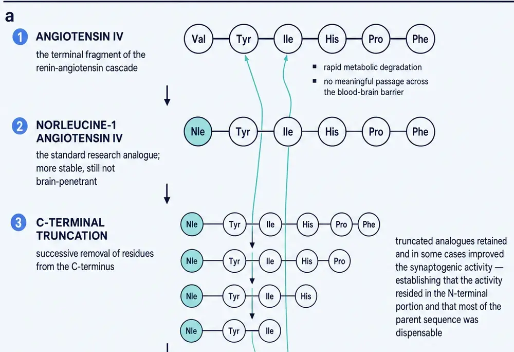
That last point deserves its own sentence, because it is the quietest anomaly in this compound's story and it goes almost unremarked in the literature that promotes it. Every structure–activity result in this lineage, from 1993 onward, says the free amino group at the front of the molecule is essential for binding the target. The Uppsala medicinal chemists demonstrated it directly: delete that amine from their macrocyclic inhibitor and the compound becomes completely inactive. Dihexa does not have one. Its N‑terminus is capped with a hexanoyl group, which is precisely what makes it survive a gut. Reviewing the field in 2020, Hallberg and Larhed stated the consequence without hedging — that dihexa's cognitive effects are not attributable to IRAP inhibition, and that no data on IRAP inhibition by dihexa exist in the literature at all (Hallberg & Larhed, 2020).
So the compound that emerged from a twenty-year programme to optimise binding at a particular site is chemically disqualified from binding it. That is not necessarily a criticism — drug programmes wander off their starting hypotheses all the time, and sometimes the wandering is the discovery. But it does mean that by 2012 dihexa needed a new target, and Part Two is the story of the one it was given.
05Making a peptide survive a stomach
The final compound was the product of a deliberate physicochemical push. The 2013 paper describes the goal in engineering terms: increase hydrophobicity, decrease hydrogen bonding, and thereby produce something that survives metabolism and crosses barriers. The chemistry that achieves it is the hexanoyl cap and the aminohexanoic amide tail — two greasy six-carbon chains bolted onto a two-residue core.
The result is a molecule that sits awkwardly against the conventional rules for drugs meant to reach the brain. Its molecular weight is 504.7, above the 500 usually taken as a soft ceiling. Its topological polar surface area is 151 Å2, where the working guideline for central-nervous-system penetration is under about 90 and the general oral guideline is under 140. It presents five hydrogen-bond donors. On paper it should not get into the brain, and the compound's own developers list its brain penetration as one of its two headline achievements.
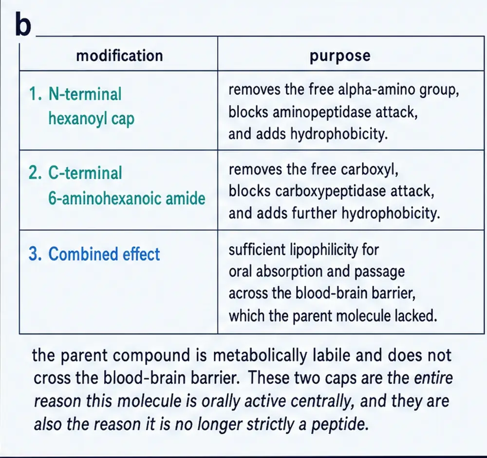
The measurement that supports the claim is a radiolabel study reported inside the 2013 paper: tritiated dihexa and carbon-14-labelled inulin were infused into rats, and the ratio of the two in brain tissue was used to estimate how much compound had crossed. That is a reasonable design, and it is the only published measurement of its kind for this molecule. It has not been independently repeated.
06Scopolamine, aged rats and a water maze
Dihexa entered the literature in a paper published online in October 2012 and in print in January 2013, in the Journal of Pharmacology and Experimental Therapeutics. Nine authors from Washington State University, led by McCoy, reported a series of metabolically stabilised angiotensin IV analogues and ended with one they had pushed furthest: N‑hexanoic‑Tyr‑Ile‑(6) aminohexanoic amide, which they named dihexa (McCoy et al., 2013).
This paper carries a Notice of Concern, issued by the journal in September 2021 and unresolved at the time of writing. It has not been retracted. Three companion papers from the same laboratory were retracted in April 2025; this one was not. Section 12 sets out exactly what was withdrawn and what was not, and why the distinction changes what this document can say.
Two behavioural models carried the efficacy claim. In the first, rats were given scopolamine, which blocks muscarinic acetylcholine receptors and produces a reliable, reversible impairment in spatial learning. In the second, the animals were simply old — twenty-four months, a stage at which a proportion of rats decline cognitively on their own. Both were tested in the Morris water maze, in which an animal must learn the position of a platform hidden just under the surface of opaque water, with the time taken to find it as the primary measure and a “probe trial” — the platform removed, the time spent searching where it used to be — as the test of whether a memory was formed rather than a swimming strategy.
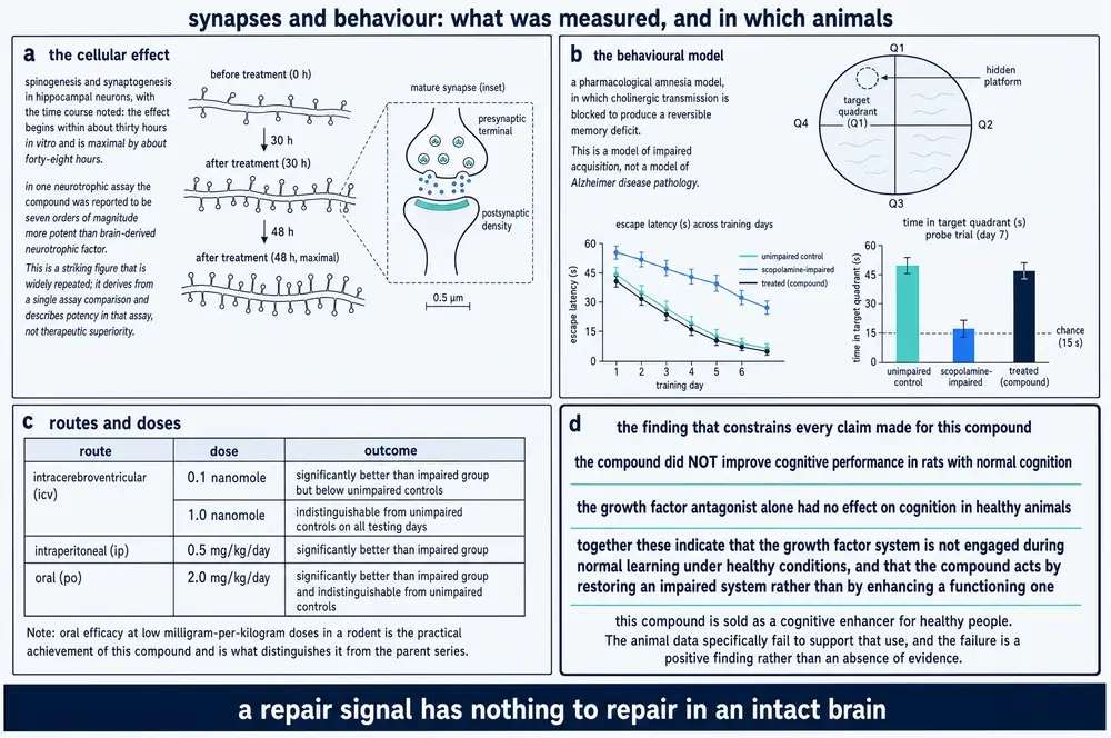
The reported outcomes were these. Given into the cerebral ventricles at 0.1 or 1.0 nanomole, dihexa improved the latency to find the platform relative to scopolamine-treated animals, and the higher dose was indistinguishable from unimpaired controls on every testing day. Given intraperitoneally at 0.5 mg/kg/day and orally at 2.0 mg/kg/day, the higher dose of each route produced water-maze performance significantly better than scopolamine-treated animals and, again, indistinguishable from controls. On the probe trial, the highest dose by every route increased time in the target quadrant, with a dose–response relationship. In twenty-four-month-old rats given 2 mg/kg/day orally, performance improved on most test days, and the paper is explicit that the aged-rat data were more variable and less robust than the scopolamine data, because not every old rat is an impaired one.
One qualification travels with all of it, and it is the qualification that the compound's later reputation ignored. In animals that were not impaired, dihexa did nothing. The Alzheimer's Drug Discovery Foundation's 2021 review of the compound states it as its summary finding: dihexa improved cognition in rat models of cognitive dysfunction, and did not improve cognitive function in rats with normal cognition. Whatever this molecule does, the published animal evidence describes the repair of a broken system, not the improvement of an intact one.
The only systematic review of this literature, published in 2018, scored the thirty-two studies it admitted on an eight-point quality scale adapted from the standard preclinical-stroke criteria. The mean and modal score was three. Only thirteen per cent described blinded outcome assessment; none monitored physiological parameters. The two dihexa studies scored five and four respectively, which places them above the field's average and still well short of what a clinical decision would need. The review's own summary judgement is that dihexa may be the most promising compound in the class for testing in humans, and its own stated limit is that it remains to be seen whether dihexa would help an animal that is not impaired (Ho & Nation, 2018).
07Spines, and where “ten million times” came from
The behavioural result was not what made dihexa famous. What made it famous was a set of photographs of dendrites.
Neurons receive most of their excitatory input on dendritic spines, small protrusions that appear, enlarge and disappear as circuits change. Counting them is a direct structural readout of synaptic connectivity, and it is one of the few measures on which a small molecule can be compared with a growth factor. In cultured rat hippocampal neurons, five days of dihexa raised the spine count to 41 per 50 micrometres of dendrite against 15 for vehicle — close to a threefold increase. A single thirty-minute application increased both spine number and spine-head width, from a mean of 0.67 to 0.80 micrometres; wider heads mean larger synapses. The new spines carried the normal molecular apparatus of a synapse — VGLUT1, synapsin, PSD‑95 — and whole-cell recordings showed a matching rise in miniature excitatory currents, which is the evidence that they were not merely present but functional.
Somewhere between that result and the public account, a number appeared that is not in the paper. On 11 October 2012, Washington State University's news office published a release headlined that a prospective Alzheimer's drug builds new brain cell connections. In it, the researchers were reported to have found dihexa seven orders of magnitude more powerful than brain-derived neurotrophic factor — in other words, that it would take ten million times as much BDNF to produce the same synapse formation.
That last degradation is worth dwelling on, because it is the mechanism by which a number becomes folklore. A press release makes a claim in units of orders of magnitude. Nine years later a peer-reviewed paper repeats it in units of multiples, cites the original work, and thereby launders a press-release figure into the citable literature with an error of six orders of magnitude in the wrong direction — and, being smaller, the wrong figure is the more believable one. Both versions circulate. Neither is a measurement reported in a paper.
None of which establishes that the underlying observation was wrong. A threefold increase in spine density in five days is a large effect, and it was measured. What it is not is a comparison with BDNF, and no such comparison appears in the primary literature this document could retrieve.
08A target arrived at backwards
By 2012 dihexa had a behavioural effect, a structural effect, and no mechanism. Its chemistry had removed the amino group that the twenty-year programme behind it had established as necessary for binding IRAP. The laboratory's answer was to propose an entirely different target.
The proposal had a precedent in the same laboratory's work on norleual, the reduced-bond analogue from the previous generation. In 2010 the group reported that norleual shares structural homology with the hinge region of hepatocyte growth factor — the linker that lets two HGF molecules pair up — and acts as a mimic of it, competitively inhibiting HGF binding to its receptor c‑Met in mouse liver membranes with a half-maximal inhibitory concentration of 3 picomolar, and blocking HGF-dependent signalling, proliferation, migration and invasion at picomolar concentrations (Yamamoto et al., 2010). That paper argued, in the same breath, that norleual's activity in cells which do not express IRAP casts doubt on the physiological significance of IRAP inhibition altogether. It carries an erratum published in 2015 and has not been retracted.
Hepatocyte growth factor is a plausible thing for a memory drug to act on. Despite the name, it is a broadly acting factor that in the nervous system guides axons, supports motor-neuron survival, and protects neurons from ischaemic injury. Its receptor, MET, is a receptor tyrosine kinase that signals through PI3K/Akt and the MAP kinase cascade — the same pathways that control spine formation. And HGF has a genuine liability as a drug: a half-life of a few minutes and essentially no brain penetration. A small molecule that made endogenous HGF work harder would solve a real problem.
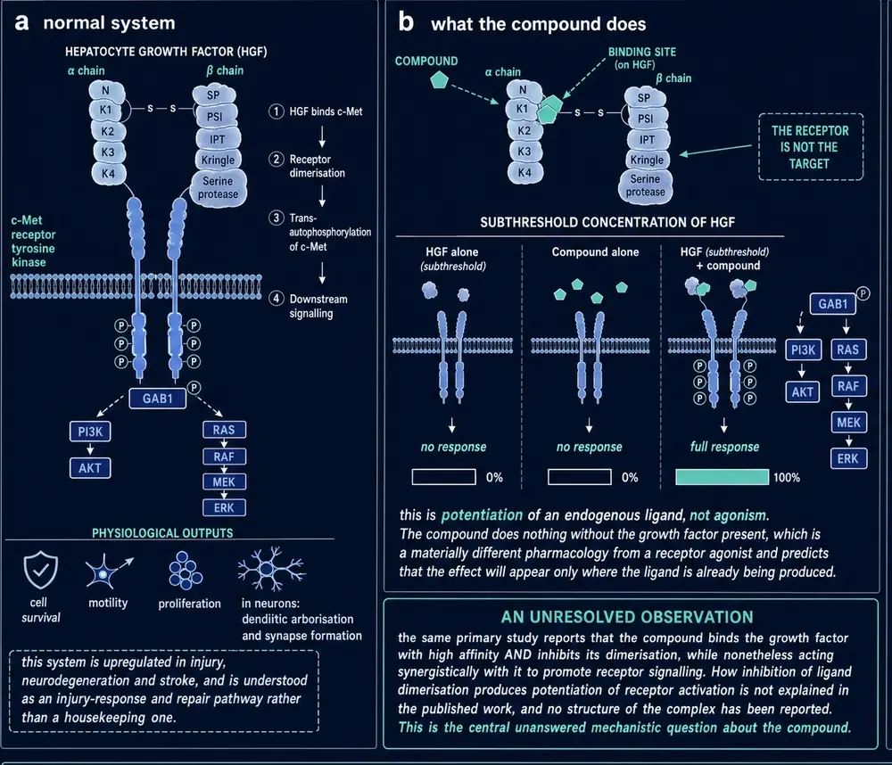
The word doing the work in that proposal is potentiator, and it is worth separating from the word it is usually confused with. An agonist binds a receptor and switches it on; its effect is bounded only by how much of it you give. A positive modulator does nothing on its own. It makes an existing signal easier to send, which means it can only act where that signal is already being sent — and hepatocyte growth factor, in the brain, is released in injury and repair rather than continuously. If the mechanism is right, the compound should therefore do nothing in a healthy animal and something in a damaged one. That is an unusually specific prediction for a drug proposal to make, it is testable, and Section 06 has already reported the test: in rats with normal cognition dihexa did nothing at all, and the antagonist that blocks the pathway also did nothing on its own. The mechanism and the behaviour agree on the point that matters most for anyone thinking of taking the compound while well.
In 2014 the group published the paper that connected dihexa to that system. Benoist and colleagues reported that dihexa binds HGF with high affinity; that dihexa and Nle1‑angiotensin IV induce phosphorylation of c‑Met in the presence of concentrations of HGF too low to do so alone; that both augment HGF-dependent cell scattering; that both induce hippocampal spinogenesis and synaptogenesis in the way HGF itself does; that these actions are blocked by an HGF antagonist and by a short hairpin RNA directed at c‑Met; and — the keystone experiment — that the procognitive effect of orally delivered dihexa in the water maze was abolished by an HGF antagonist delivered into the cerebral ventricles.
That last experiment is the strongest form of mechanistic argument available in behavioural pharmacology. A drug given by mouth produces an effect on learning; block the proposed target in the brain, and the effect disappears. If it holds, the mechanism is not merely correlated with the behaviour, it carries it.
That paper was retracted on 30 April 2025.
09What the pharmacokinetics say, and what they do not
The pharmacokinetic profile reported for dihexa is unusual enough that it deserves separate treatment, and it is the part of the 2013 paper that the later controversy has left least disturbed.
In rats, the terminal half-life was reported as 12.68 days after intravenous administration and 8.83 days after intraperitoneal administration. Those are not typographical slips for hours. A molecule of 505 daltons with two ester-free amide bonds and no obvious depot has no natural reason to persist for a fortnight, and the same paper reports a half-life in rat serum of 335.5 minutes — a figure that describes chemical stability in a tube rather than clearance from an animal, and which is roughly fifty times shorter. Nothing in the retrieved literature reconciles the two, and no independent laboratory has repeated either measurement.
Two features of the reported experiments qualify what “orally active” means here. First, the vehicle: dihexa was dissolved in dimethyl sulfoxide at one milligram per millilitre and administered in 75 per cent DMSO for the parenteral routes. DMSO is a solvent with its own pharmacology and its own capacity to alter membrane permeability, and a compound's behaviour in it is not straightforwardly its behaviour in water. Second, the oral dose that worked, 2 mg/kg/day, was the highest tested; the paper does not establish a ceiling.
The practical consequence of a long half-life is accumulation. If the rat figures are even approximately right, repeated administration does not produce a steady state within any short experiment; it produces a rising exposure. That matters for the safety question in Section 24, and it matters for reading the animal studies, because a three-month dosing study in mice at a fixed daily dose is not a study at that dose.
10A spin-out, and what it was built on
Universities commercialise discoveries, and Washington State commercialised this one. M3 Biotechnology was founded in Seattle to develop dihexa and its relatives; in 2019 it renamed itself Athira Pharma; and the company that today holds the programme is called LeonaBio. Its first chief executive was Leen Kawas, who had taken her doctorate in pharmacology at Washington State in 2011 on the work that produced the compound, and who is a co-author on the papers that established both its mechanism and the anti-cancer application of its predecessor.
What a spin-out sells is not a molecule but a package: a patent estate, a mechanism, and a body of published evidence that a licensee or an investor can read. Dihexa's package was unusually attractive. It had a striking preclinical result, a named target, a plausible disease, an oral route, and a headline number in circulation that was arresting even by the standards of the field. It also had one structural vulnerability, which is that almost all of the evidence came from one laboratory.
By 2015 that laboratory's two senior figures were publishing reviews in Progress in Neurobiology and the Journal of Alzheimer's Disease that placed dihexa at the centre of a new therapeutic approach, describing it as a first-in-class compound and carrying M3 Biotechnology affiliations alongside their university ones. Those reviews are the source of much of the compound's reputation in the secondary literature, and it is worth being clear about what they are: authoritative-sounding syntheses of the authors' own primary work, written by people with a commercial interest in it, and cited ever since by readers who did not notice the overlap.
11September 2021: four notices
On 20 June 2021 an allegation of research misconduct reached Athira's board. In September the Journal of Pharmacology and Experimental Therapeutics published four Notices of Concern, one for each of four papers from the Washington State laboratory. In October, following an independent investigation commissioned by the board, Kawas resigned as chief executive and left the board.
| Paper | What it established | Notice | Outcome |
|---|---|---|---|
| Kawas et al. 2011 | HGF hinge-region mimics with anti-Met and anticancer activity | Sept 2021 | Retracted April 2025 |
| Kawas et al. 2012 | Angiotensin IV analogues as HGF/Met modifiers | Sept 2021 | Retracted April 2025 |
| McCoy et al. 2013 | Dihexa itself — synthesis, water maze, spines | Sept 2021 | Notice stands; not retracted |
| Benoist et al. 2014 | Dihexa's mechanism — HGF/c‑Met dependence | Sept 2021 | Retracted April 2025 |
A notice of concern is not a finding. It is a journal telling its readers that questions have been raised which it cannot yet resolve, and asking them to treat the paper accordingly. For three and a half years, that was the status of the entire mechanistic case for this compound.
12April 2025: what was withdrawn, and what was not
On 30 April 2025 the journal retracted three of the four. The retraction notices are short and they are specific. Following an investigation by Washington State University, named figures in each paper — and, in one case, data submitted in a subsequent erratum — were found to contain falsified and/or fabricated data. The notices state that Leen H. Kawas and Joseph W. Harding were found to be solely responsible, and that the retraction was made at the request of the editor.
The distinction between the three that were retracted and the one that was not is the most consequential fact in this monograph, and it cuts both ways.
What was withdrawn. The mechanism. Benoist 2014 was the paper that connected dihexa to hepatocyte growth factor: the high-affinity binding, the c‑Met phosphorylation at subthreshold HGF, the shRNA experiment, and the keystone result in which an HGF antagonist delivered into the brain abolished the effect of orally delivered dihexa on learning. All of it is off the table. So is the 2012 paper that developed the analogue series as HGF/Met modifiers, and the 2011 paper that proposed the hinge-mimic concept in the first place. The 2010 norleual paper, which introduced that concept, carries an erratum and survives — but it is a paper about norleual, a different molecule, and it argues for inhibition of HGF rather than potentiation of it.
What was not withdrawn. The compound. McCoy 2013 — the synthesis, the water-maze results by three routes, the spine counts, the pharmacokinetics — carries a notice of concern and has not been retracted. Neither has Benoist 2011, the truncation study that established that the active core is three residues. Neither has any of the lineage work described in Part One: the binding site, its identification as IRAP, the structure–activity series. The scaffolding under dihexa is intact. What has been removed is the explanation of why it works.
A retracted paper is not evidence. This monograph does not cite Benoist 2014, Kawas 2012 or Kawas 2011 in support of any factual claim about what dihexa does. Where their content is described — as in Section 08 — it is described as a claim that was made and subsequently withdrawn, and the reader is told so in the same passage.
The 2013 paper is treated differently, because a notice of concern is a different instrument from a retraction and it would be as misleading to discard the paper as to rely on it uncritically. Its findings are reported with the notice stated, and wherever an independent laboratory has tested the same thing, that independent result is given precedence.
13January 2025: the settlement
Five months before the retractions, on 6 January 2025, the United States Attorney's Office for the Western District of Washington announced that Athira Pharma would pay $4,068,698 to resolve allegations under the False Claims Act. The allegation was not that the science was wrong. It was that between 1 January 2016 and 20 June 2021 the company failed to disclose, in grant applications and progress reports to the National Institutes of Health and in filings to the Department of Health and Human Services Office of Research Integrity, that allegations of image falsification had been made against its chief executive in respect of her doctoral dissertation and published papers — papers that were referenced in several of those grant applications, including one the NIH funded in 2019.
The case reached the government through a whistleblower. It was filed under the qui tam provisions of the False Claims Act by Andrew P. Mallon, who received $203,434 of the settlement. The United States Attorney's statement credits the company with notifying the NIH immediately once the full board learned of the misconduct, and records that the claims resolved by the settlement are allegations only, with no determination of liability.
Two further consequences are widely reported and are recorded here as reported rather than as verified against a primary instrument: that the independent board investigation found problems in a majority of the images in the 2011 dissertation, and that Washington State University subsequently revoked the doctorate. This document could not locate a university instrument stating the revocation, and treats it accordingly.
14Reading a literature with holes in it
What does a retraction do to the papers that cited the retracted work?
Formally, nothing. The citing papers remain in the literature, with their introductions still asserting what they asserted. In practice the effect is selective and it depends entirely on what the citation was doing. A paper that cited Benoist 2014 for the proposition that dihexa activates HGF/c‑Met — and there are several in this corpus — has lost its warrant for that sentence. A paper that used dihexa as a reagent because it behaves like an HGF agonist in a dish has lost nothing at all, because its evidence is its own result, not the citation.
That distinction organises the rest of this document. It divides the compound's literature into claims that depend on the withdrawn work and observations that do not, and the second category turns out to be larger and more interesting than the first. It contains an independent replication in a mouse model of Alzheimer's disease, a study of nerve repair in rats, an independent negative result in a model of Huntington's disease, and a body of stem-cell methodology in which dihexa is used, routinely and successfully, by laboratories that have no stake in any of this.
The most recent example is the clearest. A 2026 review of therapeutic peptides in orthopaedics devotes four sentences and a table row to dihexa, describing it as potentiating HGF and stimulating c‑Met, and suggesting it has potential for enhancing what the authors call the mind–muscle connection. It cites two sources for the compound. One is the 2015 review described above. The other is the paper that was retracted eight months before the review appeared. The review is careful in its own terms — it says plainly that clinical trials are lacking and that any use should be treated as exploratory rather than standard of care — and it is still, at the level of citation, resting on withdrawn evidence.
There is also a class of claim that survives the retraction and should not. The 2015 review in the Journal of Alzheimer's Disease describes dihexa as a first-in-class compound that facilitates the formation of new functional synaptic connections; the 2015 review in Progress in Neurobiology frames the HGF/c‑Met account as the laboratory's resolution of the receptor controversy. Both reviews rest substantially on the papers that were later withdrawn. Neither carries a notice. Both remain citable, and both continue to be cited — which is how a retracted finding goes on circulating in a literature that has formally removed it.
15Hair cells in a zebrafish
The most detailed dose–response study ever published on dihexa is not about memory. It is about deafness.
Aminoglycoside antibiotics kill the sensory hair cells of the inner ear, and in mammals those cells do not come back. Hepatocyte growth factor protects hair cells in cochlear explants, but as a drug it is hopeless — a half-life of minutes and no access to the inner ear. A stable small molecule that made endogenous HGF work harder would be a real clinical asset, and in 2015 a group working on the zebrafish lateral line tested dihexa for exactly that (Uribe et al., 2015).
The lateral line is a row of superficial sensory organs along the flank of a fish, containing hair cells structurally and functionally homologous to those of the mammalian inner ear, exposed on the skin where they can be counted in a living animal. It is a well-established screening system, and it has already delivered one otoprotective compound that translated to rodents.
What the study found is more informative than a simple positive. One micromolar dihexa protected hair cells robustly against acute neomycin and acute gentamicin, across a range of ototoxin concentrations, with no toxicity of its own. It did not protect against chronic gentamicin exposure over six hours — aminoglycosides kill hair cells by two distinguishable mechanisms, and dihexa touched only the fast one — and it did not protect against cisplatin at all. It did not reduce the amount of fluorescently tagged gentamicin entering the cells, so it is not working by blocking entry. Its protection was abolished by an antagonist that blocks HGF dimerisation, and partly attenuated by inhibitors of Akt, TOR and MEK — the pathways downstream of MET. And adding a single amino group to the N‑terminus of dihexa attenuated the protection, which is a striking result for a molecule whose N‑terminal chemistry is exactly what distinguishes it from its ancestors.
The zebrafish work is frequently offered as external corroboration of the HGF mechanism. It is not: Leen H. Kawas and Joseph W. Harding are co-authors. The behavioural and cell-biological work was done in a different laboratory on a different organism, which is genuinely valuable, but the compound, the antagonist and the mechanistic framing all came from the originating group. It should be read as an extension of that laboratory's programme into a new system, not as a test of it by outsiders.
16A mouse model of Alzheimer's disease
The clearest independent test of dihexa's central claim was published in 2021 by a group at Nanjing First Hospital, with no connection to Washington State and no commercial interest in the compound (Sun et al., 2021). They gave dihexa to APP/PS1 mice — animals carrying two human mutations that cause them to accumulate amyloid plaques and lose neurons — at 1.44 and 2.88 mg/kg, daily, from six months of age to nine.
The behavioural result reproduces the original claim. In the Morris water maze, treated animals found the platform faster on days four and five and made more crossings of the platform location on the probe trial, while swimming at the same speed as controls — which is the control that separates a memory effect from a motor one. The structural results go further than the original: Nissl staining showed more surviving cortical neurons, and synaptophysin, a presynaptic marker, was higher in treated animals. Markers of glial activation fell, as did interleukin‑1β and tumour necrosis factor α, while the anti-inflammatory interleukin‑10 rose.
The mechanistic result does not reproduce the original claim; it replaces it. The Nanjing group attributed the effects to the PI3K/Akt pathway, showed that dihexa raised PI3K and phosphorylated Akt, and demonstrated that the PI3K inhibitor wortmannin reversed the anti-inflammatory and anti-apoptotic effects. They did not measure HGF, and they did not measure MET. PI3K/Akt sits downstream of MET, so the finding is compatible with the HGF account — but it is equally compatible with a dozen other upstream events, and the study does not discriminate.
Three problems in the paper should travel with its result. Its methods section states within a single paragraph that dihexa was administered intragastrically and that it was administered intraperitoneally; the route is therefore uncertain in the primary record. Its opening finding is that dihexa raised brain angiotensin IV levels measured by immunoassay, which is difficult to credit — dihexa is not a precursor of angiotensin IV, shares only two residues with it, and cross-reactivity in the assay is the more economical explanation. And the authors themselves note a missing control: the wortmannin experiment lacks a wortmannin-only arm.
17A cut nerve, and a number that shrank a millionfold
In 2021 a group working on the problem of restoring limb function after blast injury tested dihexa in a rat model in which the sciatic nerve is transected and surgically repaired (Weiss et al., 2021). The work was done at a military medical centre with academic collaborators, and Joseph Harding is a co-author — so this is an extension of the originating programme into a new injury model rather than a test of it by outsiders. The design ran ten groups of six animals for sixteen weeks, combining mesenchymal stem cells, granulocyte colony-stimulating factor and dihexa at 2–4 mg/kg, delivered locally in a hydrogel at the repair site, systemically, and directly into the target muscle.
Sensory recovery was essentially complete by eight weeks in every group, including the vehicle controls, so the model does not discriminate there. Motor function, measured by walking footprint analysis at eight to sixteen weeks, was significantly better in animals that received stem cells plus either G‑CSF or dihexa injected into the gastrocnemius muscle, and flexion contractures were reduced. Muscle atrophy was not prevented in any group, including the treated ones. This is a modest, honestly reported positive in a combination design that cannot isolate dihexa's contribution.
The paper is included here for a second reason. Its introduction states that dihexa has neurotrophic activity seven times greater than BDNF, and cites the originating work for it. As Section 07 set out, the claim in circulation is seven orders of magnitude, it originates in a press release, and no direct comparison with BDNF appears in the primary paper. What happened here is that a figure with no measurement behind it was compressed by a factor of about 1.4 million and thereby made plausible enough to enter the peer-reviewed record as a citable fact. It is now available to be cited again.
One further result deserves its place here, because it does what none of the studies above does: it tests the mechanism directly, in neurons, in a laboratory with no connection to any of this. In 2026 a group at Bengbu Medical University, studying hippocampal neurons under oxygen–glucose deprivation, silenced hepatocyte growth factor and then applied dihexa at 100 nanomolar. It reversed the effects of the knockdown on cell viability, on pyroptosis, and on c‑Met signalling, and the reciprocal control — a c‑Met kinase inhibitor — abolished the rescue (Tang et al., 2026). That is an independent laboratory reproducing, in neural tissue and in a different system, the core mechanistic claim of the paper that was retracted. It does not restore the retracted paper. It does mean the claim that paper made is not thereby false, and this document says so because the honest position is not that dihexa's mechanism was disproved — it is that the evidence for it was withdrawn, and has begun to be rebuilt elsewhere.
18The quiet career
The largest body of work involving dihexa is not about the brain, is not about behaviour, and has nothing to do with any of the controversy. It is about livers.
Turning human pluripotent stem cells into hepatocyte-like cells is a workhorse procedure — used for drug toxicity screening, disease modelling and transplantation research — and the standard protocols depend on recombinant growth factors, of which hepatocyte growth factor is one of the most expensive and least stable. In 2015 a group at Newcastle published a protocol that replaced the growth factors with small molecules, using a Wnt agonist to drive cells to definitive endoderm, dimethyl sulfoxide to reach the hepatoblast stage, and dihexa, described simply as an HGF agonist, together with dexamethasone, for the final maturation step (Siller et al., 2015). The protocol was written up for a methods series the following year, and it worked.
What followed is the most thoroughly replicated finding this compound has. Laboratories across several countries adopted the protocol, adapted it, benchmarked it and published the results. A 2022 study using a completely small-molecule strategy reported that 0.1 micromolar dihexa in the maturation cocktail produced co-expression of the hepatoblast markers AFP and HNF4α in 95.5 per cent of cells, against 96.2 per cent for the conventional growth-factor route — equivalence, at a fraction of the cost and with none of the batch variability of a recombinant protein. Others have used it in hepatoblast expansion, in liver organoid production, in automated culture systems, in proteomic profiling and in toxicology platforms.
Two things follow from this, and they pull in opposite directions.
The first is that dihexa does something real to the HGF/MET axis. A reagent that substitutes for HGF in a demanding, multi-week differentiation protocol, in many hands, and produces cells that pass the same functional assays, is not doing nothing. This is functional evidence of HGF-pathway agonism, obtained from laboratories with no stake in the claim, and it survives the retractions completely intact because it does not depend on them. It is arguably the strongest evidence about this compound in existence.
The second is that it is not evidence about cognition, and it does not test the mechanism that was retracted. None of these papers measures HGF binding or dimerisation. They inherited the label “HGF agonist” from the very literature that has since been withdrawn, and they use it as an operational description of what the molecule does in their protocol, which is all their experiments can support. A compound can be a perfectly good growth-factor mimetic in a dish of hepatoblasts and still have no useful effect on a human brain.
19Huntington's disease, and the weight of recency
The most recent primary study of dihexa is negative, and it is independent.
In 2024 a group published a test of the compound — under its development code PNB‑0408 — in a rat model of Huntington's disease (Wells et al., 2024). Forty male Wistar rats were randomised into three groups: vehicle, the mitochondrial toxin 3‑nitropropionic acid, and 3‑nitropropionic acid plus dihexa administered alongside it. Body weight, motor function and cognition were followed for five weeks, and the brains were examined histologically afterwards.
The model worked: the toxin reduced weight gain, impaired spatial learning and memory consolidation, and produced marked motor dysfunction. Dihexa did not protect against any of it. The authors' conclusion is unambiguous — that the compound did not protect rats from the deficits induced by the toxin, and may not be an efficacious strategy in this model.
A single negative in a single model does not refute a compound; the 3‑nitropropionic acid model is a metabolic poisoning rather than a neurodegenerative disease, and a drug that promotes synaptic repair might reasonably fail against ongoing mitochondrial toxicity. But recency and independence both count, and this study has both. It is the only published attempt by an outside group to extend the compound to a new neurological indication, and it did not work.
Reproduced independently: that dihexa substitutes functionally for hepatocyte growth factor in cell differentiation, at 100 nanomolar, in laboratories on four continents; that it acts through c‑Met in hippocampal neurons; and that it improves water-maze performance in a mouse model of Alzheimer's disease, in one laboratory.
Reported once, and not independently reproduced: the original scopolamine and aged-rat behavioural results; the threefold spine increase; the multi-day half-life; brain penetration; protection of zebrafish hair cells; benefit in nerve repair as part of a combination. The last two came from studies on which the originating investigator is a co-author.
Reported and withdrawn: that dihexa binds hepatocyte growth factor, promotes its dimerisation, and requires that interaction for its effect on learning.
Tested independently and not found: protection against 3‑nitropropionic acid toxicity in a rat model of Huntington's disease.
Never studied: chronic toxicity in any species; carcinogenicity; any effect in a healthy animal or a healthy person.
20The same molecule, with a phosphate on it
In 2019 M3 Biotechnology renamed itself Athira Pharma, and the compound it took into human trials was not dihexa. It was called NDX‑1017, then ATH‑1017, and eventually received the international non-proprietary name fosgonimeton. Athira's published papers describe it as a small-molecule positive modulator of the HGF/MET system, delivered by subcutaneous injection, which is rapidly converted in plasma to an active metabolite. The metabolite is called ATH‑1001 in the 2022 paper and renamed fosgo‑AM in the papers after it.
ATH‑1001 is dihexa.
That is not an inference from the naming. It can be checked in two independent chemical registries, and it was. The United States Food and Drug Administration's substance registration system assigns dihexa the unique ingredient identifier 9WYX65A5C2 and the molecular formula C27H44N4O5; it assigns fosgonimeton the identifier H91OA9858J and the formula C27H45N4O8P. PubChem returns the same two structures under CID 129010512 and CID 156596375. The difference between the two formulas is exactly HPO3, and the structural notation shows precisely where it sits: on the phenol oxygen of the tyrosine, with every other atom and all three stereocentres unchanged. Fosgonimeton is dihexa carrying a phosphate ester. ATH‑1017, the string that appears in the trial registry, is its sodium salt.
A prodrug is not a trivial thing — it is a genuine piece of pharmaceutical engineering, and in this case a well-motivated one. Athira's own measurements, published in 2023, explain why they needed it. The free molecule has a maximum aqueous solubility below 50 micrograms per millilitre, and it does not survive the gut: 7.8 per cent remained after an hour in simulated gastric fluid and 0.1 per cent in simulated intestinal fluid. Phosphorylating the tyrosine raises solubility above 70 milligrams per millilitre, more than a thousandfold, which is enough to put a therapeutic dose into an injection volume a patient can tolerate. In rats and mice the phosphate itself is not detected in any brain region; only the released molecule crosses.
Four peer-reviewed papers describe fosgonimeton: the phase 1 report, two preclinical papers and the phase 2/3 trial report. None of them contains the word "dihexa." None contains "angiotensin," or the systematic name, or the names of the Washington State University investigators. The 2023 preclinical paper introduces the active metabolite as the best-performing of fourteen related small molecules screened against a phosphorylated-MET assay — a hit from a compound library, with no ancestry given.
This document does not claim that the omission was improper; a paper is entitled to describe the molecule in front of it. But it has a consequence the reader should hold onto. Anyone reading the fosgonimeton literature has no way of knowing that they are reading about dihexa, and anyone reading the dihexa literature has no way of knowing that it reached a phase 3 trial in another name.
21LIFT‑AD: 554 people, twenty-six weeks
The trial that tested this chemistry in people was LIFT‑AD, registered as NCT04488419, a randomised, double-blind, placebo-controlled phase 2/3 study in mild-to-moderate Alzheimer's disease across ninety sites in the United States. It screened 1,284 people, randomised 554, and dosed 549. Treatment was a daily subcutaneous injection for twenty-six weeks. The primary analysis was restricted, by a mid-trial design change, to the 287 participants not taking a cholinesterase inhibitor: 143 on fosgonimeton, 144 on placebo.
The primary endpoint was a Global Statistical Test, a single composite made by standardising each participant's change from baseline on a cognitive scale (ADAS‑Cog11) and a function scale (ADCS‑ADL23) and averaging them. A lower score means improvement.
| Endpoint at week 26 | Placebo | Fosgonimeton | Difference (SE) | p |
|---|---|---|---|---|
| Global Statistical Test (primary) | −0.13 | −0.21 | −0.08 (0.10) | 0.70 |
| ADAS‑Cog11, points | −0.39 | −1.09 | −0.70 (0.77) | 0.35 |
| ADCS‑ADL23, points | −0.02 | +0.65 | +0.67 (0.92) | 0.61 |
| Plasma neurofilament light, pg/mL | +2.95 | −0.96 | −3.91 (3.46) | 0.26 |
| Plasma p‑tau217, % change vs placebo | — | — | −13.7 % | <0.01 |
| Plasma GFAP, % change vs placebo | — | — | −9.4 % | 0.21 |
The composite did not move. Neither did either of its components. The trial had been designed on the assumption that placebo-treated participants with mild-to-moderate disease would decline measurably over six months, and they did not — both groups improved slightly on the cognitive scale from week two, peaked around week six, and drifted back. The investigators' own account attributes the null result to a population milder than intended (85 per cent were rated at the milder of the two permitted dementia stages despite nearly half qualifying as moderate on the screening test), a follow-up too short for that population, and a placebo response plausibly amplified by a daily injection.
The safety picture is the clearer half of the result. There were no deaths. The dominant finding was local: injection site reactions in 57 per cent of those on 40 mg and 73 per cent of those on 70 mg, against 14 per cent on placebo, and they drove most of the discontinuations (21.5 per cent at the higher dose against 4.6 per cent on placebo). Transient, asymptomatic eosinophilia appeared in about seven per cent of treated participants and in no placebo participant, and in more than four fifths of those cases it accompanied an injection site reaction. Five participants had serious adverse events considered possibly drug-related, including two cases of angioedema. Nothing in the record suggests a signal of the kind that would stop a programme on safety grounds.
22What a failed trial of the prodrug does, and does not, say
It is tempting to read LIFT‑AD as the verdict on dihexa, and equally tempting to read it as irrelevant to dihexa. Neither is right, and the reasons are worth separating.
What it does establish. Dihexa has now been present in human plasma, at measured concentrations, in several hundred people for six months, and the resulting safety record is unremarkable apart from injection-site effects attributable to the formulation. That is not nothing: before LIFT‑AD there was no human exposure data of any kind for this chemistry. It also establishes that the compound reaches the brain in people to some degree — the phase 1 study reported dose-related changes in quantitative electroencephalography, which is a crude but genuine indicator of central activity.
What it does not establish. It does not test dihexa taken orally, in a healthy person, at the doses sold on the research-chemical market, for the purpose those markets advertise. The trial gave a fixed 40 mg subcutaneous dose to people with an established neurodegenerative disease, and asked whether their disease progressed more slowly. A null answer to that question says nothing about whether the same molecule sharpens cognition in a person who is well — a question no trial has ever asked.
It also does not cleanly refute the mechanism, because the trial's own authors identify a limitation that cuts deeper than the population argument: there is no in vivo target-engagement data. Athira's 2024 paper states plainly that measuring transient MET phosphorylation in the relevant brain regions was not achieved, and that the molecular mechanism by which the compound binds the HGF/MET complex has not been elucidated. A trial that fails without demonstrating that the drug engaged its target has not tested the target.
There is one further wrinkle that pulls in the opposite direction from the usual "the dose was too low" objection. Athira's own animal work reports the largest effect at the lowest dose tested — 0.125 mg/kg — with efficacy declining as the dose rose, and describes the dose–response as hormetic, an inverted U. The preclinical dihexa work from Washington State had reported the same shape, and so did the independent zebrafish study in Section 15. If that is real, then a fixed 40 mg human dose chosen without exposure-matched bridging is as likely to have overshot the useful range as undershot it, and the trial cannot distinguish the two.
23What the regulator actually says
Dihexa is not approved as a medicine in any jurisdiction. It has never been the subject of a registered clinical trial under its own name: a search of ClinicalTrials.gov for dihexa, PNB‑0408, ATH‑1001 and the systematic name returns five studies, and all five are fosgonimeton.
Its most direct regulatory appearance is on a list that exists to name substances compounding pharmacies should not use. Under section 503A of the Federal Food, Drug and Cosmetic Act, a pharmacy may compound with a bulk substance only if it meets a monograph, is a component of an approved drug, or appears on a list the agency maintains. Substances nominated for that list which the agency judges to raise significant safety risks are placed in "category 2," which in practice bars them. Dihexa acetate was nominated, placed in category 2, and the nomination was subsequently withdrawn by the nominator. The agency's stated reason for the category 2 placement is the single most economical summary of this compound's evidence base:
The agency records that it has not identified any human exposure data on drug products containing dihexa acetate administered by any route, and that it therefore lacks important information about any safety issues the substance raises, including whether it would cause harm if administered to humans.
That entry was written about dihexa administered as dihexa. It remains accurate in those terms: the human exposure that exists is exposure to a phosphate prodrug given by subcutaneous injection under a protocol, which is not the same thing and was not before the agency when the nomination was assessed.
Meanwhile the compound is sold, and at least once it has been found where it was not declared. A 2025 survey of illicit and grey-market “smart drug” products seized or test-purchased by European and Australian regulators identified dihexa in a product marketed as a dietary supplement, classified by the analysts as a research chemical. That is the only record of real-world human exposure to this molecule anywhere in this monograph's corpus, and it is a chemical analysis of a product rather than any observation of a person.
Beyond that, the compound appears in this project's own vendor evidence library as a research chemical from multiple suppliers, in capsule and powder form, with certificates of analysis dated across 2023, 2025 and 2026. The research-use-only framing under which it is sold is a legal position about the transaction, not a scientific claim about the substance, and it is worth being precise about what it does and does not mean: it means no seller has asserted a medical use to a regulator, and it means no regulator has evaluated one.
24The question nobody has answered
Every account of how dihexa works — the laboratory's own, the independent replications, and the successor company's — agrees on one thing: it potentiates signalling through MET. That is the same MET whose gene is amplified in gastric and lung cancers, whose exon 14 skipping mutation defines a molecular subtype of non-small-cell lung cancer with its own approved inhibitors, and against which the pharmaceutical industry has spent two decades building drugs to block the pathway.
A molecule designed to push a receptor tyrosine kinase in the direction oncology spends its money pushing back is not thereby a carcinogen. The distinction the compound's advocates draw is real: dihexa is described as a positive modulator rather than an agonist, requiring endogenous HGF to be present, and therefore acting only where the growth factor is already being released — which in the brain means injury and repair. The Athira papers lean on the hormetic dose–response for the same reason, arguing that a system with built-in negative feedback is a system that resists being driven.
But the argument is a mechanistic prediction, not a finding, and the finding that would test it does not exist. No chronic toxicology study of dihexa has been published in any species. No carcinogenicity study. The longest published exposure in an animal is three months, in mice, in a single laboratory. The Alzheimer's Drug Discovery Foundation's 2021 review of the compound reached the same conclusion in the same words — that activation of HGF and MET could theoretically promote tumorigenesis and cancer progression, and that no study has tested it. Five years later that remains true.
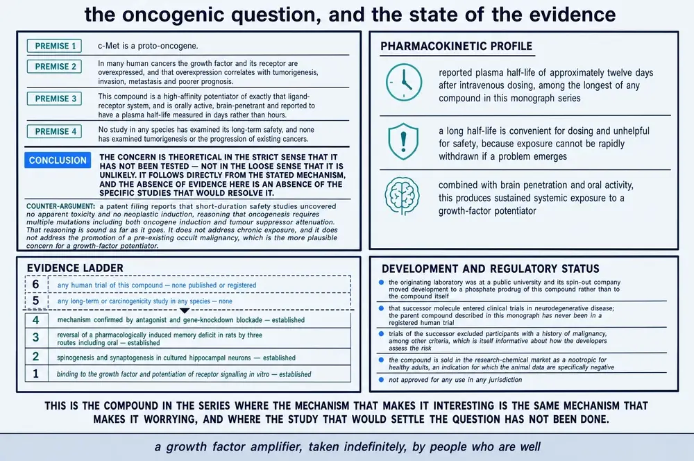
Three features of the compound make that question harder to wave away than it would be for most experimental molecules. The reported half-life in rats is measured in days rather than hours — roughly 12.7 days after intravenous and 8.8 days after intraperitoneal administration — so repeated dosing accumulates. The trial exclusion criteria written by the company that developed the chemistry bar anyone with a malignant tumour diagnosed within three years. And the population buying it on the research-chemical market is using it for months at a time, without monitoring, in the absence of the very data the exclusion criteria imply the developers thought was missing.
This document describes published research. It does not recommend human use of dihexa or of any other compound named in it, and it specifies no dose, route or schedule for any person. Every dose, concentration and duration reported above is a parameter of an experiment, stated with the species and the route it belongs to.
The compound has no approved use anywhere, no registered trial under its own name, no published chronic toxicology in any species, and — in the FDA's own assessment — no human exposure data at all. Nothing in the scientific record supports its use by a person, and this monograph should not be read as suggesting otherwise.
25References
Generated from verified NCBI records rather than from recall. Author lists, journal names, volumes, pages and identifiers are taken from the PubMed record for each citation, and the build refuses to run if any identifier fails to resolve. This series has twice shipped reference lists drafted from memory in which identifiers pointed at real but unrelated papers — and this document made the same error in its prose, naming the wrong first author for three studies until the generated list was read back against the text.
Three of the works listed below have been retracted and one carries a notice of concern. They appear because a document that discusses a retraction must identify what was retracted. They are never cited in support of a factual claim about what this compound does, and the retraction notices are listed as entries of their own so a reader can reach them directly.
- Albiston AL, McDowall SG, Matsacos D, Sim P, Clune E, Mustafa T, et al.. Evidence that the angiotensin IV (AT(4)) receptor is the enzyme insulin-regulated aminopeptidase. J Biol Chem. 2001;276(52):48623-6.
PMID 11707427 · doi:10.1074/jbc.C100512200 - Albiston AL, Morton CJ, Ng HL, Pham V, Yeatman HR, Ye S, et al.. Identification and characterization of a new cognitive enhancer based on inhibition of insulin-regulated aminopeptidase. FASEB J. 2008;22(12):4209-17.
PMID 18716029 · doi:10.1096/fj.08-112227 - Albiston AL, Fernando RN, Yeatman HR, Burns P, Ng L, Daswani D, et al.. Gene knockout of insulin-regulated aminopeptidase: loss of the specific binding site for angiotensin IV and age-related deficit in spatial memory. Neurobiol Learn Mem. 2010;93(1):19-30.
PMID 19660563 · doi:10.1016/j.nlm.2009.07.011 - Benoist CC, Wright JW, Zhu M, Appleyard SM, Wayman GA, Harding JW. Facilitation of hippocampal synaptogenesis and spatial memory by C-terminal truncated Nle1-angiotensin IV analogs. J Pharmacol Exp Ther. 2011;339(1):35-44.
PMID 21719467 · doi:10.1124/jpet.111.182220 · PMC3186286 - Benoist CC, Kawas LH, Zhu M, Tyson KA, Stillmaker L, Appleyard SM, et al.. The procognitive and synaptogenic effects of angiotensin IV-derived peptides are dependent on activation of the hepatocyte growth factor/c-met system. J Pharmacol Exp Ther. 2014;351(2):390-402.
PMID 25187433 · doi:10.1124/jpet.114.218735 · PMC4201273 - Benoist CC, Kawas LH, Zhu M, Tyson KA, Stillmaker L, Appleyard SM, et al.. Retraction notice to "The Procognitive and Synaptogenic Effects of Angiotensin IV-Derived Peptides Are Dependent on Activation of the Hepatocyte Growth Factor/c-Met System" [J Pharmacol Exp Ther 351 (2014) 390-402]. J Pharmacol Exp Ther. 2025;392(4):103567.
PMID 40312093 · doi:10.1016/j.jpet.2025.103567 · PMC13095468 - Braszko JJ, Kupryszewski G, Witczuk B, Wiśniewski K. Angiotensin II-(3-8)-hexapeptide affects motor activity, performance of passive avoidance and a conditioned avoidance response in rats. Neuroscience. 1988;27(3):777-83.
PMID 3252173 · doi:10.1016/0306-4522(88)90182-0 - De Bundel D, Smolders I, Yang R, Albiston AL, Michotte Y, Chai SY. Angiotensin IV and LVV-haemorphin 7 enhance spatial working memory in rats: effects on hippocampal glucose levels and blood flow. Neurobiol Learn Mem. 2009;92(1):19-26.
PMID 19233301 · doi:10.1016/j.nlm.2009.02.004 - Hallberg M, Larhed M. From Angiotensin IV to Small Peptidemimetics Inhibiting Insulin-Regulated Aminopeptidase. Front Pharmacol. 2020;11:590855.
PMID 33178027 · doi:10.3389/fphar.2020.590855 · PMC7593869 - Harding JW, Cook VI, Miller-Wing AV, Hanesworth JM, Sardinia MF, Hall KL, et al.. Identification of an AII(3-8) [AIV] binding site in guinea pig hippocampus. Brain Res. 1992;583(1-2):340-3.
PMID 1504842 · doi:10.1016/s0006-8993(10)80047-2 - Ho JK, Nation DA. Cognitive benefits of angiotensin IV and angiotensin-(1-7): A systematic review of experimental studies. Neurosci Biobehav Rev. 2018;92:209-225.
PMID 29733881 · doi:10.1016/j.neubiorev.2018.05.005 · PMC8916541 - Hua X, Church K, Walker W, L'Hostis P, Viardot G, Danjou P, et al.. Safety, Tolerability, Pharmacokinetics, and Pharmacodynamics of the Positive Modulator of HGF/MET, Fosgonimeton, in Healthy Volunteers and Subjects with Alzheimer's Disease: Randomized, Placebo-Controlled, Double-Blind, Phase I Clinical Trial. J Alzheimers Dis. 2022;86(3):1399-1413.
PMID 35180125 · doi:10.3233/JAD-215511 · PMC9108585 - Johnston JL, Reda SM, Setti SE, Taylor RW, Berthiaume AA, Walker WE, et al.. Fosgonimeton, a Novel Positive Modulator of the HGF/MET System, Promotes Neurotrophic and Procognitive Effects in Models of Dementia. Neurotherapeutics. 2023;20(2):431-451.
PMID 36538176 · doi:10.1007/s13311-022-01325-5 · PMC10121968 - Kawas LH, Yamamoto BJ, Wright JW, Harding JW. Mimics of the dimerization domain of hepatocyte growth factor exhibit anti-Met and anticancer activity. J Pharmacol Exp Ther. 2011;339(2):509-18.
PMID 21859930 · doi:10.1124/jpet.111.185694 - Kawas LH, McCoy AT, Yamamoto BJ, Wright JW, Harding JW. Development of angiotensin IV analogs as hepatocyte growth factor/Met modifiers. J Pharmacol Exp Ther. 2012;340(3):539-48.
PMID 22129598 · doi:10.1124/jpet.111.188136 - Kawas LH, McCoy AT, Yamamoto BJ, Wright JW, Harding JW. Retraction notice to "Development of Angiotensin IV Analogs as Hepatocyte Growth Factor/Met Modifiers" [J Pharmacol Exp Ther 340 (2012) 539-548]. J Pharmacol Exp Ther. 2025;392(4):103566.
PMID 40312092 · doi:10.1016/j.jpet.2025.103566 - Kawas LH, Yamamoto BJ, Wright JW, Harding JW. Retraction notice to "Mimics of the Dimerization Domain of Hepatocyte Growth Factor Exhibit Anti-Met and Anticancer Activity" [J Pharmacol Exp Ther 339 (2011) 509-518]. J Pharmacol Exp Ther. 2025;392(4):103568.
PMID 40312094 · doi:10.1016/j.jpet.2025.103568 - Kramár EA, Armstrong DL, Ikeda S, Wayner MJ, Harding JW, Wright JW. The effects of angiotensin IV analogs on long-term potentiation within the CA1 region of the hippocampus in vitro. Brain Res. 2001;897(1-2):114-21.
PMID 11282364 · doi:10.1016/s0006-8993(01)02100-x - Lew RA, Mustafa T, Ye S, McDowall SG, Chai SY, Albiston AL. Angiotensin AT4 ligands are potent, competitive inhibitors of insulin regulated aminopeptidase (IRAP). J Neurochem. 2003;86(2):344-50.
PMID 12871575 · doi:10.1046/j.1471-4159.2003.01852.x - Mathapati S, Siller R, Impellizzeri AA, Lycke M, Vegheim K, Almaas R, et al.. Small-Molecule-Directed Hepatocyte-Like Cell Differentiation of Human Pluripotent Stem Cells. Curr Protoc Stem Cell Biol. 2016;38:1G.6.1-1G.6.18.
PMID 27532814 · doi:10.1002/cpsc.13 - McCoy AT, Benoist CC, Wright JW, Kawas LH, Bule-Ghogare JM, Zhu M, et al.. Evaluation of metabolically stabilized angiotensin IV analogs as procognitive/antidementia agents. J Pharmacol Exp Ther. 2013;344(1):141-54.
PMID 23055539 · doi:10.1124/jpet.112.199497 · PMC3533412 - Pan T, Wang N, Zhang J, Yang F, Chen Y, Zhuang Y, et al.. Efficiently generate functional hepatic cells from human pluripotent stem cells by complete small-molecule strategy. Stem Cell Res Ther. 2022;13(1):159.
PMID 35410439 · doi:10.1186/s13287-022-02831-1 · PMC8996222 - Pan T, Tao J, Chen Y, Zhang J, Getachew A, Zhuang Y, et al.. Robust expansion and functional maturation of human hepatoblasts by chemical strategy. Stem Cell Res Ther. 2021;12(1):151.
PMID 33632328 · doi:10.1186/s13287-021-02233-9 · PMC7908723 - Porsteinsson AP, Sabbagh M, Tariot PN, Church KJ, San Martin J, Ooi KC, et al.. Fosgonimeton in mild-to-moderate Alzheimer's disease. J Alzheimers Dis Rep. 2025;9:25424823251405817.
PMID 41393340 · doi:10.1177/25424823251405817 · PMC12701236 - Rahman OF, Lee SJ, Seeds WA. Therapeutic Peptides in Orthopaedics: Applications, Challenges, and Future Directions. J Am Acad Orthop Surg Glob Res Rev. 2026;10(1).
PMID 41490200 · doi:10.5435/JAAOSGlobal-D-25-00236 · PMC12753158 - Reda SM, Setti SE, Berthiaume AA, Wu W, Taylor RW, Johnston JL, et al.. Fosgonimeton attenuates amyloid-beta toxicity in preclinical models of Alzheimer's disease. Neurotherapeutics. 2024;21(4):e00350.
PMID 38599894 · doi:10.1016/j.neurot.2024.e00350 · PMC11067346 - Sardinia MF, Hanesworth JM, Krebs LT, Harding JW. AT4 receptor binding characteristics: D-amino acid- and glycine-substituted peptides. Peptides. 1993;14(5):949-54.
PMID 8284271 · doi:10.1016/0196-9781(93)90071-n - Seyer B, Diwakarla S, Burns P, Hallberg A, Grӧnbladh A, Hallberg M, et al.. Insulin-regulated aminopeptidase inhibitor-mediated increases in dendritic spine density are facilitated by glucose uptake. J Neurochem. 2020;153(4):485-494.
PMID 31556456 · doi:10.1111/jnc.14880 - Siller R, Greenhough S, Naumovska E, Sullivan GJ. Small-molecule-driven hepatocyte differentiation of human pluripotent stem cells. Stem Cell Reports. 2015;4(5):939-52.
PMID 25937370 · doi:10.1016/j.stemcr.2015.04.001 · PMC4437467 - Stam F, Bjurling S, Zelleroth S, Badrd'din S, Gising J, Larhed M, et al.. The effects of insulin-regulated aminopeptidase inhibition with HA08 on recognition memory acquisition and spatial working memory under reversed circadian conditions in male rats. IBRO Neurosci Rep. 2025;19:854-861.
PMID 41280134 · doi:10.1016/j.ibneur.2025.10.022 · PMC12634304 - Sun X, Deng Y, Fu X, Wang S, Duan R, Zhang Y. AngIV-Analog Dihexa Rescues Cognitive Impairment and Recovers Memory in the APP/PS1 Mouse via the PI3K/AKT Signaling Pathway. Brain Sci. 2021;11(11).
PMID 34827486 · doi:10.3390/brainsci11111487 · PMC8615599 - Swanson GN, Hanesworth JM, Sardinia MF, Coleman JK, Wright JW, Hall KL, et al.. Discovery of a distinct binding site for angiotensin II (3-8), a putative angiotensin IV receptor. Regul Pept. 1992;40(3):409-19.
PMID 1438983 · doi:10.1016/0167-0115(92)90527-2 - Tang H, Gao N, Gao L, Li Y, Xia Y. Umbilical cord mesenchymal stromal cells-derived HGF inhibits STING-mediated pyroptosis to alleviate cerebral ischemia/reperfusion injury via c-Met/β-catenin/RNF5 pathway. J Transl Med. 2026;24(1).
PMID 41845461 · doi:10.1186/s12967-026-08008-1 · PMC13107589 - Uribe PM, Kawas LH, Harding JW, Coffin AB. Hepatocyte growth factor mimetic protects lateral line hair cells from aminoglycoside exposure. Front Cell Neurosci. 2015;9:3.
PMID 25674052 · doi:10.3389/fncel.2015.00003 · PMC4309183 - Vanhee C, Deconinck E, George M, Hansen A, Hackl A, Wollein U, et al.. The Occurrence of Illicit Smart Drugs or Nootropics in Europe and Australia and Their Associated Dangers: Results from a Market Surveillance Study by 12 Official Medicines Control Laboratories. J Xenobiot. 2025;15(3).
PMID 40558871 · doi:10.3390/jox15030088 · PMC12193813 - Wayner MJ, Armstrong DL, Phelix CF, Wright JW, Harding JW. Angiotensin IV enhances LTP in rat dentate gyrus in vivo. Peptides. 2001;22(9):1403-14.
PMID 11514021 · doi:10.1016/s0196-9781(01)00475-2 - Weiss JB, Phillips CJ, Malin EW, Gorantla VS, Harding JW, Salgar SK. Stem cell, Granulocyte-Colony Stimulating Factor and/or Dihexa to promote limb function recovery in a rat sciatic nerve damage-repair model: Experimental animal studies. Ann Med Surg (Lond). 2021;71:102917.
PMID 34703584 · doi:10.1016/j.amsu.2021.102917 · PMC8524106 - Wells RG, Azzam AF, Hiller AL, Sardinia MF. Effects of an Angiotensin IV Analog on 3-Nitropropionic Acid-Induced Huntington's Disease-Like Symptoms in Rats. J Huntingtons Dis. 2024;13(1):55-66.
PMID 38489193 · doi:10.3233/JHD-231507 - Wright JW, Miller-Wing AV, Shaffer MJ, Higginson C, Wright DE, Hanesworth JM, et al.. Angiotensin II(3-8) (ANG IV) hippocampal binding: potential role in the facilitation of memory. Brain Res Bull. 1993;32(5):497-502.
PMID 8221142 · doi:10.1016/0361-9230(93)90297-o - Wright JW, Kawas LH, Harding JW. The development of small molecule angiotensin IV analogs to treat Alzheimer's and Parkinson's diseases. Prog Neurobiol. 2015;125:26-46.
PMID 25455861 · doi:10.1016/j.pneurobio.2014.11.004 - Wright JW, Harding JW. The Brain Hepatocyte Growth Factor/c-Met Receptor System: A New Target for the Treatment of Alzheimer's Disease. J Alzheimers Dis. 2015;45(4):985-1000.
PMID 25649658 · doi:10.3233/JAD-142814 - Yamamoto BJ, Elias PD, Masino JA, Hudson BD, McCoy AT, Anderson ZJ, et al.. The angiotensin IV analog Nle-Tyr-Leu-psi-(CH2-NH2)3-4-His-Pro-Phe (norleual) can act as a hepatocyte growth factor/c-Met inhibitor. J Pharmacol Exp Ther. 2010;333(1):161-73.
PMID 20086056 · doi:10.1124/jpet.109.161711 · PMC2846015 - Yeatman HR, Albiston AL, Burns P, Chai SY. Forebrain neurone-specific deletion of insulin-regulated aminopeptidase causes age related deficits in memory. Neurobiol Learn Mem. 2016;136:174-182.
PMID 27713012 · doi:10.1016/j.nlm.2016.09.017 - [No authors listed]. Notice of Concern: McCoy AT, Benoist CC, Wright JW, Kawas LH, Bule-Ghogare JM, Zhu M, Appleyard SM, Wayman GA, and Harding JW (2013) Evaluation of Metabolically Stabilized Angiotensin IV Analogs as Procognitive/Antidementia Agents, J Pharmacol Exp Ther, 344: 141-154; DOI: https://doi.org/10.1124/jpet.112.199497. J Pharmacol Exp Ther. 2021;378(3):313.
PMID 34551989 · doi:10.1124/jpet.112.199497concern - [No authors listed]. Notice of Concern: Benoist CC, Kawas LH, Zhu M, Tyson KA, Stillmaker L, Appleyard SM, Wright JW, Wayman GA, and Harding JW (2014) The Procognitive and Synaptogenic Effects of Angiotensin IV-Derived Peptides Are Dependent on Activation of the Hepatocyte Growth Factor/c-Met System, J Pharmacol Exp Ther, 351: 390-402; DOI: https://doi.org/10.1124/jpet.114.218735. J Pharmacol Exp Ther. 2021;378(3):311.
PMID 34551987 · doi:10.1124/jpet.114.218735concern
Sources without a PubMed record
Regulatory instruments, registries, chemical databases and press material have no PubMed record and are listed separately, so that the generated list above remains wholly machine-verified.
- U.S. Food and Drug Administration. Certain Bulk Drug Substances for Use in Compounding that May Present Significant Safety Risks — category 2, and substances nominated but withdrawn (entry: dihexa acetate). FDA, Center for Drug Evaluation and Research; page last modified 22 April 2026.
https://www.fda.gov/drugs/human-drug-compounding/certain-bulk-drug-substances- - U.S. Department of Justice, U.S. Attorney's Office, Western District of Washington. Bothell biopharmaceutical company pays $4 million to resolve allegations it relied on falsified academic papers to obtain federal grant. Press release, 6 January 2025; United States ex rel. Mallon v. Athira Pharma, Inc., No. 2:21-cv-853-RSL (W.D. Wash.).
https://www.justice.gov/usao-wdwa/pr/bothell-biopharmaceutical-company-pays-4- - Alzheimer's Drug Discovery Foundation. Cognitive Vitality Report: Dihexa. ADDF Aging and Alzheimer's Prevention Program; last updated 13 August 2021.
https://www.alzdiscovery.org/uploads/cognitive_vitality_media/Dihexa_1.pdf - Washington State University News. Prospective Alzheimer's drug builds new brain cell connections. University news release, 11 October 2012 — the origin of the claim that the compound is seven orders of magnitude more powerful than BDNF.
https://archive.news.wsu.edu/news/2012/10/11/prospective-alzheimer%C2%92s-drug - National Library of Medicine. ClinicalTrials.gov record NCT04488419 (LIFT-AD), with posted results; and NCT04491006, NCT04831281, NCT04886063, NCT05511558. Registry records retrieved and verified 2 August 2026.
https://clinicaltrials.gov/study/NCT04488419 - National Center for Biotechnology Information. PubChem Compound CID 129010512 (dihexa) and CID 156596375 (fosgonimeton): molecular formula, exact mass, isomeric SMILES and computed physicochemical descriptors. PubChem, retrieved 2 August 2026.
https://pubchem.ncbi.nlm.nih.gov/compound/129010512 - U.S. Food and Drug Administration, Global Substance Registration System. Substance records DIHEXA (UNII 9WYX65A5C2) and FOSGONIMETON (UNII H91OA9858J): molecular formulas and structures. FDA GSRS, retrieved 2 August 2026.
https://gsrs.ncats.nih.gov/ginas/app/ui/substances/9WYX65A5C2
26How this document was assembled
The corpus was built against project 05, the Therapeutic Peptide Research Library. Two features of this compound shaped the method: its name is a chemical prefix rather than a coined word, and its own literature is the smallest in this series.
The identity problem, in numbers. Every file with a document extension in the project's stores was opened — 52,490 of them — and its extracted text searched. 32 contained the string; 1 were refused as chemical homographs; 31 were admitted. In the retrieved literature the same gate refused a further 23 articles. Those refusals are the point of the exercise, and they are worth naming, because a plain word boundary does not catch them. A chemical name split across a line at typesetting time keeps its hyphen in the database record, so 1,2-dihexa-decanoyl-sn-glycero-3-phosphoethanolamine passes a boundary test exactly as cleanly as dihexa-treated does. Four of the five false hits in a bare PubMed query survive that way; a fifth is a melanocyte-stimulating-hormone radiopeptide written DOTA-diHexa(NC-NC)-amide, with this compound's exact spelling and capitalisation. The matcher therefore refuses a hyphen followed by a chemical continuation and a parenthesis that abuts the name, and it was tested against all five before it was trusted.
A sixth shape defeats even that, and was found only in the full-text sweep: a paper on botanical pesticides prints l-(+)-ascorbic acid 2,6-dihexa three times, because a mass-spectrometry library truncated the name mid-word. The string is boundaried, unhyphenated and completely wrong. Nothing structural can catch it, so a long document must additionally show subject-matter corroboration near the name; an indexed record, being one focused statement about one paper, does not.
The source-kind problem. Of the 31 admitted local files, 4 were peer-reviewed scientific full texts. The remainder were vendor certificates of analysis and seller-page captures, and this project's own generated dossier layer. For most compounds in this series that commercial tier dominates — fifty to one on one recent build. Here it does not, and the small absolute number is itself informative about how narrow this compound's market is.
The external harvest, in four named arms. The compound's own PubMed surface is eighteen records, of which twelve are about the molecule — the smallest core surface in this series. Harvesting only those would have produced a document with nothing to say about the forty years of angiotensin biology and thirty years of growth-factor biology it sits on top of. The query therefore ran four arms, reported separately here so a reader can see which evidence is about dihexa and which is context: the compound itself; its lineage, meaning angiotensin IV as a cognitive agent and the AT4/IRAP receptor question; the mechanism, meaning the HGF/MET system in the nervous system; and the successor, fosgonimeton, which is a different molecule and is counted as one. Together they returned 1918 records, of which 1688 survived a relevance screen.
| Harvest arm | What it covers | Records |
|---|---|---|
| Core | dihexa, PNB-0408, the systematic name | 12 |
| Successor | fosgonimeton, ATH-1017, ATH-1105 — a different molecule | 8 |
| Predecessors | norleual, Nle1-AngIV and the other analogues | 17 |
| Lineage | angiotensin IV, AT4, IRAP, as a cognition question | 398 |
| Mechanism | HGF and MET in the nervous system | 1,253 |
Because PubMed indexes only titles, abstracts and MeSH terms, a second route searched PubMed Central's full text and returned 73 matches, of which 57 were invisible to the first route — a large proportion for a compound this size, and the reason the stem-cell methodology literature in Section 18 appears in this document at all. Stage 03 fetched the union: 732 documents.
The far-side screen. Of those 732 fetched documents, 669 never named the compound in their retrieved body — they are the context arms, returned because they are about angiotensin IV or about MET rather than about dihexa. A further 23 named something else by the same string, 14 mentioned it below the substantive-use threshold, and 4 carried no retrievable body text. That leaves 22 articles that actually discuss the compound.
Merging the local and fetched sets by PMCID and removing the 4 documents present in both gives the reading corpus this monograph is written from: 22 unique scientific full texts, roughly 310 printed-page equivalents, together with the complete 1688-record metadata layer covering the four arms.
| Stage | What it does | Result |
|---|---|---|
| 01b | Targeted scan of the project's document stores | 52,490 files opened |
| 01g | Classification of local hits by source kind | 4 of 31 are literature |
| 02 | PubMed harvest, date-partitioned, four named arms | 1918 records |
| 02b | PubMed Central full-text search | 73 matches |
| 02d | Assignment of every record to one named arm | 5 arms, no overlap |
| 02e | Publication-integrity check against PubMed itself | 8 notices found |
| 03 | Open-access full-text retrieval of the union | 732 documents |
| 03c | Identity gate and substantive-use screen | 22 retained |
| 04 | Keyed union, de-duplication, inventory | 22 unique full texts |
| 05 | Reference list from verified NCBI records | 45 citations |
| 06 | Assembly of this document | 1 deliverable |
Figures and commissioned artwork
Seventeen figures run as one series: twelve authored in SVG from values traceable to the evidence dossier, and five commissioned crops. Every colour in an authored figure resolves through the series token set rather than a fixed value, so the same file is correct on paper and on a screen in either theme.
Five plates were supplied against a caption list describing five. The caption list was identity-checked against the images before anything else was done with it, and it passes — each caption describes the plate it belongs to. Three plates were admitted whole or in part; two panels and one inset were withheld. A value-by-value audit recording the outcome for every number and every claim printed on every plate is in the project's artwork mapping file. In summary:
| Withheld | Why |
|---|---|
| The primary-structure panel | Both residues drawn as β‑homologues — an extra carbon in the backbone, the side chain hanging off the stereocentre — so the molecule depicted is an isomer of dihexa. The module labels immediately beneath it call both residues canonical amino acids, which as drawn neither is. |
| The identity block | States “no disulfide, no ring, no charge at physiological pH” for a molecule containing a benzene ring, drawn in the plate's own structure panel. Every other identifier in the block is correct and is carried by the authored identity figure. |
| A structure inset in the lineage plate | Draws the C‑terminal cap without its terminal primary amide, labels it as a hexylamide, and labels the N‑terminal cap as heptanoyl. It contradicts the other plate in the same set about the same functional group. |
| The blockade-experiment panel | All three experiments are from a paper retracted in April 2025, and the panel presents them under the heading “experiments that established causality.” Sections 08 and 12 say that evidence has been withdrawn; shipping the panel would make this document contradict itself. |
One trap in that audit is worth stating on its own, because the standing check does not catch it. The series requires the atoms in a generated structure drawing to be counted and compared with the printed formula. That test passes on the withheld structure panel: moving a carbon from a side chain into the backbone is an isomerisation, so the drawing still derives C27H44N4O5 exactly and the printed mass is right for it. An atom count cannot detect a connectivity error. Connectivity has to be read.
Where a plate was admitted with a value this document could not confirm, the caption says so in place rather than the number being dropped: the spine time course on the behaviour plate, the potency comparison with brain-derived neurotrophic factor, the binding site marked on the growth factor, and the patent counter-argument on the safety plate are each labelled. In the other direction, one plate was corroborated by checking: its observation that the developing company moved to a phosphate prodrug rather than to the compound itself is the conclusion Section 20 reaches independently from the chemical registries.
27Evidence handling
Findings are labelled by the kind of study that produced them, in the sentence that reports them, and the species is named every time. On this compound that discipline is not decorative. There is no clinical evidence about dihexa at all, so every efficacy statement in this document is a statement about rats, mice, zebrafish or cultured cells.
Retracted sources. Three papers central to this compound were retracted in April 2025 after a university investigation found falsified or fabricated data. This document does not rely on them for any factual claim. Where their content appears, it is described as a claim that was made and withdrawn, and the retraction is named in the same passage. The paper that introduced dihexa carries a notice of concern and has not been retracted; it is treated as a source whose findings are reported with the notice attached, and wherever an independent laboratory has tested the same thing, the independent result is given precedence.
Sources that could not be read in full. The two most important primary papers — the 2013 synthesis and behavioural report, and the 2014 mechanism paper — are author-manuscript deposits whose PubMed Central records carry the abstract and the integrity notices but no body text, and Europe PMC holds neither. Their numbers reach this document through their own structured abstracts and through the Alzheimer's Drug Discovery Foundation's 2021 review, which extracted them independently. Where a value is second-hand in that way, the text says so. A corpus figure must never imply that a paper was read in full when it could not be retrieved.
Three molecules, kept apart. Dihexa, angiotensin IV and fosgonimeton share a chemical descent, a mechanism claim and a literature, and the terms that identify one identify all three. Every finding in this document names the molecule that was actually studied. The consequence is the largest single risk of misreading this compound: fosgonimeton has a completed phase 2/3 trial in 554 people and dihexa has no registered trial at all, and the two are related as prodrug and released moiety — a relationship that is real, that is stated wherever it matters, and that does not make one molecule's trial record the other's.
Recency. Where an older positive and a newer null disagree, this document says which is which and why. The most recent primary study of the compound, published in 2024 by a laboratory with no connection to its originators, found no protection in a rat model of Huntington's disease. The most recent mechanistic study, published in 2026 by another independent group, supports the c-Met account the retracted paper had proposed. Both are reported, and neither is allowed to stand for the whole.

Continue exploring