
Thymosin β4 The actin buffer that was mistaken for a hormone, and the fragment that is mistaken for the protein
This molecule has been misnamed twice, and the thing most widely sold under its street name is not the molecule at all. It was christened a thymic hormone in 1981 and shown not to come from the thymus in 1982. It was correctly identified a decade later, by a different field, as the protein that holds roughly half the unpolymerised actin inside almost every human cell. And the product sold as TB-500 is, in every published analysis, a seven-residue fragment of the forty-three-residue protein the research literature is actually about. Everything interesting here — including everything genuinely promising — sits inside that triple gap between the name, the biology and the vial.
Amounts appear only as parameters that a study actually used, always with the species and duration attached. No human use, dose, route or schedule is recommended anywhere in this document.
01A peptide from a calf’s thymus
In the 1970s the thymus was the most interesting gland in immunology. It was known that removing it from a newborn animal wrecked the immune system, and known that the damage could be partly undone by injecting extracts of thymus tissue back in. The obvious inference was that the thymus was an endocrine organ like the thyroid or the adrenal — that it made hormones, that those hormones instructed developing white blood cells, and that somebody would eventually isolate them and put names on them.
The laboratory that set out to do this most systematically belonged to Allan Goldstein, and the material it worked from was a crude, partially purified calf thymus extract called thymosin fraction 5. Fraction 5 was not one substance. It was a soup, and the programme consisted of pulling individual peptides out of the soup and asking what each one did. The peptides were sorted by their isoelectric point — the pH at which a molecule carries no net charge — into alpha, beta and gamma groups, and numbered in order of discovery. The fourth acidic one to be characterised became thymosin beta 4.
In 1981 Teresa Low, Shu-Kuang Hu and Goldstein published its complete amino acid sequence (Low et al., 1981). Forty-three residues. Molecular weight 4,982. Isoelectric point 5.1. An acetyl group capping the front end of the chain. The same year, the peptide was made from scratch by chemical synthesis, which confirmed the sequence was right (Wang et al., 1981).
The title of the 1981 paper describes it as a thymic hormone. The evidence offered was that the peptide induced an enzyme called terminal deoxynucleotidyl transferase in immature mouse thymus cells, both in a dish and in a living animal — an animal in vivo and in vitro result that was read, reasonably enough at the time, as a hormone acting on lymphoid stem cells to push them along the path to becoming T cells.
That reading survived about twelve months.
02The athymic mouse problem
In 1982 Ewald Hannappel, working with B. L. Horecker, measured how much thymosin beta 4 was present in tissues that were not the thymus. The answer was: a great deal, nearly everywhere. Spleen, brain, lung, liver, heart muscle. The richest source was not thymus at all but peritoneal macrophages — scavenging immune cells that live in the abdominal cavity (Hannappel et al., 1982a; animal in vivo).
Then came the observation that settled it. The peptide was abundant in the tissues of nude mice — a mutant strain born without a functioning thymus. A hormone secreted by an organ cannot be plentiful in an animal that has no such organ. In the paper, the authors write the word “thymosin” in quotation marks. One year after the molecule was named, the people working on it were already flagging the name as provisional.
Supporting results arrived quickly. Macrophages and spleen cells were shown to synthesise the peptide themselves rather than absorb it from circulation (Xu et al., 1982; animal in vitro). Close relatives kept turning up — thymosins beta 8 and beta 9 in the same calf thymus fraction (Hannappel et al., 1982b), then beta 10, then a version in trout liver — which made it clear this was a widespread family of small cytoplasmic peptides rather than a thymic endocrine system. Modern surveys confirm the picture that work sketched: the peptide and its relative thymosin beta 10 are expressed across essentially every human organ throughout development (Faa et al., 2024; human tissue).
The most quietly damning result came in 1984, when the rat gene was cloned. Proteins destined for secretion are made with a short leader sequence, a molecular address label that routes them out of the cell. Thymosin beta 4 has no such leader (Wodnar-Filipowicz et al., 1984; in vitro). At the level of its own gene, it is not built to be exported. Whatever it was doing, it was doing it inside cells.
03Fx
The second act begins somewhere else entirely, among people who did not care about the thymus.
Cell biologists studying blood platelets had a puzzle. Platelets are packed with actin, the protein that forms the filaments cells use to hold their shape and to move. In a resting platelet, more than half of that actin sits unpolymerised — loose single molecules, not filaments. This is thermodynamically strange: at the concentrations present, actin should spontaneously assemble. Something was holding it back. Whatever it was showed up on their gels as an unremarkable acidic 5-kilodalton peptide, and because nobody knew what it was, they called it Fx.
In 1991 Daniel Safer, Marshall Elzinga and Vivianne Nachmias sequenced Fx. It was thymosin beta 4. Not similar to it — the paper’s title says indistinguishable from it. Authentic thymosin beta 4, tested side by side, bound actin monomers one-to-one and blocked them from polymerising, exactly as Fx did. The paper notes, almost in passing, that this peptide had been “thought to be a thymic hormone” (Safer et al., 1991; human in vitro).
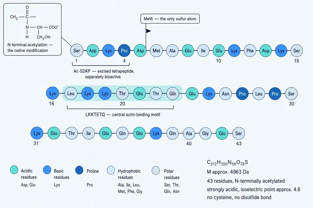
The numbers came immediately after, and they are worth sitting with. Thymosin beta 4 binds a single actin monomer with a dissociation constant of 0.4–0.7 µM; the affinity does not depend on calcium; it binds only free monomers, never the ends or sides of an existing filament. Resting human platelets contain roughly 280 µM monomeric actin and 560 µM thymosin beta 4 — two molecules of peptide for every molecule of actin it is holding (Weber et al., 1992; human in vitro). In resting human neutrophils the cytoplasmic concentration is about 149 µM, and it accounts for the majority of the free actin pool (Cassimeris et al., 1992; human in vitro).
Those are not the concentrations of a hormone. Hormones work at picomolar and nanomolar levels, in vanishing quantities, on receptors tuned to detect them. This peptide is present at a hundred micromolar and above. It is one of the most abundant proteins in the cell. It is not a signal. It is infrastructure.
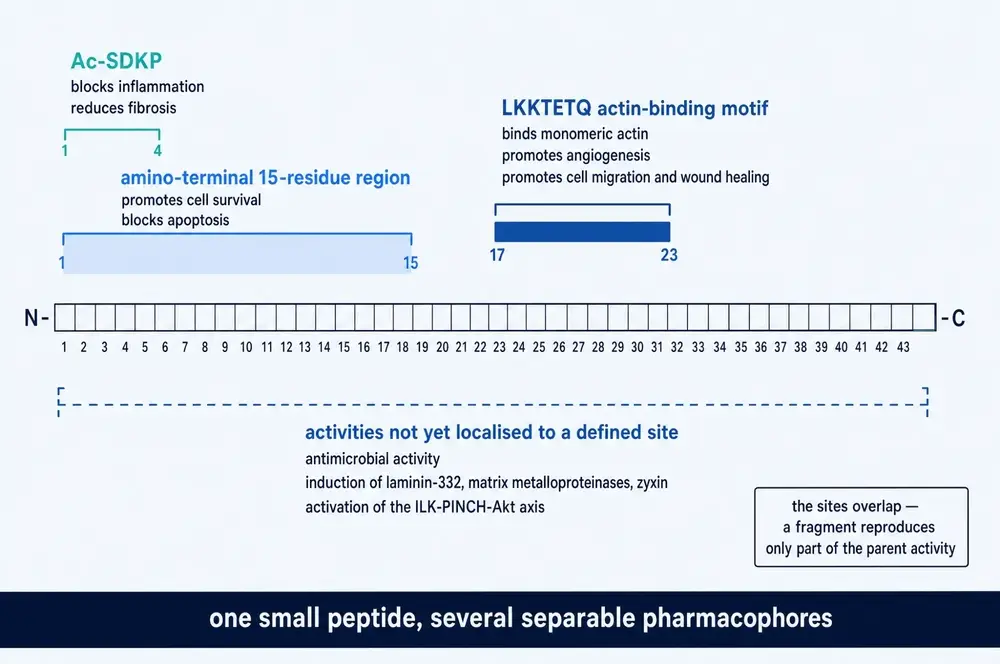
The functional map in Figure 2 explains something that will matter a great deal later in this document. The activities attributed to this molecule do not sit in one place along the chain. Residues 1–4 carry anti-inflammatory and anti-fibrotic effects. The first fifteen residues, which contain those four, carry anti-apoptotic effects. Residues 17–23 — the sequence LKKTETQ — carry actin binding, angiogenesis and cell migration (Van Troys et al., 1996; in vitro). The regions overlap and are not co-extensive, which means a fragment of this peptide reproduces some of its parent’s activity and not the rest. Hold that thought.
04A molecule with no shape of its own
There is one more structural fact that makes thymosin beta 4 unusual, and it took another two decades to pin down.
Most proteins fold into a definite three-dimensional shape and keep it. This one does not. Free in solution it is intrinsically disordered — a floppy chain sampling an enormous range of conformations with no stable core, no persistent helix, no sheet. Computational analysis puts its disorder at about 83 per cent, and a hydration-based nuclear magnetic resonance method independently estimates that only around 22 per cent of the chain adopts ordered secondary structure at any moment (Bokor et al., 2021; in vitro and in silico).
Structure appears only when it binds. On contact with an actin monomer, two short amphipathic helices crystallise out of the disorder — one near the front of the chain, one near the back — while the LKKTETQ segment between them stays extended and lies flat across the actin surface.
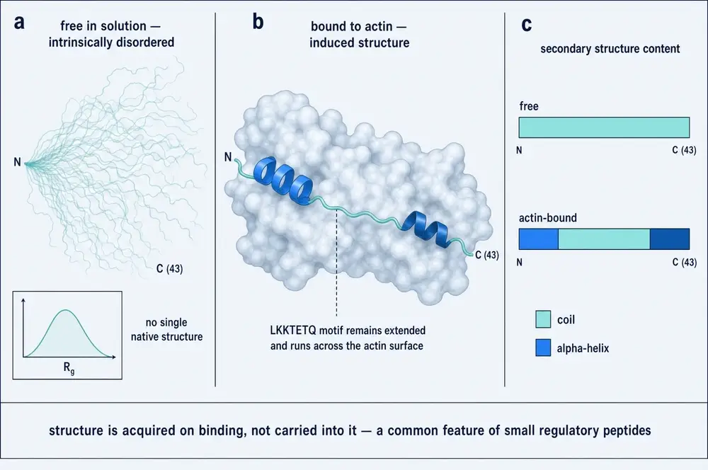
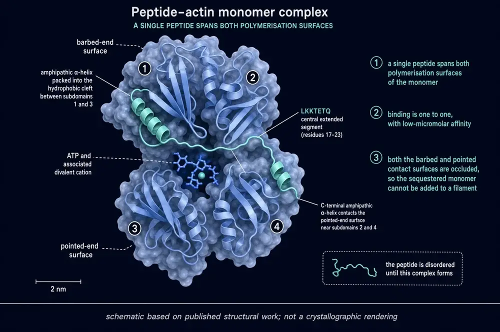
Figure 4 shows why this arrangement sequesters rather than caps. An actin monomer joins a filament through two different faces: the barbed face and the pointed face. A single molecule of thymosin beta 4 is long enough to drape across the monomer and cover both. The front helix wedges into the hydrophobic cleft between subdomains 1 and 3; the rear helix touches subdomains 2 and 4; the LKKTETQ segment runs across the middle (Irobi et al., 2004; in vitro structural). With both faces occluded, the captured monomer cannot be added to a filament at either end. It is not slowed down. It is taken out of the game.
The affinity has been measured repeatedly and lands in the low micromolar range — 0.5 ± 0.1 µM by isothermal titration calorimetry, with an independent solution-NMR docking model agreeing with the crystal structure to within 0.89 Å (Huang et al., 2016; in vitro). Full-length structures solved in 2014 added a second conformation with a more open nucleotide cleft, which explains how a sequestered monomer can still be handed off to profilin (Xue et al., 2014; in vitro).
05Why a cell needs a monomer bank
To see why any of this matters, consider the problem a cell actually has.
A crawling cell — a white blood cell chasing a bacterium, a skin cell closing a wound — moves by building actin filaments at its leading edge, fast, exactly where it needs them, and dismantling them behind. The build has to happen in seconds. That means the raw material must already be present, in bulk, throughout the cytoplasm, before the cell decides where to push.
But actin is a self-assembling protein. Above a threshold called the critical concentration — roughly 0.1–0.2 µM at the fast end of a filament, 0.7 µM at the slow end — free monomers polymerise on their own. Cells carry 100–500 µM of the stuff: several hundred times over the line. Left alone, the cytoplasm would set solid like a gel.
It does not, because most of that actin is held. Thymosin beta 4 is the holder. Modelling of live-cell measurements puts the partition at roughly 2 µM genuinely free, about 10 µM bound to profilin, and about 88 µM bound to thymosin beta 4 (Kiuchi et al., 2011; human cells in vitro). The cell keeps a large, safe, non-polymerising reserve and draws on it on demand.
The counterpart to sequestration is delivery. Profilin also binds actin monomers, but instead of withholding them it accelerates their nucleotide exchange and hands them to the growing end of a filament. The two proteins compete for the same pool, and the balance between them sets how fast a cell can build. Because both bind and release quickly, a modest local rise in profilin activity can override a large excess of thymosin beta 4 in that spot — which is how a cell turns a uniform, cell-wide reservoir into a filament in one specific place (Goldschmidt-Clermont et al., 1992; in vitro). The peptide also prefers ATP-loaded actin to ADP-loaded actin by roughly fiftyfold, giving the cell a second lever (Carlier et al., 1993; in vitro).
This is not a theory built only from test tubes. Mice engineered without the gene have platelets with reduced free actin, excess filamentous actin, impaired assembly on activation, abnormally large and scarce platelets, and altered bleeding and thrombosis (Scheller et al., 2022; animal in vivo). Remove the buffer and the equilibrium it maintains visibly breaks.
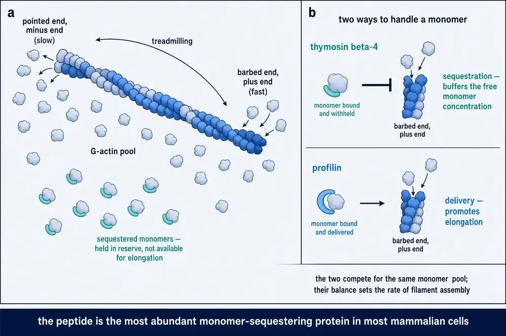
So the molecule has now been misnamed twice over. It is not from the thymus, and it is not a hormone. It is the most abundant actin-monomer-sequestering protein in most mammalian cells — a piece of cytoplasmic plumbing that happened to be discovered by immunologists, given an immunological name, and correctly identified ten years later by people studying platelets. Every therapeutic claim made for it since rests on the second identity, not the first.
06Four residues that connect it to blood-pressure drugs
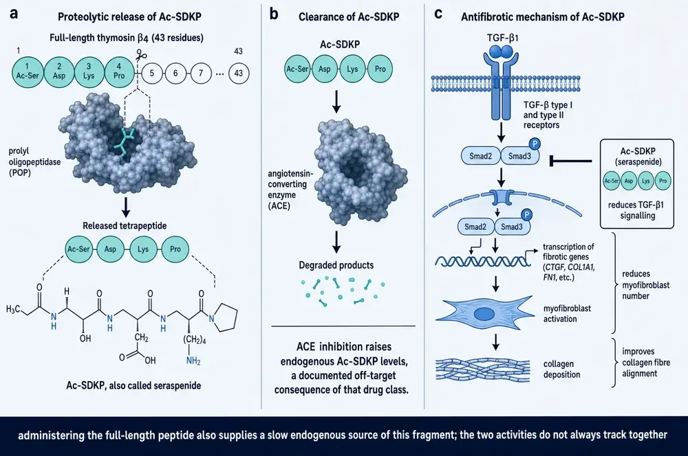
Cut the first four residues off thymosin beta 4 and you get a tetrapeptide: N-acetyl-seryl-aspartyl-lysyl-proline, written Ac-SDKP and sometimes called seraspenide. It is not a laboratory curiosity. The body makes it, from this peptide, continuously, and it has a pharmacology of its own.
Getting it out requires two enzymes working in sequence, and the order matters. The 43-residue chain is first cut by a metalloprotease called meprin-alpha into intermediate fragments shorter than about thirty residues. Only then can prolyl oligopeptidase act, because that enzyme is structurally incapable of handling a substrate longer than roughly thirty residues; it makes the final cut after the proline at position 4 and liberates the tetrapeptide. Incubating the full-length peptide with either enzyme alone releases nothing. Only the pair works, and mice lacking meprin-alpha have lower baseline kidney Ac-SDKP and cannot generate it from thymosin beta 4 in tissue (Kumar et al., 2016; animal in vivo and in vitro).
Then comes the part that connects this obscure fragment to one of the most widely prescribed drug classes in the world.
Ac-SDKP is destroyed almost exclusively by angiotensin-converting enzyme — ACE, the target of ramipril, lisinopril, enalapril and the rest. ACE has two catalytic domains. The C-domain does the famous job, converting angiotensin I to angiotensin II and thereby raising blood pressure. The N-domain does something almost entirely different: it degrades Ac-SDKP. Ordinary ACE inhibitors block both. So every patient taking an ACE inhibitor for hypertension or heart failure is also, incidentally, accumulating a peptide fragment of thymosin beta 4 (Cavasin et al., 2004; animal in vivo).
The effect is not subtle. In rats, captopril raised plasma Ac-SDKP from 3.1 to 15.1 nmol/L — roughly fivefold. Co-administering a meprin-alpha inhibitor cut that rise to 6.1 nmol/L, proving the drug works by sparing a peptide the body is already making rather than by stimulating new production (Kumar et al., 2016; animal in vivo).
This has a consequence that runs in both directions. Part of the anti-fibrotic benefit long credited to ACE inhibition — the observation that these drugs reduce scarring in heart and kidney beyond what blood-pressure lowering explains — appears to be Ac-SDKP accumulation, and a neutralising antibody against Ac-SDKP has been shown to abolish that benefit without touching blood pressure (Wang et al., 2022; review of animal work). Conversely, and this is the practically important direction: any study of thymosin beta 4 in fibrosis conducted in subjects taking ACE inhibitors is measuring two overlapping interventions at once.
07What it does at the level of a cell
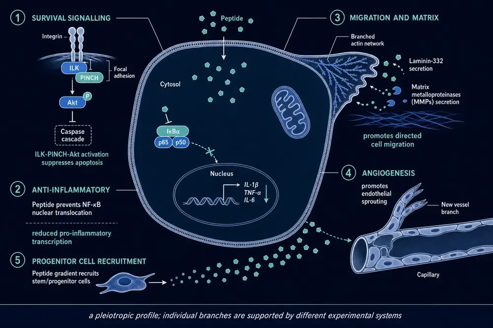
Beyond holding actin, thymosin beta 4 has been reported to do a great many things. The honest summary is that it is pleiotropic — it touches several unrelated systems — and that the different branches rest on different experimental evidence of quite different quality.
The best-supported branches, in rough order of how directly they have been demonstrated:
Anti-inflammatory action through NF-kappa-B. NF-kappa-B is the master switch for inflammatory gene transcription; normally it is held inactive in the cytoplasm and released into the nucleus on an inflammatory signal. Thymosin beta 4 binds the RelA/p65 subunit directly, blocks its entry into the nucleus and prevents it settling on target gene promoters — and it does this independently of its actin-binding activity, which is the strongest available evidence that this is a genuine second function rather than a downstream consequence of cytoskeletal change (Qiu et al., 2011; in vitro).
Survival signalling through ILK–PINCH–Akt. The peptide forms a complex with integrin-linked kinase and the adaptor PINCH at focal adhesions, activating Akt and suppressing the caspase cascade that executes programmed cell death (Bock-Marquette et al., 2004; animal in vivo and in vitro). The dependence is testable and has been tested: blocking Akt with wortmannin abolishes the protective effect in a mouse heart (Sopko et al., 2011; animal in vivo).
Migration and matrix remodelling. Releasing actin at the leading edge frees the peptide to bind ILK, which drives production of matrix metalloproteinases and laminin-332 — so the same event that supplies the building material for a protrusion also licenses the cell to digest a path through the matrix ahead of it (Tantos et al., 2013; in vitro).
Angiogenesis. Endothelial sprouting is the most frequently reported downstream effect and the one most often invoked to explain benefit in wounds and ischaemic tissue. It is well replicated but almost entirely gain-of-function — add peptide, see vessels — rather than demonstrated by removing the peptide and watching sprouting fail.
Autophagy and phagocytosis. In chronic granulomatous disease models, thymosin beta 4 promotes a form of phagocytosis that depends on stabilising the oxygen-sensing factor HIF-1-alpha; silencing HIF-1-alpha abolishes the effect, and silencing the peptide itself makes fungal lung infection worse (Renga et al., 2019; animal in vivo and human cells in vitro). This is a proper loss-of-function result in both directions.
Two cautions belong here rather than in a late safety section. First, the molecule has no identified cell-surface receptor. Its non-actin partners — PINCH, ILK, stabilin-2 — bind it weakly, with dissociation constants of 630 µM, 380 µM and 2.8 mM respectively, forming loose “fuzzy” complexes in which neither partner properly folds (Tantos et al., 2013; in vitro). Those are very weak interactions. How an extracellular peptide reaches these intracellular partners at all remains unresolved. A further activity sits outside the actin story entirely: the peptide chelates iron and blocks ferroptosis, an iron-dependent form of cell death, in a macrophage line (Lachowicz et al., 2022; in vitro) — a genuinely separate chemistry whose relevance to anything therapeutic is not yet established.
Second, this pleiotropy cuts both ways. A molecule that promotes cell migration, angiogenesis, matrix digestion and resistance to programmed cell death has, in one sentence, described four of the six classical hallmarks of cancer. That is not an insinuation; it is the reason Part Five exists.
08Repair: the cornea and the skin
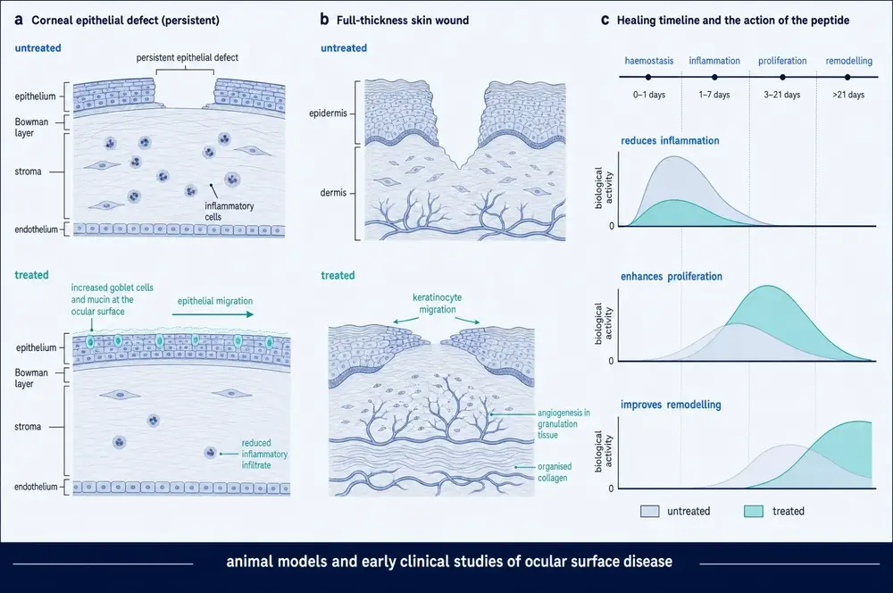
If the cell-level story is right, the tissues where it should show up most clearly are those that repair by having cells crawl across a gap. Two qualify: the surface of the eye and the skin.
In corneal models the findings are consistent across laboratories. Thymosin beta 4 accelerates re-epithelialisation after chemical and mechanical injury, promotes hemidesmosomal adhesion, and increases laminin-332 (Sosne et al., 2007; review of animal work). It suppresses apoptosis in injured corneal cells: in ethanol-injured human corneal keratocytes, 1 µg/mL raised viability from 61 to 88 per cent and cut the apoptotic fraction from 0.35 to 0.15 (in vitro), with the corresponding mouse experiment reducing the residual epithelial defect at day 3 from 61 to 21 per cent (animal in vivo). It reduces inflammatory cytokines and NF-kappa-B activation in dry-eye models, and it raises conjunctival goblet cell number and mucin. In bacterial keratitis it restores corneal nerve density and length to levels indistinguishable from uninfected controls when added to an antibiotic (Ebrahim et al., 2026; animal in vivo). There is even a plausible reason the eye should be responsive: expression of the gene is reduced in corneas from donors with diabetic retinopathy, alongside other repair factors (Saghizadeh et al., 2005; human tissue).
In skin, the pattern is the same and the mechanism is specific in an instructive way. In a mouse full-thickness wound model, topical thymosin beta 4 significantly accelerated closure at days 9 and 11, increased blood-vessel area at days 3 and 5, and increased collagen deposition at days 3 and 5 (animal in vivo). In the same study, across every concentration from 0.01 to 10 µg/mL, it had no effect whatsoever on fibroblast proliferation (in vitro) (Jing et al., 2019).
That null result is the useful one. The peptide does not make cells divide. It makes them move, and it brings blood supply with them. Everything reported downstream is consistent with a molecule that accelerates the migratory and vascular phases of repair without touching the mitotic one.
09Fibrosis, and a mechanism it shares with its own fragment
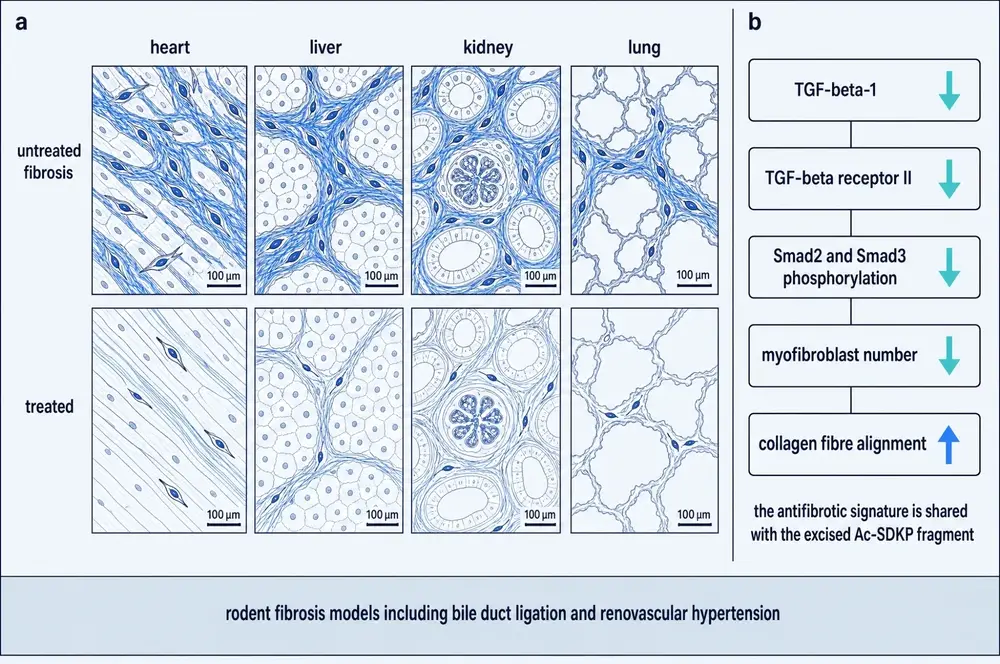
Repair that goes too far becomes scar. Fibrosis — the replacement of working tissue with disorganised collagen — is the common end-stage of chronic injury in the heart, liver, kidney and lung, and it is where thymosin beta 4 has been tested most widely in animals.
The finding is unusually consistent: administered thymosin beta 4 is anti-fibrotic in essentially every organ where it has been tried. Kidney, in unilateral ureteral obstruction and in glomerular disease; liver, in bile-duct ligation and carbon tetrachloride models; lung, in bleomycin injury; colon, in chemical colitis; even uterus, in an adhesion model (all animal in vivo). The mechanism converges on the same axis every time: less TGF-beta-1, fewer TGF-beta receptors, less Smad2 and Smad3 phosphorylation, fewer myofibroblasts, better-aligned collagen.
The size of the effect is real but moderate. In a mouse kidney obstruction model, the full-length peptide reduced late-stage fibrosis by about 35 per cent; Ac-SDKP alone reduced it by about 12 per cent (Zuo et al., 2013; animal in vivo). In gene-therapy form, adeno- associated virus delivering the peptide cut albuminuria in a mouse kidney injury model from 1,448 to 214 µg per 24 hours (Mason et al., 2022; animal in vivo).
There is, however, a genuine unresolved contradiction underneath the tidy picture, and it concerns endogenous peptide rather than administered peptide. In the kidney, mice lacking the gene do worse: knockouts develop far heavier albuminuria and inflammation in nephritis (Vasilopoulou et al., 2016), and in angiotensin-II hypertension they suffer worse kidney and heart damage and around 40 per cent mortality against zero in wild-type animals (Kumar et al., 2018; both animal in vivo). Endogenous peptide there is protective.
In the liver, the opposite. Activated hepatic stellate cells — the cells that lay down liver scar — switch the gene on as they activate, and deleting it specifically in those cells protects mice against fibrosis, while re-introducing it by viral vector reverses that protection (Kim et al., 2015; Kim et al., 2023; animal in vivo). A separate group reports the reverse direction in the same disease, finding the peptide reduced in human fibrotic liver and protective on knockdown (Zhu et al., 2017). Both lines are competent. The field has not reconciled them.
10An exerkine, and the negative result that matters most
One 2021 study deserves separate treatment, because it is the single piece of work in this entire literature that most directly tests what the consumer market claims.
Researchers profiling proteins secreted by contracting muscle cells found that thymosin beta 4 was the most upregulated secreted protein in the medium of exercising myotubes. They then measured it in people, and found it rose acutely in the plasma of exercising humans — irrespective of whether the subject was insulin-resistant and irrespective of exercise mode (Gonzalez-Franquesa et al., 2021; human observational). It is, genuinely, an exercise-released factor.
In the same paper, the same team gave it to mice. It did not ameliorate the metabolic consequences of diet-induced obesity. It did not enhance muscle regeneration in vivo (animal in vivo). It did increase osteoblast proliferation and neurite outgrowth in culture — which, as the authors themselves note, is consistent with its classification by the World Anti-Doping Agency as a prohibited growth factor.
11TB-500 is not thymosin beta 4
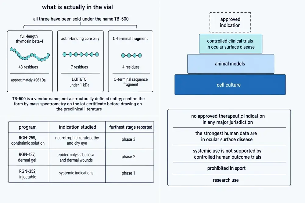
Everything in this document so far concerns a 43-residue protein. The product sold under the street name TB-500 is, in every independent analysis that has been published, something else.
The question was first forced not by a regulator or a physician but by horse-racing chemistry. A veterinary preparation called TB-500 appeared on the market, and laboratories responsible for equine doping control needed to know what was in it so they could test for it. In 2012 the Hong Kong Jockey Club laboratory reported the answer: the active ingredient is the peptide LKKTETQ with a synthetic acetyl group on its front end — seven residues, the actin-binding motif at positions 17 to 23 of thymosin beta 4, and nothing else (Ho et al., 2012).
The same year, an entirely separate group in Belgium reached the same conclusion by high-resolution Orbitrap mass spectrometry, then synthesised Ac-LKKTETQ independently to confirm the match (Esposito et al., 2012). Two laboratories, two continents, two methods, one answer.
This matters because of a habit of argument that is universal in the consumer literature: cite a study of thymosin beta 4, then apply its conclusion to TB-500. Almost the entire preclinical evidence base described in Parts One and Two — the cardiac work, the corneal programme, the fibrosis models, the neurological studies — used the full-length peptide or its gene. Transferring those results to a seven-residue fragment assumes that the fragment reproduces the parent’s activity. Figure 2 shows why that assumption is unsafe: the activities are distributed along the chain and overlap, and this fragment carries one of them.
There is a second problem, and it is about manufacturing rather than biology. In 2023 a French laboratory analysed products sold over the internet under the names TB500, TB1000 and SGF1000. Their conclusion was that the contents of the TB500 and TB1000 products were “not systematically consistent” with what the labels described, and that SGF1000 was mainly sheep extracellular matrix and blood proteins with the purported active ingredient present only in vanishing dilution. They characterise these straightforwardly as misbranded and adulterated drugs, produced without official control (Delcourt et al., 2023).
So the position is not simply that TB-500 is a fragment rather than the protein. It is that TB-500 is a trade name, not a defined chemical entity, and that what a given vial contains is an empirical question about that vial.
12And the fragment may not be the active thing either
In 2024 a Korean group did the experiment that follows logically from all of this: they gave TB-500 to rats, tracked what it turned into, and tested each breakdown product for the activity the parent is sold for (Rahaman et al., 2024; animal in vivo and in vitro).
Ac-LKKTETQ does not last. In serum and in living rats it is chewed back from the far end. The dominant species in the first six hours was Ac-LK — two residues. Ac-LKK persisted longest, still detectable at 72 hours. Then they ran wound-healing assays on fibroblasts with the parent and each metabolite side by side. Of all of them, only Ac-LKKTE produced significant wound-healing activity relative to control.
The authors’ own conclusion is that the wound-healing activity previously reported for TB-500 may belong to a metabolite rather than to the compound administered. Nothing in the study was cytotoxic, which is reassuring as far as it goes. But it leaves the chain of inference in an awkward state: a protein is studied, a fragment of it is sold under the protein’s reputation, and the fragment’s activity may in turn belong to a fragment of the fragment.
13Prohibited, unapproved, and detectable
Thymosin beta 4 and its synthetic fragments are prohibited in sport. The World Anti-Doping Agency, the Fédération Équestre Internationale and the International Federation of Horseracing Authorities all ban them, and no thymosin beta 4 product is authorised as a medicine in any major jurisdiction (Delcourt et al., 2025). The 2021 exerkine paper notes the WADA classification explicitly as a prohibited growth factor (Gonzalez-Franquesa et al., 2021).
Detection is well established. Acetylated LKKTETQ and its metabolites can be confirmed at 0.02 ng/mL in equine plasma and 0.01 ng/mL in equine urine, demonstrated in samples from horses given a single dose containing 10 mg of the peptide (Ho et al., 2012; animal in vivo). For the full-length peptide, a 2025 population study established that endogenous concentrations in racehorses do not vary significantly with sex, age or breed, and that a non-natural synthesis impurity can be detected after administration — giving testers a way to distinguish an injected product from the animal’s own peptide (Delcourt et al., 2025).
14What has never been tested
Which brings us to the plainest statement in this document.
Eighteen human clinical trials of thymosin beta 4 are registered on ClinicalTrials.gov, enrolling roughly 2,200 participants between them across two decades. Every one of them used full-length thymosin beta 4 or a recombinant version of it. Not one tested the seven-residue fragment that is actually sold.
Figure 11 also shows something that no summary of the published literature reveals on its own. The three largest trials ever conducted with this molecule — ARISE-1, ARISE-2 and ARISE-3, together enrolling 1,618 people with dry eye disease — all completed, the last of them in 2021, and none has posted results. No approval followed. A reader working only from journal articles would not know these trials existed.
15What counts as evidence here
Thymosin beta 4 has been proposed as a treatment for myocardial infarction, dry eye, neurotrophic keratopathy, venous ulcers, epidermolysis bullosa, stroke, traumatic brain injury, multiple sclerosis, diabetic neuropathy, kidney fibrosis, liver fibrosis, pulmonary fibrosis, colitis, sepsis, hair loss and Alzheimer’s disease. That list is not evidence of versatility. It is what a literature looks like when a molecule is abundant, cheap to obtain, easy to add to a culture dish, and involved in a process — cell migration — that participates in nearly every disease. The standard reviews of the field present that breadth as promise (Xing et al., 2021; Dai et al., 2021); it is at least as reasonable to read it as a warning about specificity.
The useful question is not where has it shown an effect but where does the evidence reach human beings.
16The heart: the claim
The largest therapeutic claim ever made for this molecule is cardiac, and it was made in three steps.
In 2004, Ildiko Bock-Marquette and Deepak Srivastava reported in Nature that thymosin beta 4 promoted the migration and survival of heart muscle cells, formed a complex with PINCH and integrin-linked kinase, activated Akt, and — after coronary artery ligation in mice — improved cardiac function (Bock-Marquette et al., 2004; animal in vivo).
In 2007, Nicola Smart and Paul Riley reported, also in Nature, that the peptide was essential to coronary vessel development in mice and could wake up quiescent adult epicardium — the thin outer layer of the heart — sending cells out of it that differentiated into fibroblasts, smooth muscle and endothelium (Smart et al., 2007; animal in vivo).
In 2011, the same group went further. Priming mice with the peptide before inducing a heart attack, they used genetic lineage tracing to report that epicardium-derived cells became bona fide new cardiomyocytes — structurally integrated, electrically coupled, beating in time with the existing muscle, and demonstrably not the product of cell fusion (Smart et al., 2011; animal in vivo). If true and translatable, this was the thing cardiology had been looking for since the 1990s: a drug that regrows heart muscle.
Supporting work was strong in places. In pigs, embryonic endothelial progenitor cells reduced infarct size from 54 to 38 per cent of the area at risk; knocking down thymosin beta 4 in those cells abolished the protection entirely, taking infarct size to 62 per cent; and giving the peptide alone reproduced the benefit at 37 per cent (Hinkel et al., 2008; animal in vivo). That is a proper necessity-and-sufficiency experiment in a large animal, and it holds up.
17The heart: what actually replicated
The regeneration claim did not.
Start with the arithmetic, which the originating group itself published. The conversion efficiency — the fraction of traced progenitors that became cardiomyocytes — was 0.59 per cent, about 193 new cells per heart. In their own review they describe this as “certainly insufficient” against the roughly 109 cardiomyocytes lost in a human myocardial infarction (Smart et al., 2012). Figure 13 puts those two numbers on the same axis, which is the only honest way to look at them.
Then the protocol. The 2011 result required priming: seven days of daily peptide before the heart attack. No patient receives a drug the week before an unpredictable event. When the same group treated mice after infarction — the clinically meaningful design — the epicardium thickened and other benefits appeared, but the resulting cells did not migrate into the myocardium and did not express cardiomyocyte markers; some became myofibroblasts instead (Zhou et al., 2012; animal in vivo).
Then independent reassessment. Using a genetic Islet-1 reporter rather than antibody staining, and running the same priming protocol, one group found a single Islet-1-positive cell outside the expected regions across roughly 2,500 sections from eight treated hearts — against two such cells in five controls (Weinberger et al., 2018; animal in vivo). No reactivation.
Then the knockouts. Both global and heart-specific deletion of the gene produced mice with normal cardiac development, no fibrosis, normal echocardiography to twelve months, and no coronary vascular deficit (Banerjee et al., 2012; animal in vivo) — directly contradicting an earlier knockdown study that had reported embryonic lethality and severe vascular defects, and which the later authors attribute to off-target effects of the knockdown reagent.
Then large-animal systemic dosing. In pigs undergoing global ischaemia and reperfusion on cardiopulmonary bypass, intravenous peptide before and during reperfusion produced no significant effect on apoptosis, troponin release, myocardial blood flow or ejection fraction — 56.7 against 61.9 per cent, p=0.45 (Stark et al., 2016; animal in vivo).
18The heart: the human record
Two phase 1 safety studies exist and both are reassuring on tolerability. Synthetic peptide given intravenously to healthy volunteers at single doses of 42, 140, 420 or 1,260 mg, then daily for fourteen days, produced infrequent mild-to-moderate adverse events, no dose-limiting toxicity and no serious adverse events (Ruff et al., 2010; human in vivo). A recombinant version tested in 54 healthy Chinese volunteers at 0.05 to 25.0 µg/kg, and in 30 more at up to 5.0 µg/kg daily for ten days, gave the same result with no accumulation on repeat dosing (Wang et al., 2021; human in vivo).
The efficacy record is thinner and more recent. In 2025 a randomised, placebo-controlled, double-blind trial gave recombinant human thymosin beta 4 to 96 patients with ST-elevation myocardial infarction after primary angioplasty. Infarct area at 90 days was significantly reduced in the subgroup receiving their first dose within eight hours of the procedure (n=43). Across the full randomised population of 96, the difference was not significant. The authors say so plainly and call for further rigorous randomised studies (Zhang et al., 2025; human in vivo).
That is a negative trial with a positive subgroup. The eight-hour window was not the randomised comparison; it is a subset defined after the fact by how quickly each patient happened to be dosed. Subgroups of this kind generate hypotheses. The next trial in the programme — a 189-patient phase 2c — had not begun recruiting when this document was compiled. No mortality or major-adverse-cardiac-event data exist for this molecule in humans at all.
One human dataset does connect the peptide to cardiac outcome, and it points the other way. In a 657-person case-control study, plasma thymosin beta 4 was elevated in heart failure with preserved ejection fraction against controls (1,401 against 985 ng/mL), the difference driven entirely by women; and among women with heart failure, a concentration above 2,000 ng/mL predicted worse two-year survival (adjusted hazard ratio 1.668, 95% CI 1.033–2.691) (Drum et al., 2017; human observational). This measures the body’s own peptide, not an administered drug, and it is a subgroup finding — but it is a reminder that more of this molecule in the circulation is not self-evidently a good thing.
19The eye, where it went furthest — and stopped
The ophthalmic programme is the most advanced clinical use of thymosin beta 4 and the most instructive, because the published literature and the trial registry tell noticeably different stories.
The published record reads well. A 2015 phase 2 trial in nine patients with severe dry eye reported a 35.1 per cent reduction in ocular discomfort against vehicle (p=0.0141) and a 59.1 per cent reduction in corneal fluorescein staining (p=0.0108) at day 56 (Sosne et al., 2015a; human in vivo). A 2022 paper titled as a phase III trial in neurotrophic keratopathy reported complete healing in 6 of 10 treated patients against 1 of 8 on placebo (Sosne et al., 2022; human in vivo).
Read the numbers rather than the framing and the picture changes.
That 6-versus-1 healing result carries p=0.0656. The paper states explicitly that the primary efficacy endpoint was not met with statistical significance, describing it instead as a strong efficacy trend, and leads with secondary and post-hoc analyses that did cross the threshold. The trial is SEER-1, and the registry records it as terminated after 18 of a planned 46 patients, because recruitment in a rare disease was too slow. An independent Cochrane review reanalysed the same 18-patient dataset in 2025 and obtained a relative risk of 9.00 with a 95 per cent confidence interval from 0.57 to 141.88 — an interval that spans no effect — and graded the evidence low certainty (Kruoch et al., 2025).
The same pattern appears in dry eye. A 72-patient phase 2 trial using a controlled adverse environment model missed both co-primary endpoints — corneal staining p=0.2586, ocular discomfort p=0.2210 — and its positive claims rest entirely on secondary and post-hoc analyses (Sosne et al., 2015b; human in vivo).
And then the three large trials that should settle the question. ARISE-1, ARISE-2 and ARISE-3 enrolled 317, 601 and 700 patients respectively. All completed. None posted results. No approval has been granted in any indication, anywhere, and a 2025 paper from within the field states that full FDA approval remains pending (Nguyen et al., 2025).
20Skin, and the shape of the dose–response
Three dermal trials were registered: venous stasis ulcers (72 patients, completed), pressure ulcers (72, completed) and epidermolysis bullosa (30, terminated). The claim that circulates — that a topical gel healed chronic ulcers about a month faster than placebo — appears in the literature reviewed here only as a secondary citation inside another paper’s introduction, without enrolment numbers, dose or p-values. The one primary publication is a study-design and interim-conduct report describing 21 patients enrolled in the lowest-dose group, and it reports no efficacy outcome at all (Guarnera et al., 2007; human in vivo). The same review that repeats the “one month faster” claim also notes that patient response was inconsistent and that a way to distinguish responders from non-responders would be needed.
The animal and cell work, by contrast, is coherent — and it contains the single most practically useful finding in this document.
The dose–response is not monotonic. It is bell-shaped. In endothelial cell assays, activity rose to a peak at or below 10 µg/mL, fell sharply above it, and had disappeared entirely at 100 µg/mL. In rats, a mid concentration of 0.25 mg/mL outperformed a high concentration of 0.5 mg/mL, and the authors note that earlier work had also found peak activity at intermediate concentrations (Xu et al., 2013; in vitro and animal in vivo). An entirely independent study reached the same shape: in a fibroblast scratch assay, the optimum was 1 µg/mL, and raising the concentration to 10 µg/mL produced a negative effect on migration — for native thymosin beta 4 and for every variant tested (Jing et al., 2019).
Two laboratories, different species, different assays, same result: past a modest optimum, more of this peptide does less, and eventually nothing. Whatever else is true, the intuition that a larger amount should produce a larger effect is contradicted by the preclinical data.
A practical footnote sits behind all of this. Making intact thymosin beta 4 is difficult — small peptides are readily chewed up by proteases during production, and several groups report outright failure to produce the full-length molecule by genetic engineering. The workarounds in the literature are revealing: fusing two copies into a dimer (Xu et al., 2013), or stringing four copies together and growing them in tobacco plants (Janarthini et al., 2016; in vitro and animal in vivo). A molecule that is awkward to manufacture correctly is a molecule whose unregulated supply is worth treating sceptically.
21Hair, and why cashmere goats are not the evidence they look like
Thymosin beta 4 does influence hair follicles. In mice, transgenic overexpression accelerated regrowth after depilation to 11 days against 13 in wild-type, while knockout mice took 16 days; overexpressors had 5.65-fold higher messenger RNA, 2.33-fold higher VEGF and denser hair shafts (Gao et al., 2015; animal in vivo).
The striking numbers in this literature, however, come from somewhere the casual reader would not expect. A CRISPR knock-in cashmere goat carrying the gene at a safe-harbour locus under a hair-follicle-specific promoter yielded 815.7 g of cashmere — a 74.5 per cent increase over controls — with a 28.4 per cent higher secondary-to-primary follicle ratio (Li et al., 2019; animal in vivo).
22The nervous system: a good animal story with no human chapter
The neurological literature is, on its own terms, among the better work on this molecule — and it is entirely preclinical.
In rat stroke models, delayed treatment improved neurological scores by 24–35 per cent across three tests, with a modelled optimum around 3.75 mg/kg and, notably, no change in lesion volume — function improving without the infarct shrinking, which points to repair rather than protection (Morris et al., 2014; animal in vivo). The mechanism is oligodendrocyte generation and remyelination, supported in vitro by a p38-MAPK-dependent pathway and reproduced in a multiple sclerosis model. In rat traumatic brain injury the effect splits by timing: treatment at six hours reduced lesion volume dose-dependently and improved spatial learning (Xiong et al., 2012), while treatment delayed to 24 hours did not touch lesion volume yet still improved function and increased hippocampal neurogenesis (Xiong et al., 2011; both animal in vivo) — protection early, repair late. In Alzheimer’s models, overexpression reduced amyloid plaque burden in mice, and exogenous peptide reduced amyloid and raised mature neuron density in human stem-cell-derived brain organoids (Zeng et al., 2025; animal in vivo and human cells in vitro).
Two findings raise the quality of this work above the usual. In diabetic peripheral neuropathy, a neutralising antibody against Tie2 abolished the benefit on nerve conduction, vascular density and nerve fibre density — a genuine loss-of-function test of the proposed mechanism (Wang et al., 2019; animal in vivo). And in zebrafish, regeneration of the Mauthner axon was reduced by knocking out the gene, rescued by restoring it in single cells, and enhanced by overexpression — but a mutant lacking the actin-binding region did nothing, and an actin-polymerisation inhibitor blocked the effect entirely (Song et al., 2024; animal in vivo). That resolves a long-running argument about whether the peptide helps axons by sequestering actin or by enabling its polymerisation, in favour of polymerisation.
One result deserves emphasis for how strange it is. In aged rats, the same dose and schedule that improved function without shrinking infarcts in young animals did the opposite: it halved infarct volume and produced no functional improvement at all (Morris et al., 2017; animal in vivo). Age did not weaken the effect. It inverted the mechanism.
Against all of that: there is no human interventional neurological evidence whatsoever. Not a randomised trial, not an open-label study, not a case series with clinical endpoints. The only human data are observational — a cross-sectional study finding salivary thymosin beta 4 elevated in Alzheimer’s disease (Contini et al., 2021; human observational) — and a healthy-volunteer safety trial from 2010 that was never followed by an efficacy study in any neurological indication.
23Immunity and infection, including one clear signal of harm
The anti-inflammatory pharmacology is real and mechanistically well supported (section 07). In animals it translates into survival benefits: 80 per cent 72-hour survival in mouse endotoxaemia against untreated controls (Ye et al., 2026), and better than 50 per cent survival in a chronic granulomatous disease model of fungal lung infection at a time when all untreated animals had died (Renga et al., 2019; both animal in vivo).
The original immunological claim, though, has not aged well. The one modern study in this corpus that tested thymosin peptides head to head on dendritic cell maturation found that thymosin alpha 1 drove maturation and that thymosin beta 4 did not (Yao et al., 2007; human cells in vitro). Forty-five years on, the function the molecule was named for remains the one function nobody has convincingly re-demonstrated.
A related claim needs the same discipline. Thymosin beta 4 released from thrombin-stimulated human platelets kills bacteria and fungi in a dish (Tang et al., 2002; in vitro), and it is routinely described as an antimicrobial peptide on that basis. But tested directly against Pseudomonas aeruginosa across a full concentration range it showed no direct bactericidal activity at all; what it did instead was upregulate the host’s own antimicrobial peptides and inflammation-resolving lipid pathways (Carion et al., 2020; in vitro). The benefit in infection models is host-modulatory, not antiseptic — a distinction that matters, because the two imply very different things about what this molecule could be used for.
And there is a harm signal that belongs here rather than in a footnote. In a 2025 study of stress-induced gut dysfunction, mast-cell-derived and exogenously administered thymosin beta 4 disrupted intestinal tight junctions, suppressed a protective signalling axis, and increased bacterial translocation and dysbiosis — while rats engineered to lack the peptide were protected from stress-induced barrier failure (Sun et al., 2025; animal in vivo, with human tissue). Here the peptide is not the repair molecule. It is the lesion.
That result does not overturn the anti-inflammatory literature. It bounds it. This is a context-dependent molecule whose direction of effect changes with the tissue and the trigger, and any account that presents it as uniformly protective is describing a subset of the evidence.
24The oncology problem, stated precisely
A molecule that promotes cell migration, angiogenesis, matrix digestion and resistance to programmed cell death raises an obvious question. The literature has an answer to part of it, and a conspicuous silence where the rest should be. Both halves need saying exactly.
What is established
The gene is overexpressed in many human tumours, and it predicts worse outcomes. In glioma, TMSB4X is an independent adverse prognostic factor in both a public dataset and an independent patient cohort — multivariable hazard ratio 1.225 (95% CI 1.014–1.481, p=0.035), and 1.345 (95% CI 1.064–1.700, p=0.013) for progression-free survival in the validation cohort (Li et al., 2025; human observational). In 143 colorectal cancers, high expression correlated with lymphovascular invasion, depth of invasion, nodal and distant metastasis and stage, and independently predicted worse recurrence-free (p=0.001) and overall survival (p=0.005) (Lee et al., 2017; human observational). Head-and-neck squamous carcinoma and non-small-cell lung cancer show the same direction (Chi et al., 2017; Yang et al., 2023). In thyroid cancer it is expressed across every subtype and undetectable in normal or benign thyroid tissue.
In animals, changing how much of it a tumour cell makes changes how that tumour behaves. This is the part that goes beyond correlation. In a mouse fibrosarcoma model, transfecting the gene into weakly tumorigenic, non-metastatic cells turned them into tumour-forming cells that produced numerous lung metastases; transfecting an antisense construct into malignant cells suppressed both tumour formation and metastasis, with vector controls behaving like the parent lines (Kobayashi et al., 2002; animal in vivo). Deleting the gene in melanoma cells with TALEN nucleases significantly reduced lung metastasis after tail-vein injection (Lee et al., 2015; animal in vivo). Knockdown reduced proliferation and invasion in head-and-neck cancer and suppressed lymph-node metastasis in an orthotopic model (Chi et al., 2017; animal in vivo).
It is not universal. In multiple myeloma the direction reverses: expression is significantly lower in myeloma cells than in normal plasma cells across 298 patients (p<0.001), overexpression reduced proliferation and increased apoptosis sensitivity, mice given overexpressing cells survived longer (88.9 against 65.9 days, p<0.05), and below-median expression predicted shorter event-free survival in 209 transplant patients (Caers et al., 2010; human observational and animal in vivo). It is underexpressed in acute myeloid leukaemia. In prostate cancer, overexpression in the surrounding fibroblasts — rather than in the cancer cells — reduced tumour cell migration.
What is not established
Here is the precise gap, and it is the most important single statement in this document.
Everything above concerns endogenous expression: how much of the peptide a tumour cell manufactures for itself, or how a tumour behaves when its own gene is switched up or down. None of it is the same experiment as administering the peptide from outside to a living body over time and counting tumours.
That experiment has not been done. Searching this corpus:
- There is no chronic or lifetime carcinogenicity bioassay of exogenous thymosin beta 4 or of TB-500 — the standard two-year rodent study that regulators require before approving a drug for long-term use.
- There is no human pharmacovigilance dataset and no cohort study of cancer incidence in anyone exposed to it, whether in a trial or through the unregulated market.
- There is no animal study in which the peptide alone — without a co-administered carcinogen and without a transgenic background — was given systemically over an extended period with tumour incidence, latency or multiplicity as the primary endpoint.
The closest thing that exists is a study in which tumour-bearing mice received the peptide intraperitoneally at 15 mg/kg over 21 days. It found a significant fall in a plasma adhesion molecule that normally restrains cancer cells crossing blood-vessel walls, and its authors describe the peptide as a tumour inducer in that context. But its endpoints were plasma protein concentrations and cell-culture migration assays. It did not report tumour volume, tumour weight or metastatic burden (Bednarek et al., 2020; animal in vivo). It is a plausible mechanism, not a measured outcome.
One study did combine transgenic overexpression with a chemical carcinogen and found larger melanoma xenografts in the engineered mice (Lee et al., 2021; animal in vivo). That is germline overexpression plus an established carcinogenic stressor, not peptide administration, and it required both to show an effect.
It is also not established that it does not — because the study that would answer the question has never been run. The theoretical concern is not speculative hand-waving: it rests on a large, consistent human expression literature and on matched gain- and loss-of-function animal experiments showing that the amount of this protein present causally changes how tumour cells proliferate, invade and metastasise.
Absence of a carcinogenicity study is not evidence of safety. It is an absence, and it is the single largest gap in what is known about this molecule.
One further detail is worth recording because of who noticed it. The head-and-neck cancer group whose knockdown experiments showed reduced proliferation and metastasis explicitly named the commercial product in their paper, observing that the 17–23 segment is the main ingredient in TB-500 and that it is used for doping in human and equine sport. They raised it as work that ought to be done. They did not do it, and nobody else has (Chi et al., 2017).
25What would actually settle it
It is easy to end a document like this by saying more research is needed. More useful is to name the specific experiments whose results would change what a reasonable person concludes.
A chronic carcinogenicity bioassay of the administered peptide. Two years, rodent, systemic dosing, tumour incidence and latency as primary endpoints, no co-administered carcinogen, no transgenic background. This is a standard, unglamorous, expensive study. Until it exists, the oncology question stays open no matter how many mechanism papers accumulate.
A head-to-head comparison of the 43-mer, Ac-LKKTETQ and Ac-LKKTE. Same model, same molar doses, same endpoints. The entire consumer market rests on an assumption of equivalence between these three that has never been tested directly, and the one metabolism study that looked found the activity may sit with the third of them (Rahaman et al., 2024).
Posted results for ARISE-1, ARISE-2 and ARISE-3. Sixteen hundred and eighteen people took part in those trials. Whatever the outcome, it is the largest body of human efficacy evidence that exists for this molecule, and it has been sitting unpublished since 2021.
A properly powered myocardial infarction trial with the treatment window as the randomised comparison. The 2025 STEMI result is interesting precisely because the eight-hour subgroup separated. Randomise on it and the question is answerable.
Any human neurological study at all. Four decades of rodent work, a completed safety trial, and not one efficacy study in stroke, brain injury or demyelination.
Thymosin beta 4 holds no marketing approval in any indication in any major jurisdiction. It is prohibited in sport. The material sold as TB-500 is not thymosin beta 4, is not a defined chemical entity, and has never been the subject of a registered human clinical trial.
26References
- Banerjee I, Zhang J, Moore-Morris T, Lange S, Shen T, Dalton ND, et al.. Thymosin beta 4 is dispensable for murine cardiac development and function. Circulation research. 2012;110(3):456-64. PMID 22158707 · doi · PMC3739283
- Bednarek R, Selmi A, Wojkowska D, Karolczak K, Popielarski M, Stasiak M, et al.. Functional inhibition of F11 receptor (F11R/junctional adhesion molecule-A/JAM-A) activity by a F11R-derived peptide in breast cancer and its microenvironment. Breast cancer research and treatment. 2020;179(2):325-335. PMID 31650345 · doi · PMC6987052
- Bock-Marquette I, Saxena A, White MD, Dimaio JM, Srivastava D. Thymosin beta4 activates integrin-linked kinase and promotes cardiac cell migration, survival and cardiac repair. Nature. 2004;432(7016):466-72. PMID 15565145 · doi
- Bokor M, Tantos Á. Secondary Structures of Proteins: A Comparison of Models and Experimental Results. Journal of proteome research. 2021;20(3):1802-1808. PMID 33620224 · doi · PMC8028322
- Caers J, Hose D, Kuipers I, Bos TJ, Van Valckenborgh E, Menu E, et al.. Thymosin β4 has tumor suppressive effects and its decreased expression results in poor prognosis and decreased survival in multiple myeloma. Haematologica. 2010;95(1):163-7. PMID 19833631 · doi · PMC2805724
- Carion TW, Ebrahim AS, Alluri S, Ebrahim T, Parker T, Burns J, et al.. Antimicrobial Effects of Thymosin Beta-4 and Ciprofloxacin Adjunctive Therapy in Pseudomonas aeruginosa Induced Keratitis. International journal of molecular sciences. 2020;21(18). PMID 32961846 · doi · PMC7555736
- Carlier MF, Jean C, Rieger KJ, Lenfant M, Pantaloni D. Modulation of the interaction between G-actin and thymosin beta 4 by the ATP/ADP ratio: possible implication in the regulation of actin dynamics. Proceedings of the National Academy of Sciences of the United States of America. 1993;90(11):5034-8. PMID 8506348 · doi · PMC46648
- Cassimeris L, Safer D, Nachmias VT, Zigmond SH. Thymosin beta 4 sequesters the majority of G-actin in resting human polymorphonuclear leukocytes. The Journal of cell biology. 1992;119(5):1261-70. PMID 1447300 · doi · PMC2289708
- Cavasin MA, Rhaleb NE, Yang XP, Carretero OA. Prolyl oligopeptidase is involved in release of the antifibrotic peptide Ac-SDKP. Hypertension (Dallas, Tex. : 1979). 2004;43(5):1140-5. PMID 15037553 · doi · PMC4677773
- Chi LH, Chang WM, Chang YC, Chan YC, Tai CC, Leung KW, et al.. Global Proteomics-based Identification and Validation of Thymosin Beta-4 X-Linked as a Prognostic Marker for Head and Neck Squamous Cell Carcinoma. Scientific reports. 2017;7(1):9031. PMID 28831179 · doi · PMC5567379
- Contini C, Olianas A, Serrao S, Deriu C, Iavarone F, Boroumand M, et al.. Top-Down Proteomics of Human Saliva Highlights Anti-inflammatory, Antioxidant, and Antimicrobial Defense Responses in Alzheimer Disease. Frontiers in neuroscience. 2021;15:668852. PMID 34121996 · doi · PMC8189262
- Dai B, Sha RN, Yuan JL, Liu DJ. Multiple potential roles of thymosin β4 in the growth and development of hair follicles. Journal of cellular and molecular medicine. 2021;25(3):1350-1358. PMID 33393222 · doi · PMC7875905
- Delcourt V, Garcia P, Chabot B, Barnabé A, Bouscarel M, Loup B, et al.. TB500/TB1000 and SGF1000: A scientific approach for a better understanding of misbranded and adulterated drugs. Drug testing and analysis. 2023;15(4):458-464. PMID 36482504 · doi
- Delcourt V, Garcia P, Chabot B, Aber N, Pescher M, Cacault M, et al.. Equine Doping Controls of Thymosin β 4: A Population Study and Strategy for Misuse Detection. Drug testing and analysis. 2025;17(7):1071-1077. PMID 39314109 · doi
- Drum CL, Tan WKY, Chan SP, Pakkiri LS, Chong JPC, Liew OW, et al.. Thymosin Beta-4 Is Elevated in Women With Heart Failure With Preserved Ejection Fraction. Journal of the American Heart Association. 2017;6(6). PMID 28611096 · doi · PMC5669175
- Ebrahim AS, Liu L, Carion TW, Banga L, Alhasan G, Al Rashidi M, et al.. Reparative Outcomes in Corneal Infection: Linking Adjunctive Tβ4 Treatment to Nerve Regeneration and Visual Function. Investigative ophthalmology & visual science. 2026;67(6):22. PMID 42283548 · doi · PMC13274770
- Esposito S, Deventer K, Goeman J, Van der Eycken J, Van Eenoo P. Synthesis and characterization of the N-terminal acetylated 17-23 fragment of thymosin beta 4 identified in TB-500, a product suspected to possess doping potential. Drug testing and analysis. 2012;4(9):733-8. PMID 22962027 · doi
- Faa G, Messana I, Coni P, Piras M, Pichiri G, Piludu M, et al.. Thymosin β4 and β10 Expression in Human Organs during Development: A Review. Cells. 2024;13(13). PMID 38994967 · doi · PMC11240739
- Gao X, Liang H, Hou F, Zhang Z, Nuo M, Guo X, et al.. Thymosin Beta-4 Induces Mouse Hair Growth. PloS one. 2015;10(6):e0130040. PMID 26083021 · doi · PMC4470810
- Gladka MM, Johansen AKZ, van Kampen SJ, Peters MMC, Molenaar B, Versteeg D, et al.. Thymosin β4 and prothymosin α promote cardiac regeneration post-ischaemic injury in mice. Cardiovascular research. 2023;119(3):802-812. PMID 36125329 · doi · PMC10153422
- Goldschmidt-Clermont PJ, Furman MI, Wachsstock D, Safer D, Nachmias VT, Pollard TD. The control of actin nucleotide exchange by thymosin beta 4 and profilin. A potential regulatory mechanism for actin polymerization in cells. Molecular biology of the cell. 1992;3(9):1015-24. PMID 1330091 · doi · PMC275662
- Gonzalez-Franquesa A, Stocks B, Borg ML, Kuefner M, Dalbram E, Nielsen TS, et al.. Discovery of thymosin β4 as a human exerkine and growth factor. American journal of physiology. Cell physiology. 2021;321(5):C770-C778. PMID 34495765 · doi
- Guarnera G, DE Rosa A, Camerini R. Thymosin beta-4 and venous ulcers: clinical remarks on a European prospective, randomized study on safety, tolerability, and enhancement on healing. Annals of the New York Academy of Sciences. 2007;1112:407-12. PMID 17495250 · doi
- Hannappel E, Xu GJ, Morgan J, Hempstead J, Horecker BL. Thymosin beta 4: a ubiquitous peptide in rat and mouse tissues. Proceedings of the National Academy of Sciences of the United States of America. 1982;79(7):2172-5. PMID 6954532 · doi · PMC346152
- Hannappel E, Davoust S, Horecker BL. Thymosins beta 8 and beta 9: two new peptides isolated from calf thymus homologous to thymosin beta 4. Proceedings of the National Academy of Sciences of the United States of America. 1982;79(6):1708-11. PMID 6952223 · doi · PMC346049
- Hannappel E, Iavarone F, Castagnola M. Thymosin fraction 5 re-evaluated after 35 years by high-resolution mass spectrometry. Expert opinion on biological therapy. 2018;18(sup1):199-203. PMID 30063862 · doi
- Hinkel R, El-Aouni C, Olson T, Horstkotte J, Mayer S, Müller S, et al.. Thymosin beta4 is an essential paracrine factor of embryonic endothelial progenitor cell-mediated cardioprotection. Circulation. 2008;117(17):2232-40. PMID 18427126 · doi · PMC2672916
- Ho EN, Kwok WH, Lau MY, Wong AS, Wan TS, Lam KK, et al.. Doping control analysis of TB-500, a synthetic version of an active region of thymosin β₄, in equine urine and plasma by liquid chromatography-mass spectrometry. Journal of chromatography. A. 2012;1265:57-69. PMID 23084823 · doi
- Huang S, Umemoto R, Tamura Y, Kofuku Y, Uyeda TQ, Nishida N, et al.. Utilization of paramagnetic relaxation enhancements for structural analysis of actin-binding proteins in complex with actin. Scientific reports. 2016;6:33690. PMID 27654858 · doi · PMC5031973
- Irobi E, Aguda AH, Larsson M, Guerin C, Yin HL, Burtnick LD, et al.. Structural basis of actin sequestration by thymosin-beta4: implications for WH2 proteins. The EMBO journal. 2004;23(18):3599-608. PMID 15329672 · doi · PMC517612
- Janarthini R, Wang X, Chen L, Gao L, Zhao L. A Tobacco-Derived Thymosin β4 Concatemer Promotes Cell Proliferation and Wound Healing in Mice. BioMed research international. 2016;2016:1973413. PMID 27493953 · doi · PMC4963596
- Jing J, Sun X, Zhou C, Zhang Y, Shen Y, Zeng X, et al.. Cloning, Expression and Effects of P. americana Thymosin on Wound Healing. International journal of molecular sciences. 2019;20(19). PMID 31590392 · doi · PMC6801668
- Keplinger AJ, Srinivasan PA, Christensen SM, Suarez C, Ruthenburg AJ. A revised model of nuclear actin import: Importin 9 competes with cofilin, profilin, and RanGTP for actin binding. The Journal of biological chemistry. 2026;302(2):111123. PMID 41478570 · doi · PMC12860355
- Kim CE, Kleinman HK, Sosne G, Ousler GW, Kim K, Kang S, et al.. RGN-259 (thymosin β4) improves clinically important dry eye efficacies in comparison with prescription drugs in a dry eye model. Scientific reports. 2018;8(1):10500. PMID 30002412 · doi · PMC6043477
- Kim J, Wang S, Hyun J, Choi SS, Cha H, Ock M, et al.. Hepatic stellate cells express thymosin Beta 4 in chronically damaged liver. PloS one. 2015;10(3):e0122758. PMID 25826335 · doi · PMC4380456
- Kim J, Lee C, Han J, Jeong H, Wang S, Choi YH, et al.. Targeted Deletion of Thymosin Beta 4 in Hepatic Stellate Cells Ameliorates Liver Fibrosis in a Transgenic Mouse Model. Cells. 2023;12(12). PMID 37371128 · doi · PMC10297343
- Kim JE, Han D, Jeong JS, Moon JJ, Moon HK, Lee S, et al.. Multisample Mass Spectrometry-Based Approach for Discovering Injury Markers in Chronic Kidney Disease. Molecular & cellular proteomics : MCP. 2021;20:100037. PMID 33453410 · doi · PMC7950200
- Kiuchi T, Nagai T, Ohashi K, Mizuno K. Measurements of spatiotemporal changes in G-actin concentration reveal its effect on stimulus-induced actin assembly and lamellipodium extension. The Journal of cell biology. 2011;193(2):365-80. PMID 21502360 · doi · PMC3080261
- Kobayashi T, Okada F, Fujii N, Tomita N, Ito S, Tazawa H, et al.. Thymosin-beta4 regulates motility and metastasis of malignant mouse fibrosarcoma cells. The American journal of pathology. 2002;160(3):869-82. PMID 11891186 · doi · PMC1867176
- Kruoch Z, Choo AY, Kemp A, Gonzales M, Yim TW, McCann P, et al.. Medical and surgical interventions for neurotrophic keratopathy. The Cochrane database of systematic reviews. 2025;12(12):CD015723. PMID 41347649 · doi · PMC12679690
- Kumar N, Nakagawa P, Janic B, Romero CA, Worou ME, Monu SR, et al.. The anti-inflammatory peptide Ac-SDKP is released from thymosin-β4 by renal meprin-α and prolyl oligopeptidase. American journal of physiology. Renal physiology. 2016;310(10):F1026-34. PMID 26962108 · doi · PMC4889319
- Kumar N, Liao TD, Romero CA, Maheshwari M, Peterson EL, Carretero OA. Thymosin β4 Deficiency Exacerbates Renal and Cardiac Injury in Angiotensin-II-Induced Hypertension. Hypertension (Dallas, Tex. : 1979). 2018;71(6):1133-1142. PMID 29632102 · doi · PMC5945311
- Lachowicz JI, Pichiri G, Piludu M, Fais S, Orrù G, Congiu T, et al.. Thymosin β4 Is an Endogenous Iron Chelator and Molecular Switcher of Ferroptosis. International journal of molecular sciences. 2022;23(1). PMID 35008976 · doi · PMC8745404
- Lee JW, Ryu YK, Ji YH, Kang JH, Moon EY. Hypoxia/reoxygenation-experienced cancer cell migration and metastasis are regulated by Rap1- and Rac1-GTPase activation via the expression of thymosin beta-4. Oncotarget. 2015;6(12):9820-33. PMID 25888632 · doi · PMC4496400
- Lee JW, Thuy PX, Han HK, Moon EY. Di-(2-ethylhexyl) phthalate-induced tumor growth is regulated by primary cilium formation via the axis of H2O2 production-thymosin beta-4 gene expression. International journal of medical sciences. 2021;18(5):1247-1258. PMID 33526986 · doi · PMC7847613
- Lee SY, Park MJ, Lee HK, Son HJ, Kim CN, Kim JH, et al.. Increased Expression of Thymosin β4 Is Independently Correlated with Hypoxia Inducible Factor-1α (HIF-1α) and Worse Clinical Outcome in Human Colorectal Cancer. Journal of pathology and translational medicine. 2017;51(1):9-16. PMID 27744656 · doi · PMC5267536
- Li S, Fan T, Hao Z, Zhang Z, Han X, Li J, et al.. The Prognostic Significance of TMSB4X in Glioma Patients. International journal of general medicine. 2025;18:2923-2940. PMID 40502330 · doi · PMC12152311
- Li X, Hao F, Hu X, Wang H, Dai B, Wang X, et al.. Generation of Tβ4 knock-in Cashmere goat using CRISPR/Cas9. International journal of biological sciences. 2019;15(8):1743-1754. PMID 31360116 · doi · PMC6643211
- Low TL, Hu SK, Goldstein AL. Complete amino acid sequence of bovine thymosin beta 4: a thymic hormone that induces terminal deoxynucleotidyl transferase activity in thymocyte populations. Proceedings of the National Academy of Sciences of the United States of America. 1981;78(2):1162-6. PMID 6940133 · doi · PMC319967
- Mason WJ, Jafree DJ, Pomeranz G, Kolatsi-Joannou M, Rottner AK, Pacheco S, et al.. Systemic gene therapy with thymosin β4 alleviates glomerular injury in mice. Scientific reports. 2022;12(1):12172. PMID 35842494 · doi · PMC9288454
- Morris DC, Cui Y, Cheung WL, Lu M, Zhang L, Zhang ZG, et al.. A dose-response study of thymosin β4 for the treatment of acute stroke. Journal of the neurological sciences. 2014;345(1-2):61-7. PMID 25060418 · doi · PMC4177939
- Morris DC, Cheung WL, Loi R, Zhang T, Lu M, Zhang ZG, et al.. Thymosin β4 for the treatment of acute stroke in aged rats. Neuroscience letters. 2017;659:7-13. PMID 28864242 · doi · PMC5624853
- Nguyen J, Verma S, Vuong VT, Queener H, Coulson-Thomas VJ, Gesteira TF. Engineered Tandem Thymosin Peptide Promotes Corneal Wound Healing. Investigative ophthalmology & visual science. 2025;66(14):31. PMID 41235866 · doi · PMC12636994
- Peng H, Chai Y, Gong C, Hao Y, Peng Y, Zhang W, et al.. PEGylated thymosin β4 is a thiol-site-specific prodrug treating myocardial infarction in vivo. Bioengineering & translational medicine. 2026;11(4):e70144. PMID 42394899 · doi · PMC13327619
- Qiu P, Wheater MK, Qiu Y, Sosne G. Thymosin beta4 inhibits TNF-alpha-induced NF-kappaB activation, IL-8 expression, and the sensitizing effects by its partners PINCH-1 and ILK. FASEB journal : official publication of the Federation of American Societies for Experimental Biology. 2011;25(6):1815-26. PMID 21343177 · doi · PMC3101037
- Rahaman KA, Muresan AR, Hasan ML, Joung YK, Min H, Son J, et al.. Detection and quantification of the metabolite Ac-Tβ1-14 in in vitro experiments and urine of rats treated with Ac-Tβ4: A potential biomarker of Ac-Tβ4 for doping tests. Drug testing and analysis. 2023;15(11-12):1454-1467. PMID 37515313 · doi
- Rahaman KA, Muresan AR, Min H, Son J, Han HS, Kang MJ, et al.. Simultaneous quantification of TB-500 and its metabolites in in-vitro experiments and rats by UHPLC-Q-Exactive orbitrap MS/MS and their screening by wound healing activities in-vitro. Journal of chromatography. B, Analytical technologies in the biomedical and life sciences. 2024;1235:124033. PMID 38382158 · doi
- Renga G, Oikonomou V, Moretti S, Stincardini C, Bellet MM, Pariano M, et al.. Thymosin β4 promotes autophagy and repair via HIF-1α stabilization in chronic granulomatous disease. Life science alliance. 2019;2(6). PMID 31719116 · doi · PMC6851533
- Ruff D, Crockford D, Girardi G, Zhang Y. A randomized, placebo-controlled, single and multiple dose study of intravenous thymosin beta4 in healthy volunteers. Annals of the New York Academy of Sciences. 2010;1194:223-9. PMID 20536472 · doi
- Safer D, Elzinga M, Nachmias VT. Thymosin beta 4 and Fx, an actin-sequestering peptide, are indistinguishable. The Journal of biological chemistry. 1991;266(7):4029-32. PMID 1999398
- Saghizadeh M, Kramerov AA, Tajbakhsh J, Aoki AM, Wang C, Chai NN, et al.. Proteinase and growth factor alterations revealed by gene microarray analysis of human diabetic corneas. Investigative ophthalmology & visual science. 2005;46(10):3604-15. PMID 16186340 · doi · PMC1459105
- Scheller I, Beck S, Göb V, Gross C, Neagoe RAI, Aurbach K, et al.. Thymosin β4 is essential for thrombus formation by controlling the G-actin/F-actin equilibrium in platelets. Haematologica. 2022;107(12):2846-2858. PMID 34348450 · doi · PMC9713564
- Skruber K, Read TA, Vitriol EA. Reconsidering an active role for G-actin in cytoskeletal regulation. Journal of cell science. 2018;131(1). PMID 29321224 · doi · PMC5818056
- Smart N, Risebro CA, Melville AA, Moses K, Schwartz RJ, Chien KR, et al.. Thymosin beta4 induces adult epicardial progenitor mobilization and neovascularization. Nature. 2007;445(7124):177-82. PMID 17108969 · doi
- Smart N, Bollini S, Dubé KN, Vieira JM, Zhou B, Davidson S, et al.. De novo cardiomyocytes from within the activated adult heart after injury. Nature. 2011;474(7353):640-4. PMID 21654746 · doi · PMC3696525
- Smart N, Bollini S, Dubé KN, Vieira JM, Zhou B, Riegler J, et al.. Myocardial regeneration: expanding the repertoire of thymosin β4 in the ischemic heart. Annals of the New York Academy of Sciences. 2012;1269:92-101. PMID 23045976 · doi · PMC4084412
- Song Z, Han A, Hu B. Thymosin β4 promotes zebrafish Mauthner axon regeneration by facilitating actin polymerization through binding to G-actin. BMC biology. 2024;22(1):244. PMID 39443925 · doi · PMC11515629
- Sopko N, Qin Y, Finan A, Dadabayev A, Chigurupati S, Qin J, et al.. Significance of thymosin β4 and implication of PINCH-1-ILK-α-parvin (PIP) complex in human dilated cardiomyopathy. PloS one. 2011;6(5):e20184. PMID 21625516 · doi · PMC3098280
- Sosne G, Qiu P, Kurpakus-Wheater M. Thymosin beta 4: A novel corneal wound healing and anti-inflammatory agent. Clinical ophthalmology (Auckland, N.Z.). 2007;1(3):201-7. PMID 19668473 · PMC2701135
- Sosne G, Dunn SP, Kim C. Thymosin β4 significantly improves signs and symptoms of severe dry eye in a phase 2 randomized trial. Cornea. 2015;34(5):491-6. PMID 25826322 · doi
- Sosne G, Ousler GW. Thymosin beta 4 ophthalmic solution for dry eye: a randomized, placebo-controlled, Phase II clinical trial conducted using the controlled adverse environment (CAE™) model. Clinical ophthalmology (Auckland, N.Z.). 2015;9:877-84. PMID 26056426 · doi · PMC4445951
- Sosne G, Kleinman HK, Springs C, Gross RH, Sung J, Kang S. 0.1% RGN-259 (Thymosin ß4) Ophthalmic Solution Promotes Healing and Improves Comfort in Neurotrophic Keratopathy Patients in a Randomized, Placebo-Controlled, Double-Masked Phase III Clinical Trial. International journal of molecular sciences. 2022;24(1). PMID 36613994 · doi · PMC9820614
- Stark CK, Tarkia M, Kentala R, Malmberg M, Vähäsilta T, Savo M, et al.. Systemic Dosing of Thymosin Beta 4 before and after Ischemia Does Not Attenuate Global Myocardial Ischemia-Reperfusion Injury in Pigs. Frontiers in pharmacology. 2016;7:115. PMID 27199757 · doi · PMC4853610
- Sun YS, Bai XQ, Sun KD, Li J, Liu L, Chen YY, et al.. Thymosin β4 released by mast cells under stress conditions impairs intestinal epithelial barrier via IL22RA1/JAK1/STAT3 signaling in irritable bowel syndrome. World journal of gastroenterology. 2025;31(42):111706. PMID 41278163 · doi · PMC12635723
- Tang YQ, Yeaman MR, Selsted ME. Antimicrobial peptides from human platelets. Infection and immunity. 2002;70(12):6524-33. PMID 12438321 · doi · PMC132966
- Tantos A, Szabo B, Lang A, Varga Z, Tsylonok M, Bokor M, et al.. Multiple fuzzy interactions in the moonlighting function of thymosin-β4. Intrinsically disordered proteins. 2013;1(1):e26204. PMID 28516021 · doi · PMC5424802
- Van Troys M, Dewitte D, Goethals M, Carlier MF, Vandekerckhove J, Ampe C. The actin binding site of thymosin beta 4 mapped by mutational analysis. The EMBO journal. 1996;15(2):201-10. PMID 8617195 · PMC449934
- Vasilopoulou E, Kolatsi-Joannou M, Lindenmeyer MT, White KE, Robson MG, Cohen CD, et al.. Loss of endogenous thymosin β4 accelerates glomerular disease. Kidney international. 2016;90(5):1056-1070. PMID 27575556 · doi · PMC5073078
- Vitriol EA, McMillen LM, Kapustina M, Gomez SM, Vavylonis D, Zheng JQ. Two functionally distinct sources of actin monomers supply the leading edge of lamellipodia. Cell reports. 2015;11(3):433-45. PMID 25865895 · doi · PMC4418508
- Wang L, Chopp M, Szalad A, Lu X, Lu M, Zhang T, et al.. Angiopoietin-1/Tie2 signaling pathway contributes to the therapeutic effect of thymosin β4 on diabetic peripheral neuropathy. Neuroscience research. 2019;147:1-8. PMID 30326249 · doi · PMC6462258
- Wang SS, Wang BS, Chang JK, Low TL, Goldstein AL. Synthesis of thymosin beta 4. International journal of peptide and protein research. 1981;18(4):413-5. PMID 7030998
- Wang W, Jia W, Zhang C. The Role of Tβ4-POP-Ac-SDKP Axis in Organ Fibrosis. International journal of molecular sciences. 2022;23(21). PMID 36362069 · doi · PMC9655242
- Wang X, Liu L, Qi L, Lei C, Li P, Wang Y, et al.. A first-in-human, randomized, double-blind, single- and multiple-dose, phase I study of recombinant human thymosin β4 in healthy Chinese volunteers. Journal of cellular and molecular medicine. 2021;25(17):8222-8228. PMID 34346165 · doi · PMC8419156
- Weber A, Nachmias VT, Pennise CR, Pring M, Safer D. Interaction of thymosin beta 4 with muscle and platelet actin: implications for actin sequestration in resting platelets. Biochemistry. 1992;31(27):6179-85. PMID 1627561 · doi
- Weinberger F, Nicol P, Starbatty J, Stubbendorff M, Becher PM, Schrepfer S, et al.. No effect of thymosin beta-4 on the expression of the transcription factor Islet-1 in the adult murine heart. Pharmacology research & perspectives. 2018;6(3):e00407. PMID 29864245 · doi · PMC5986028
- Wodnar-Filipowicz A, Gubler U, Furuichi Y, Richardson M, Nowoswiat EF, Poonian MS, et al.. Cloning and sequence analysis of cDNA for rat spleen thymosin beta 4. Proceedings of the National Academy of Sciences of the United States of America. 1984;81(8):2295-7. PMID 6201851 · doi · PMC345045
- Xing Y, Ye Y, Zuo H, Li Y. Progress on the Function and Application of Thymosin β4. Frontiers in endocrinology. 2021;12:767785. PMID 34992578 · doi · PMC8724243
- Xiong Y, Mahmood A, Meng Y, Zhang Y, Zhang ZG, Morris DC, et al.. Treatment of traumatic brain injury with thymosin β₄ in rats. Journal of neurosurgery. 2011;114(1):102-15. PMID 20486893 · doi · PMC2962722
- Xiong Y, Zhang Y, Mahmood A, Meng Y, Zhang ZG, Morris DC, et al.. Neuroprotective and neurorestorative effects of thymosin β4 treatment initiated 6 hours after traumatic brain injury in rats. Journal of neurosurgery. 2012;116(5):1081-92. PMID 22324420 · doi · PMC3392183
- Xu GJ, Hannappel E, Morgan J, Hempstead J, Horecker BL. Synthesis of thymosin beta 4 by peritoneal macrophages and adherent spleen cells. Proceedings of the National Academy of Sciences of the United States of America. 1982;79(13):4006-9. PMID 6955787 · doi · PMC346565
- Xu TJ, Wang Q, Ma XW, Zhang Z, Zhang W, Xue XC, et al.. A novel dimeric thymosin beta 4 with enhanced activities accelerates the rate of wound healing. Drug design, development and therapy. 2013;7:1075-88. PMID 24109178 · doi · PMC3792846
- Xue B, Leyrat C, Grimes JM, Robinson RC. Structural basis of thymosin-β4/profilin exchange leading to actin filament polymerization. Proceedings of the National Academy of Sciences of the United States of America. 2014;111(43):E4596-605. PMID 25313062 · doi · PMC4217450
- Yang Z, Luo J, Zhang M, Zhan M, Bai Y, Yang Y, et al.. TMSB4X: A novel prognostic marker for non-small cell lung cancer. Heliyon. 2023;9(11):e21505. PMID 38027718 · doi · PMC10663839
- Yao Q, Doan LX, Zhang R, Bharadwaj U, Li M, Chen C. Thymosin-alpha1 modulates dendritic cell differentiation and functional maturation from human peripheral blood CD14+ monocytes. Immunology letters. 2007;110(2):110-20. PMID 17532057 · doi · PMC1986709
- Ye Y, Yang X, Liu Y, Zhao J, Chen T, Xing Y, et al.. Recombinant Human Thymosin β4 Attenuates Endotoxemia-Induced ALI and EAE by Suppressing Inflammatory and Oxidative Responses. Biomolecules. 2026;16(6). PMID 42352234 · doi · PMC13296540
- Zeng PM, Sun XY, Li Y, Wu WD, Huang J, Cao DD, et al.. Thymosin beta 4 as an Alzheimer disease intervention target identified using human brain organoids. Stem cell reports. 2025;20(9):102601. PMID 40816274 · doi · PMC12447315
- Zhang J, Zhang ZG, Morris D, Li Y, Roberts C, Elias SB, et al.. Neurological functional recovery after thymosin beta4 treatment in mice with experimental auto encephalomyelitis. Neuroscience. 2009;164(4):1887-93. PMID 19782721 · doi · PMC2784109
- Zhang Y, Dong Q, Bian X, Qiao Z, Cui C, Yang N, et al.. Recombinant human thymosin beta 4 improves ischemic cardiac dysfunction in mice and patients with acute ST-segment elevation myocardial infarction after reperfusion. Cardiovascular research. 2025;121(17):2747-2758. PMID 41229390 · doi
- Zhou B, Honor LB, Ma Q, Oh JH, Lin RZ, Melero-Martin JM, et al.. Thymosin beta 4 treatment after myocardial infarction does not reprogram epicardial cells into cardiomyocytes. Journal of molecular and cellular cardiology. 2012;52(1):43-7. PMID 21907210 · doi · PMC3664360
- Zhu L, Cheng M, Liu Y, Yao Y, Zhu Z, Zhang B, et al.. Thymosin-β4 inhibits proliferation and induces apoptosis of hepatic stellate cells through PI3K/AKT pathway. Oncotarget. 2017;8(40):68847-68853. PMID 28978161 · doi · PMC5620301
- Zuo Y, Chun B, Potthoff SA, Kazi N, Brolin TJ, Orhan D, et al.. Thymosin β4 and its degradation product, Ac-SDKP, are novel reparative factors in renal fibrosis. Kidney international. 2013;84(6):1166-75. PMID 23739235 · doi · PMC3830708
27How this document was assembled
This monograph was produced by a seven-stage reproducible pipeline drawing on project 05, the Therapeutic Peptide Research Library. Every quantitative claim traces to an evidence dossier assembled before drafting began.
| Stage | What it did | Output |
|---|---|---|
| 01b · Local discovery | Opened and text-searched every document-extension file across 21 library stores | 45,807 files → 755 raw matches |
| 02 · Publication record | NCBI E-utilities harvest of the complete indexed literature | 1,314 records |
| 03 · Full-text acquisition | Fetched every open-access full text carrying a PubMed Central identifier | 448 texts · 2,095,983 words |
| 04 · Classification | De-duplicated and classified local assets by kind of source | 553 assets |
| 05 · References | Reference list generated from verified NCBI records | 101 references |
| 06–08 · Build | Plate encoding, HTML assembly, PDF render | 3 deliverables |
What the corpus turned out to be
The local scan opened 45,807 files and returned 755 raw matches, collapsing to 553 after de-duplication. Classified by what kind of source each one actually is:
| Class | Assets | Page equivalents |
|---|---|---|
| Peer-reviewed scientific full text | 97 | 1,527 |
| Vendor catalogue and product-page archive | 269 | 1,361 |
| Consumer web content captured for training | 124 | 2,199 |
| Bulk acquisition corpus | 54 | 2,404 |
| Internal dossier / knowledge base / regulatory | 9 | 791 |
| Total local | 553 | 8,284 |
Only 97 of 553 local hits were science. That ratio is itself a finding: a keyword search of a peptide library returns mostly commerce. For this compound the commercial material is not merely irrelevant but actively misleading, because most of it describes a seven-residue fragment under a name belonging to a 43-residue protein.
Because the local snapshot was thin relative to the size of the published field, stage 02 queried PubMed directly, returning 1,314 indexed records, of which 449 carry a PubMed Central identifier. Stage 03 retrieved 448 open-access full texts totalling 2,095,983 words. Removing the 75 documents present in both sets gives the reading corpus this monograph is written from:
28Evidence handling
Study type is declared in the sentence that reports a finding, not in a distant methods note. Human in vivo, human observational, animal in vivo, in vitro, in silico and review are used consistently and are not interchangeable.
Three distinctions are enforced throughout, because collapsing any of them is how this literature is most often misread:
- Endogenous expression — how much peptide a tissue makes on its own — is not evidence about administering peptide. In the liver and in the kidney these point in opposite directions.
- Transgenic overexpression — a lifelong genetic modification — is not peptide dosing. Most of the striking hair-follicle numbers come from engineered goats.
- A met primary endpoint is not a favourable trend, a secondary analysis or a post-hoc subgroup. Where a published paper frames a missed primary comparison as a positive result, both the framing and the number are reported.
Conflicting evidence is presented as conflict. Where a newer result does not simply supersede an older one — the liver fibrosis contradiction, the two directions of the gut-barrier effect, the reversal of stroke mechanism with age — the disagreement is stated and left open rather than resolved by preference.
Recency is weighted but not decisive. Newer results carry more weight where methods have genuinely improved — genetic lineage tracing over antibody staining, registry records over published framing — and less where a single recent paper contradicts a well-replicated body of work.
References are generated from verified NCBI records, never from recall. Author lists, journals, volumes, pages and identifiers come from the retrieved record. The build refuses to run if any cited identifier is unresolved.
Figures. The document runs one continuous figure series in document order. Figures 1–10 are commissioned illustrations prepared for this monograph; Figures 11–17 are charts authored here, encoding values traceable to the evidence dossier. No third-party published figure is reproduced anywhere in this document. Where a figure shows a schematic rather than measured data, its caption says so.
Continue exploring