
Oxytocin The molecule that built peptide chemistry, and the biology of birth, milk and attachment
Oxytocin is nine amino acids long, and two of those nine are the only thing separating it from vasopressin, the hormone that governs how much water the body keeps. It was the first peptide hormone whose sequence was worked out and then rebuilt from scratch in a laboratory, and that achievement — not the biology — is what won the 1955 Nobel Prize in Chemistry. Since then the molecule has acquired a second career, as the love hormone of magazine covers and the active ingredient in a fifteen-year research programme aimed at autism. The scientific interest of oxytocin in 2026 lies in the gap between those two careers. It is simultaneously one of the best-established drugs in obstetrics, sitting at the centre of a Cochrane network meta-analysis of 122 trials in 121,931 women, and one of the least substantiated in psychiatry, where the founding trust experiment has now failed replication twice and the largest autism trial found a difference of 0.2 points. Both facts are about the same nine amino acids, and the reason they can both be true is a measurement: about seven parts in a thousand of a nasal dose reach the bloodstream.
Findings are labelled by the kind of study that produced them, in the sentence that reports them. A result in a vole is called a result in a vole. For this compound the labelling matters more than usual, because the animal literature is unusually rich and unusually influential: the popular understanding of oxytocin rests substantially on rodent work, and the human work that followed has not reproduced all of it. Where a result comes from people, the study, its size and its duration are given.
A note on doses. This document reports doses administered in registered trials, exposures recorded in observational cohorts, and the quantity named in a World Health Organization recommendation, because those are facts about the research. It recommends nothing. No dose, route or schedule in this document is offered for use by any person.
01The smallest interesting molecule in endocrinology
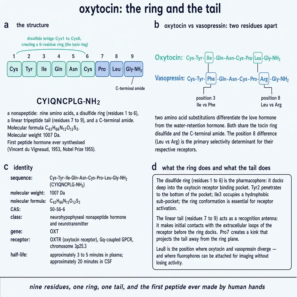
Most hormones that get written about are large. Growth hormone is 191 amino acids. Insulin is two chains held together by disulfide bridges. The gut peptide behind the current generation of obesity drugs is thirty residues long and uses almost its entire length to grip its receptor. Oxytocin is nine residues and weighs 1,007 grams per mole (Uvnäs-Moberg et al., 2019). Written out, it is Cys-Tyr-Ile-Gln-Asn-Cys-Pro-Leu-Gly-NH2: a six-residue ring closed by a disulfide bond between the first and sixth cysteines, and a three-residue tail that finishes not in a free acid but in an amide (Camerino, 2023).
That is the whole molecule. It is small enough to write on the back of a hand, and small enough that the entire modern discipline of peptide chemistry could begin with it, which is what happened.
Now the fact that organises everything else in this document. Change two of those nine residues — isoleucine at position three to phenylalanine, leucine at position eight to arginine — and you have vasopressin, whose job is to tell the kidney to hold on to water (Camerino, 2023). Two substitutions out of nine separate a hormone of birth and milk from a hormone of thirst and blood pressure. Nothing else in mammalian endocrinology places two such different physiologies so close together in sequence space.
The consequences of that proximity run through the rest of this monograph, and they are not merely a curiosity. Almost every hard problem in the oxytocin literature is, at bottom, a selectivity problem or a measurement problem descended from it. Drugs built to block one receptor block the other. Antibodies raised against one peptide see the other. A peptide given at high dose to contract a uterus also, unavoidably, tells a kidney to retain water. When a molecule differs from its sibling by two positions, the biology cannot be kept tidy and neither can the assays.
It is worth pausing on the units, because they are peculiar and they appear in every clinical sentence written about this drug. Oxytocin is dosed in international units rather than in milligrams, a convention inherited from the era when the material was a pituitary extract of unknown composition and could only be standardised by what it did to a strip of uterine muscle. One international unit is 1.67 micrograms (Uvnäs-Moberg et al., 2019). The ten international units that appear in the World Health Organization's recommendation are therefore about seventeen millionths of a gram, which is a striking quantity of material to stand between a woman and the leading cause of maternal death worldwide.
02Where it is made, and how it leaves
Oxytocin is not made in the pituitary gland, although that is where it is stored and where it enters the blood. It is made in the brain, in two clusters of large neurones in the hypothalamus: the supraoptic nucleus and the paraventricular nucleus. The OXT gene encodes not the finished peptide but a prepropeptide, in which the nine residues sit attached to a carrier protein called neurophysin I; the two are packaged together into secretory granules, and the granules travel down the axon to the posterior pituitary (Camerino, 2023).
This is a distribution system, not a factory floor. The peptide is synthesised at one end of a cell and released at the other, centimetres away, and the store held in the posterior pituitary is large enough that the gland can release far more in a few minutes than the neurones could make in that time.
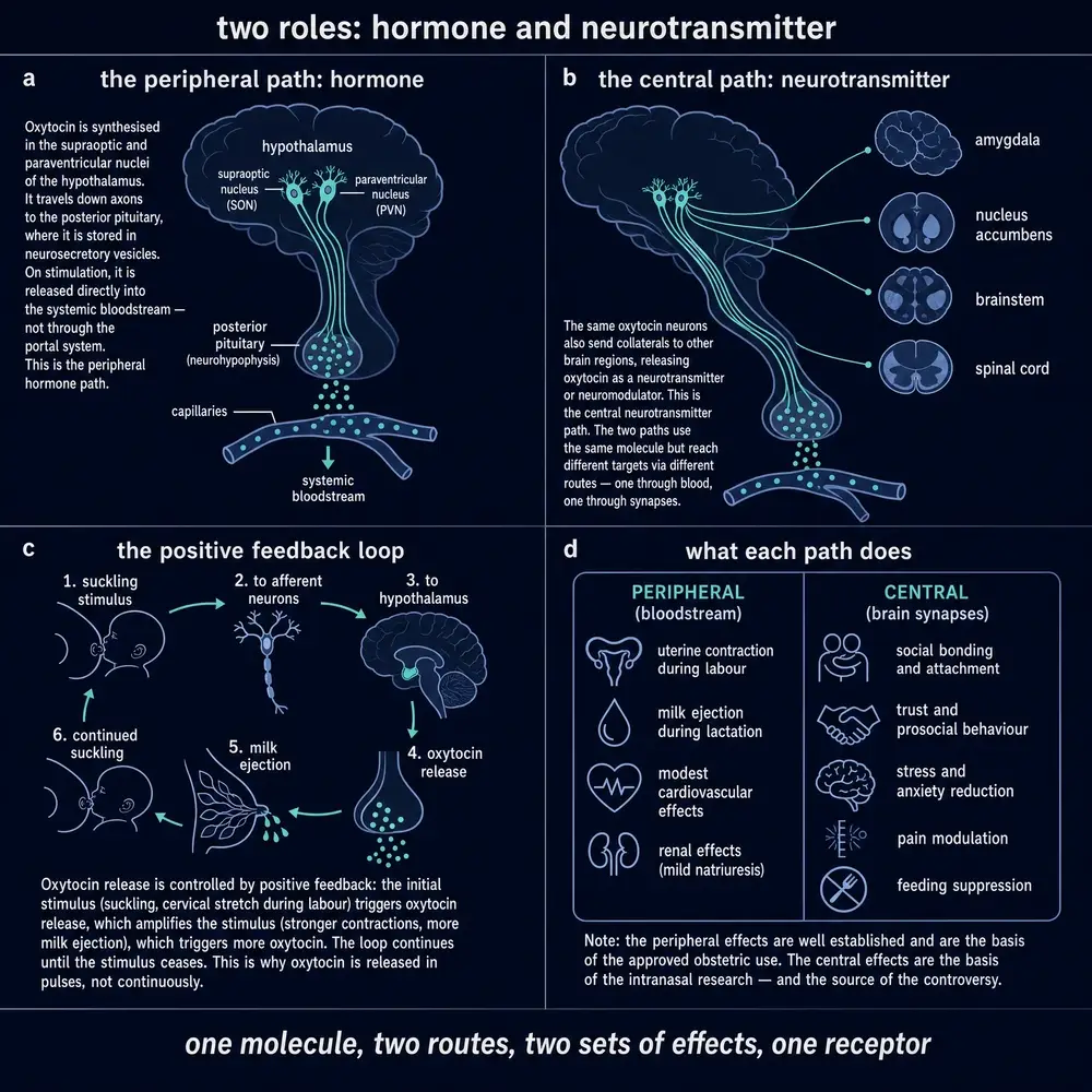
class="hf dark" so the mat stays navy in both editions.The second route out is the one that matters for everything in Part Four. The same neurones release oxytocin from their dendrites, back into the brain tissue around them, and that released peptide acts on the neurones that released it. The effect is self-reinforcing: dendritic release excites the cell, which fires, which releases more. In a lactating animal this produces something remarkable to watch on an electrode. The oxytocin cells, which otherwise fire irregularly and independently, synchronise into brief coordinated bursts, and each burst pushes a pulse of hormone into the blood. That description comes from electrode recordings in anaesthetised suckled rats, which is animal in vivo work (Leng et al., 2016). The system is pulsatile by design, and the pulses are generated by a positive feedback loop that runs inside the brain rather than by a clock somewhere else.
Two things follow. The first is that oxytocin release into the blood and oxytocin release into the brain are driven by the same cells but are not the same event, and Section 14 describes the experiment that proves they can be separated. The second is that a hormone released in pulses cannot be characterised by a single blood concentration, which is a large part of why the measurement literature on this molecule is in the state it is in.
031895 to 1955: how a gland extract became a sequence
The story begins with an extract and a dog. In 1895, George Oliver and Edward Sharpey-Schafer injected material extracted from the pituitary gland into dogs and found that it raised blood pressure (Camerino, 2023). That result launched the entire neurohypophysial research programme, and it is worth noting at the outset that the activity they saw was not oxytocin's. It belonged to the other peptide in the extract.
What followed over the next two decades was a slow untangling of one preparation into two hormones. Henry Dale reported that a substance from the posterior pituitary made the uterus contract, drove lactation, and reduced urine output (Camerino, 2023). Three effects, one extract, and — as later purification would show — two molecules. The pressor and antidiuretic activity separated out as vasopressin; the uterotonic and milk-ejection activity separated out as oxytocin, from the Greek for swift birth.
Dale's uterine-contraction report is conventionally placed in 1906, and that date appears in a great many secondary accounts. The historical review read for this document names Dale and describes what he reported but does not give the year, and no primary Dale paper was read here. The date is therefore omitted rather than borrowed. The same discipline applies to the sequence: the review that carries this history says 1950 in one place and 1955 in another, and the widely repeated date of 1953 could not be confirmed from anything read for this build. What the verified record supports is stated in the paragraph below, and no more.
The person who ended the ambiguity was Vincent du Vigneaud. Working through the early 1950s, his group determined the sequence of oxytocin and then made it — not extracted it, made it, from constituent amino acids in a laboratory — and published the consolidated account of the isolation, structure and synthesis in 1955 (du Vigneaud, 1955). His own retrospective, published in Science the following year, is titled for the thread that ran through his whole career: the trail of sulfur research, from insulin to oxytocin (du Vigneaud, 1956). The Nobel Prize in Chemistry followed in 1955 (Camerino, 2023).
It is worth being exact about what the synthesis proved, because the prize was for chemistry and not for physiology. Before du Vigneaud, a hormone was an activity: something in an extract that did something to a tissue, standardised in units defined by the assay. After him, a hormone was a structure, and a structure can be written down, checked, and made again by anyone with the reagents. Every peptide drug that exists — every insulin analogue, every incretin, every research peptide in the library this document was built from — descends from the demonstration that this is possible, and oxytocin is where it was demonstrated. The historical review read here frames it as the first polypeptide hormone to be sequenced and synthesised, and that framing is the review's rather than an independent finding of this document (Camerino, 2023).
One residue of the older world survives in current practice, and it dates the field more precisely than any anniversary does. The World Health Organization's recommendation for preventing postpartum haemorrhage, which Section 12 takes apart, is written in international units and not in milligrams. That is the vocabulary of the extract era described above, in which potency could only be expressed as what a preparation did to a tissue, and it has been carried forward unchanged into a molecule that has been synthesised to a known structure for seventy years. Nothing turns on it at the bedside. What it records is the order in which this compound was understood: the pharmacology arrived first and the chemistry came afterwards to explain it, rather than the other way round. That order is the reason so much of what follows is an argument about measurement. It runs through Part Four in a more consequential form, where a behavioural literature four decades old is still measuring what oxytocin does without a settled account of where the molecule it is measuring actually goes.
The seventy years since divide cleanly into three phases, and the timeline above shows the shape of them. The first was clinical and quick: a defined molecule that could be manufactured became a drug given to a substantial fraction of everyone born, and it has stayed one. The second was neuroscientific and began in the 1990s, when comparative work in voles suggested that where the receptor sits in a brain might predict how a species behaves. The third is the one this document is being written in the middle of: a fifteen-year attempt to turn that suggestion into a treatment, conducted largely with a nasal spray, which has not worked, and a simultaneous run of structural biology which has been spectacular. Part Two takes the structures first, because everything in Parts Three and Four is easier to weigh once the receptor is on the table.
04One receptor, and why it took until 2020 to see it
There is exactly one oxytocin receptor. That is unusual and it is convenient: much of the confusion in this field is about where the peptide goes and how much of it arrives, not about which of several receptor subtypes it hits. The receptor, OXTR, is a class A G-protein-coupled receptor — the large, well-studied family that also contains the receptors for adrenaline and serotonin — and it signals principally through the Gq and G11 proteins, which raise intracellular calcium and make smooth muscle contract (Meyerowitz et al., 2022).
Given how ordinary that description sounds, and how long the peptide has been available in pure form, the obvious question is why nobody had a picture of the receptor until 2020. Three reasons recur in the structural papers. There were no selective small molecules to stabilise it. The binding site turned out to be unusually large and unusually exposed to solvent, which is a difficult thing to crystallise. And the receptor behaved inconsistently in vitro in a way that nobody had explained — sensitive to how the membrane was prepared and to what was in the buffer (Waltenspühl et al., 2020). That last point turned out not to be a nuisance. It was the biology.
05The inactive picture, 2020
The first structure came from Waltenspühl and colleagues in 2020: an X-ray crystal structure of the human oxytocin receptor bound to retosiban, a non-peptide antagonist, solved in a lipidic cubic phase at 3.20 Ångström resolution (Waltenspühl et al., 2020). This is human in vitro structural work on purified protein, and it is worth knowing what it cost.
The wild-type receptor would not crystallise, so it was rebuilt until it would. Two rounds of directed evolution in yeast produced eight stabilising mutations. A stretch of the third intracellular loop was cut out and replaced with a glycogen synthase domain borrowed from an archaeon. Thirty residues were removed from the C terminus, and two further residues were mutated to alanine. The object that finally gave up a diffraction pattern was, in a precise sense, not the human oxytocin receptor. It was a receptor engineered until it could be photographed, and every interpretation drawn from it carries that caveat.
What it showed was worth the trouble. The binding pocket is enlarged and open to solvent compared with other peptide-binding receptors of the same class, with the outer tips of three helices displaced outward. Retosiban sits in it held by two faces: a polar interface involving glutamine and lysine residues, and a hydrophobic face made of isoleucines and a phenylalanine. The authors then took each contact away in turn and measured what it cost, which converts a picture into numbers.
Removing one glutamine cost sixteen hundred-fold in binding affinity. That is the kind of number that tells you the contact is not decoration. Two other findings from the same structure are less immediately dramatic and turned out to be more important. One cholesterol molecule was resolved in a groove on the outside of the helical bundle, between helices four and five, capped by the second extracellular loop; from there it positions a tyrosine that in turn hydrogen-bonds a glutamine and organises the whole polar network of the binding site. And two negatively charged residues sit at the extracellular tips of the first and second helices — residues that occur in no other receptor family — which the authors nominated as the structural explanation for a long-standing observation that this receptor's affinity for its agonist depends on magnesium (Waltenspühl et al., 2020).
Both were proposals. The structure was of an antagonist-bound, inactive, heavily engineered receptor, and it could not see a magnesium ion because in that state there is nothing for one to coordinate to.
06The active picture, 2022, and what a working receptor looks like
Two years later Meyerowitz and colleagues solved the other state, and did it the hard way: cryo-electron microscopy of the wild-type human receptor, with no thermostabilising mutations, bound to oxytocin itself and coupled to its G protein (Meyerowitz et al., 2022). This is human in vitro structural work again, but on the unmodified protein doing the thing it exists to do.
The same paper measured what the receptor's signalling actually looks like across the G-protein repertoire. It is a strong activator of Gq, G11 and G15 and a weak activator of the Gi/o family, and oxytocin drives the G11 response with a half-maximal effective concentration of 3.7 nanomolar, with a 95 per cent confidence interval from 2.7 to 5.3. That is the single cleanest potency figure in the material read for this document, and it is worth holding on to, because Part Four is largely a discussion of whether an administered dose ever reaches a concentration like that anywhere it matters.
Three things appear in the active state that were invisible in the inactive one. The first is a capping complex over the top of the bound peptide: an arginine from the receptor's own N-terminal segment, a phenylalanine and a second arginine from the first extracellular loop arrange themselves into a cation–π–cation sandwich, while the second extracellular loop swings open. Prior work cited in the same paper reports that removing that first arginine costs roughly two thousand-fold in oxytocin binding affinity. In the new measurements, mutating the phenylalanine moved potency from 3.7 to 18 nanomolar and mutating the arginine moved it to 88.
The second is the activation switch itself, and it is unexpectedly literal. Oxytocin's tyrosine at position two reaches across the pocket and makes a hydrogen bond to the backbone of the seventh transmembrane helix. That bond breaks the helix — a short stretch of it simply stops being a helix — and that local unwinding is what the rest of the receptor detects. The authors present this as the structural difference between full agonism by oxytocin and partial agonism by its analogues, which do not make the contact (Meyerowitz et al., 2022).
The third is the magnesium. The structure resolves a coordination complex formed between peptide and receptor with a magnesium ion at its centre, and the paper shows that the identity of a single conserved residue determines whether a receptor in this family requires the cation as a cofactor at all. The 2020 proposal was right, and it took an active-state structure to see it.
The two structures were solved three years apart, by different groups, using different methods, on differently prepared protein — one an engineered antagonist complex by X-ray crystallography, the other a wild-type agonist complex by cryo-electron microscopy. Both found a sterol occupying the same extrahelical pocket between the fourth and fifth helices. Nobody was looking for agreement; the agreement is what makes the pair trustworthy, and it is a better argument for the cholesterol site being real than either paper makes on its own.
07A receptor that needs a metal and a sterol
Most drug targets can be studied in a defined buffer. This one cannot. Normal agonist binding at the oxytocin receptor requires a divalent cation coordinated between the peptide and the protein, and a cholesterol molecule wedged into a groove on the outside of the helix bundle (Waltenspühl et al., 2020; Meyerowitz et al., 2022). Neither is a detail of the preparation. Both are part of the receptor.
The consequence is worth stating plainly, because it is the least discussed contributor to a reproducibility problem that is usually blamed on psychology. Membrane cholesterol content varies between cell lines, between preparations, and between tissues. Magnesium concentration varies between buffers. A receptor whose agonist affinity depends on both will give different numbers in different laboratories for reasons that have nothing to do with the experiment being run, and until 2022 nobody could say why. Some of the field's inconsistency is chemistry.
08Same receptor, different answers: biased agonism
The idea that a receptor is a switch, and that anything which turns it on produces the same downstream result, is convenient and wrong. A receptor is better thought of as a hub with several outputs, and different ligands can drive different subsets of them. Oxytocin pharmacology contains an unusually clean demonstration of this, and it involves a drug in routine clinical use.
Carbetocin is a long-acting oxytocin analogue, given in some countries in place of oxytocin to prevent bleeding after caesarean section. Passoni and colleagues characterised it in human cells using bioluminescence resonance energy transfer biosensors, which report on individual signalling arms separately — human in vitro work (Passoni et al., 2016). Four findings, and they compound.
Carbetocin activates the oxytocin receptor but not the two vasopressin receptors tested, where it may in fact act as an antagonist; so it is more selective than oxytocin, not less. At the oxytocin receptor it activates only the Gq arm, leaving other pathways alone. On that arm it is a partial agonist, producing a submaximal response however much of it is present. And after it binds, the receptor is internalised by a route that does not use β-arrestin, and is not recycled back to the cell surface afterwards.
The authors' conclusion is the one that matters, and it generalises well beyond this molecule: carbetocin should not be expected to reproduce oxytocin's effects in the brain. An analogue is not a slow-release version of its parent. It is a different drug that happens to bind the same protein, and the phrase “an oxytocin analogue” carries no guarantee about anything except where it binds.
Atosiban, which appears repeatedly in later sections as an experimental tool, carries the mirror-image problem. It is a competitive antagonist, and it blocks both oxytocin and vasopressin receptors (Chen et al., 2021). That dual action is why an experiment showing that atosiban changes some physiological variable cannot, on its own, establish that oxytocin signalling was responsible — a caveat that Sections 22 and 23 have to apply more than once.
09Ancient machinery
The oxytocin and vasopressin receptors are two branches of one family, and the family is old. A phylogenetic and chromosomal analysis across jawed vertebrates concludes that the receptor genes expanded by gene duplication rather than by independent convergence on similar structures, and the pattern of duplication can be traced against the large-scale genome duplications early in vertebrate history (Ocampo Daza & Larhammar, 2021). Related work has examined how variation in these receptor genes maps onto human social behaviour (Theofanopoulou et al., 2022).
What that history supports, and what it does not, is worth separating. It supports a simple ordering claim: the machinery is ancient and its original business was osmotic regulation and reproduction. The behavioural roles that dominate the modern literature were layered onto equipment built for water balance and birth, not the other way round. Carter and colleagues push the argument further, situating the peptide in the physiology of oxidative metabolism and framing it as part of an evolutionary solution to the demands of oxygen-dependent life (Carter et al., 2022).
It does not support the just-so story that the reader has probably met, in which evolution produced a bonding hormone. Nothing in a duplication history tells you what a receptor does in a living animal, and the temptation to read adaptive purpose backwards out of a gene tree should be resisted here as everywhere. The most useful thing the evolutionary record contributes to this document is a warning about expectations: a system this old, this widely expressed and this deeply embedded in basic physiology is unlikely to have one function, and Section 22 shows that it does not.
10What actually happens during labour
Ask what oxytocin does in childbirth and the textbook answer is that it makes the uterus contract. That is true, and it is not what the measurements show happening. The most careful synthesis available is a systematic review of maternal plasma oxytocin during physiological childbirth, which screened 4,039 records and found twenty studies that met its criteria — human in vivo measurement studies, nineteen of the twenty using radioimmunoassay (Uvnäs-Moberg et al., 2019).
The shape it describes is this. Baseline plasma oxytocin rises three- to fourfold over the course of pregnancy. Superimposed on that rising baseline are pulses, which grow in frequency, duration and amplitude from late pregnancy through labour, reaching a maximum of roughly three pulses every ten minutes towards the end. A further three- to fourfold rise occurs at birth itself, and pulses continue through the third stage as the placenta is delivered. Oxytocin was also raised in cerebrospinal fluid during labour, so the central and peripheral systems are active together.
The shape of the curve is well evidenced — rising baseline, intensifying pulses, a peak at birth, continuing third-stage pulses — because it is consistent across twenty studies. The concentrations are not, and this document does not print them. Nineteen of those twenty studies measured oxytocin by radioimmunoassay, and a 2025 pharmacokinetic study using mass spectrometry found that immunoassay reads systematically lower than a specific method on the same samples (Shafer et al., 2025). An absolute plasma oxytocin concentration in this literature is a statement about the assay at least as much as about the patient. The figure below is therefore drawn as a shape and labelled as one.
Now the finding that should change the reader's mental model, and which is easy to miss because it is a negative. The plasma peaks did not line up in time with individual uterine contractions (Uvnäs-Moberg et al., 2019). The uterus is not being conducted beat by beat by a hormone arriving from the pituitary. Whatever is timing individual contractions, it is not the arrival of a pulse in the blood.
That implies something about which variable actually changes during labour. It is not principally the hormone. It is the tissue: the density of oxytocin receptors in the myometrium rises enormously towards term, and a uterus that has become exquisitely sensitive will respond to concentrations that would have done nothing to it weeks earlier. The hormone is closer to a permissive signal than to a conductor's baton, and this matters clinically, because it is the reason a tiny quantity of infused peptide can drive a labour and the reason the same quantity does nothing at all before term.
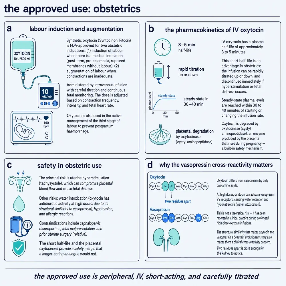
One further number from the same review is useful for reading everything that follows: an infusion of up to about ten milliunits per minute produces plasma concentrations similar to those of physiological labour, and doubling the rate roughly doubles the measured concentration (Uvnäs-Moberg et al., 2019). Clinical oxytocin, given at these rates, is not a pharmacological curiosity; it is within the range the body produces on its own. What it does not reproduce is the pattern. An infusion is continuous where the physiology is pulsatile, and nothing in the read record tells us what that difference costs.
11Postpartum haemorrhage, and the honest ranking
Bleeding after childbirth is the leading cause of maternal death worldwide, and the intervention that prevents most of it is a few micrograms of this peptide given at the moment of delivery. This is the strongest evidence base oxytocin has, and it is worth reporting precisely, because what it shows is more interesting than the reputation.
The synthesis is a Cochrane network meta-analysis of uterotonic agents, searched to February 2024, covering 122 randomised trials in 121,931 women (Gallos et al., 2025). A network meta-analysis compares drugs that were never tested head to head by chaining together the comparisons that were made, and this one is unusually careful: of 196 trials in the previous version of the review, 112 survived a formal trustworthiness screen, and the rest were excluded. Risk of bias remains substantial — for the main outcome, 49 of 81 trials were rated at high risk. Oxytocin is the reference against which everything else in the network is measured.
Against oxytocin, ergometrine plus oxytocin reduces blood loss of 500 mL or more, with a relative risk of 0.76 and a confidence interval from 0.64 to 0.90, and the review grades that evidence high certainty. Misoprostol plus oxytocin probably does the same, with a relative risk of 0.70, graded moderate. Carbetocin makes little or no difference, graded high. Misoprostol alone is worse for severe bleeding, with a relative risk of 1.24 for loss of a litre or more, graded low. For blood transfusion the combinations again come out ahead: misoprostol plus oxytocin has a relative risk of 0.40, ergometrine plus oxytocin 0.73.
Translated out of relative risks, which mislead almost everyone: of a thousand women given oxytocin at a vaginal birth, about 83 would bleed 500 mL or more, against 63 on ergometrine plus oxytocin and 58 on misoprostol plus oxytocin. About 89 per thousand on oxytocin would need a second uterotonic drug, and about 15 per thousand would need a transfusion, against 6 on misoprostol plus oxytocin (Gallos et al., 2025).
In the review's own ranking statistic, oxytocin comes fourth — behind both combinations and fractionally behind carbetocin, which it is statistically indistinguishable from.
So why is it first-line everywhere? Not because it is the most effective molecule in the network, and this document is not going to pretend otherwise. It is first-line because effectiveness is one input among several. Ergometrine raises blood pressure and causes vomiting, and cannot be given to women with hypertensive disease, which in many settings is a large fraction of them. Misoprostol causes shivering and fever. Carbetocin costs more and, in the form that does not need refrigeration, is a recent arrival in most of the world. Oxytocin is cheap, universally available, and has a side-effect profile clinicians have a century of familiarity with. “Recommended” is a statement about a whole profile, and reading it as “best” misreads the evidence in a way the evidence itself does not support.
12Ten international units, and what the guideline does not know
The World Health Organization's recommendation is short: oxytocin, ten international units, intramuscular or intravenous, is the recommended uterotonic for preventing postpartum haemorrhage, for all births. Where a woman giving birth vaginally already has intravenous access, slow intravenous administration of the ten units is preferred to intramuscular. For caesarean section the same quantity is recommended with no stated preference between routes. These are guideline parameters attributed to their source and are reported here as facts about the guideline; nothing in this section is a direction for use.
What makes the recommendation worth a section of its own is what the Guideline Development Group says about it in the same document. The trials underlying the intravenous question all used ten units — and gave them over anything from one minute to forty, diluted in anything from one millilitre to a litre of saline. No direct comparative evidence exists on which speed or which dilution is better. The group's preference for slow, dilute administration rests on observational studies in caesarean section suggesting that rapid intravenous injection produces harmful haemodynamic effects, not on a trial that compared the two.
Read that again with the scale attached. This is among the most widely administered medicines in the world. The recommended quantity is settled and evidenced. How to put it in — over one minute or over forty, in a millilitre or in a litre — is not, and the guideline says so in its own remarks rather than leaving a reader to discover it. That is a gap in the evidence base, not a gap in the guideline, and Section 24 lists it among the things this document could not find an answer to.
13More is not better
Three recent human observational studies, all published in 2025 or 2026, all point the same way: larger cumulative exposure to oxytocin during labour and delivery is associated with worse outcomes, not better ones.
A retrospective human cohort of 1,996 caesarean sections over fifteen years, with complete haemoglobin data for 1,504, examined intraoperative prophylactic dose against measured blood loss (Busłowicz et al., 2026). Before labour had started, doses above 13 IU were associated with 315 mL more adjusted blood loss than the reference exposure. After labour had started, the same high exposure was associated with 170 mL more, while a 3 IU exposure was associated with 116 mL less. No consistent dose–response benefit appeared anywhere in the cohort.
A retrospective human study of induced labours, sampling 504 of 3,598, found that increasing total oxytocin dose was associated with greater risk of caesarean delivery and of postpartum haemorrhage, with the excess caesarean risk appearing from a cumulative 5 IU upward (Payrastre et al., 2025). Nulliparity and cervical ripening were co-factors. No association with neonatal morbidity appeared in this sample.
A third retrospective human study, of 500 mother–neonate pairs, found neonatal hyperbilirubinaemia in 10 per cent overall, but distributed steeply by exposure tertile: 2.98 per cent in the low-dose group, 8.98 per cent in the moderate group, and 18.18 per cent in the high group, with an adjusted odds ratio for high dose of 7.9 (Liang et al., 2025).
Three cohorts, three countries, one direction. It would be easy to conclude that oxytocin exposure causes these harms, and the authors of the largest of them do not conclude it. The reason is confounding by indication, and it is not a technicality: a woman who receives 15 IU of oxytocin is, by definition, a woman whose labour was not going well. High dose is a marker of difficult labour, atonic uterus and prolonged delivery, all of which independently predict bleeding. The association is real and the causal claim is not established by any of these designs.
What none of them can do is separate the drug from the reason for the drug. That would take a randomised comparison of dose regimens, which does not exist in anything read for this document. Until it does, the honest summary is that higher cumulative intrapartum exposure travels with worse outcomes, that the direction is consistent across recent cohorts, and that the mechanism is undetermined.
There is one more record here, and it is not epidemiology. A qualitative study interviewed fifteen midwives across ten hospitals, using a descriptive phenomenological method (Xiao et al., 2025). What they described was clinical alertness set against systemic vulnerability, professional judgement set against limited authority, and accumulated experience set against an absence of clear guidelines — with missed care recurring specifically around oxytocin infusion for induction and augmentation. This cannot quantify risk and does not try to. It names a mechanism the cohorts cannot see: the hazard in this drug is not molecular toxicity but titration under time pressure without a protocol, and that is the same gap the guideline itself admits to in Section 12.
A widely repeated claim holds that synthetic oxytocin given in labour interferes with maternal attachment and breastfeeding. A prospective case–control study in humans, comparing 75 exposed women with 78 unexposed at term low-risk vaginal births, found no difference in maternal or cord-blood oxytocin, no difference in early breastfeeding attitude, and no difference in maternal attachment scores (Özdemir et al., 2025). It is small, single-centre and used immunoassay, so it does not settle the question — but it is a direct null and it is reported here rather than omitted.
The mechanistic worry has animal support. In prairie voles — animal in vivo work — DNA methylation at four sites in the oxytocin receptor gene — sites conserved with the human gene — normally predicts receptor expression in the nucleus accumbens; that relationship disappears at term pregnancy and returns immediately after birth, and giving exogenous oxytocin to model an induced labour altered the switch (Perkeybile et al., 2025). That is a mechanism in a rodent. It has no human replication in this corpus, and the human null above sits directly beside it.
14Milk, and the reflex that runs on positive feedback
Milk is not sucked out of a breast. It is pushed, by a layer of contractile cells wrapped around the milk-producing alveoli, and what makes them contract is a pulse of oxytocin. The reflex is one of the most thoroughly characterised pieces of neuroendocrinology there is, and it has been mapped at the level of individual neurones firing.
The picture, from electrode recordings in anaesthetised suckled rats, is the positive-feedback loop described in Section 02: suckling drives oxytocin release from the dendrites of magnocellular neurones, that dendritic oxytocin excites the same neurones, and the population synchronises into brief bursts that each deliver a pulse of hormone to the gland — animal in vivo electrophysiology (Leng et al., 2016). Blocking oxytocin centrally stops milk transfer.
The same paper contains the experiment this document has been building towards, and it is a small, decisive piece of work. The peptide oxytocin antagonist F-792 was given to lactating rats by three routes at three doses. At 7 micrograms per kilogram intravenously, it blocked the mammary gland's response to oxytocin. At 0.2 micrograms given directly into the brain, it blocked the milk-ejection reflex. And at 100 micrograms per kilogram intravenously — fourteen times the dose that abolished the gland's response — it blocked milk ejection at the gland while leaving the suckling-induced burst firing of identified paraventricular oxytocin neurones completely intact, recorded in five neurones across five animals.
It is worth being explicit about why that design settles anything, because the result is easy to state and easy to misread. Three routes at three doses is not thoroughness for its own sake; each arm removes one way of being wrong. The low intravenous dose establishes that the antagonist reaches the gland and works there. The tiny central dose establishes that the same molecule, placed directly into the brain, is enough to abolish the reflex, so the reflex has a central component and this antagonist can block it. The fourteen-fold intravenous dose then asks the only question that remains: if enough antagonist had crossed from blood into brain to matter, the burst firing should have stopped. It did not stop. The gland went quiet and the neurones carried on firing.
A systemically administered peptide antagonist does not enter the brain in useful quantity. The doses quoted above are experimental parameters in anaesthetised rats and are reported as such; the finding they establish is structural rather than pharmacological. Central and peripheral oxytocin are separate compartments, and in this animal in vivo preparation that separation is demonstrated rather than assumed (Leng et al., 2016).
Everything in Part Four depends on that sentence. If a peptide cannot readily cross from blood into brain, then a peptide given into a nose has to get into the brain some other way, or it has to work from the periphery, or it has to not work. Which of those three is true is the central unresolved question of the modern oxytocin literature, and Section 18 is about the attempts to answer it.
15The uterus, closer up
Two recent studies add mechanism to the clinical picture, and both are worth a paragraph because they show how much is still being learned about the tissue this drug was built for.
The first concerns fuel. Human myometrial cells in primary culture — human in vitro work — run on oxidative phosphorylation while quiescent and shift towards glycolysis when stimulated with oxytocin (Prifti et al., 2026). Blocking glucose oxidation reduced both basal oxygen consumption and contractility; blocking fatty-acid or glutamine oxidation did not, except that in oxytocin-stimulated cells glutamine blockade did reduce oxygen consumption, implying the tissue recruits an additional fuel under contractile load. Uterine contraction is metabolically expensive, and the substrate the muscle uses changes when the hormone arrives.
The second concerns time of day, and it carries three evidence tiers in one paper, which this document keeps separate (Van-Quynh Duong et al., 2026). In mice — animal in vivo — uterine oxytocin receptor expression varies with time of day under the control of the circadian regulator BMAL1, and both smooth-muscle-specific knockouts of that regulator and mice with diet-induced gestational diabetes show reduced uterine contractility in response to oxytocin. In an immortalised human myometrial cell line — human in vitro — time of day likewise affected oxytocin-induced contractility. And in a retrospective review of 2,367 patients at or beyond 39 weeks undergoing induction — human observational — morning induction was associated with labour roughly an hour and a half shorter than midnight induction in controls, and roughly seven hours shorter in patients with gestational diabetes.
The mouse work is mechanistic and the human work is retrospective and association-only, so nothing here establishes that scheduling an induction changes an outcome. What the pair does establish is that the tissue's responsiveness to this drug is not a constant, and that at least two of the things that modulate it — circadian phase and metabolic state — are not currently accounted for anywhere in how the drug is given.
16The voles
In 1992 Thomas Insel and Lawrence Shapiro published a comparison that changed what people thought oxytocin was for. They took four species of vole — two that form lasting pair bonds and two that do not — and mapped where the oxytocin receptor sits in each brain, using autoradiography with a radiolabelled ligand. This is animal in vitro work on brain sections, not a behavioural experiment (Insel & Shapiro, 1992).
In the monogamous prairie vole, receptor density was highest in the prelimbic cortex, the bed nucleus of the stria terminalis, the nucleus accumbens, midline thalamic nuclei and the lateral amygdala. In the polygamous montane vole, those regions showed little binding; the receptors were somewhere else entirely, concentrated in the lateral septum, the ventromedial hypothalamus and the cortical amygdala. The same contrast held for a second monogamous–polygamous pair, pine and meadow voles.
The reason this result carried, rather than being one more species difference among many, is the control. The authors also mapped benzodiazepine and mu-opioid receptors in the same brains, and those distributions showed no comparable species difference. So the finding is not that monogamous and polygamous voles have different brains in general. It is that this particular receptor is distributed differently, in regions associated with reward and social memory, in the species that pair-bond.
And one further observation, easy to overlook, does more work than the rest. The montane vole is affiliative only after giving birth — and its receptor distribution changed within 24 hours of parturition. A map that shifts inside a day, in step with a change in behaviour, is harder to dismiss as an incidental species difference than a static comparison would be.
Be precise about what this is. It is an anatomical correlation in animals. It shows that receptor distribution tracks social organisation; it does not show that receptor distribution causes it, and Insel and Shapiro did not claim otherwise. Three decades of popular writing about the love hormone rest on a foundation that is, in its original form, a careful piece of comparative neuroanatomy with a well-chosen control.
17What the animal work does and does not establish
The causal work came later, and a good deal of it is excellent. The question it has been organised around is one that Grinevich and Neumann put plainly in a review of the field: how does a single neuropeptide produce so many different effects? Their answer is that it does not act as one thing — oxytocin neurones diverge anatomically, project to many targets, act on different cell types in different regions, and recruit several intracellular pathways (Grinevich & Neumann, 2021). Three animal in vivo studies illustrate what that looks like in practice, and none of them supports the idea that the peptide installs a behaviour.
In mice, oxytocin enables maternal behaviour by rebalancing inhibition in the left auditory cortex, so that pup distress calls become behaviourally salient — the peptide changes what the animal notices, and the behaviour follows (Marlin et al., 2015). In the nucleus accumbens, social reward requires oxytocin and serotonin acting together, with oxytocin acting on presynaptic terminals to permit a serotonin-dependent form of synaptic depression (Dölen et al., 2013). And a 2026 multisite recording study found that oxytocin suppressed baseline activity across prefrontal cortex, auditory cortex and the basolateral amygdala while selectively strengthening low-frequency coupling from auditory cortex to prefrontal cortex (Jung et al., 2026). The peptide improved the temporal coordination of information flow without amplifying the signal — which is a very different description from turning up a social dial.
Recent work has extended this in several directions at once. Oxytocin receptor neurones in the paraventricular thalamus modulate sociability and fear extinction bidirectionally in mice, while the same manipulation in prefrontal cortex leaves sociability untouched — animal in vivo chemogenetics (Yamamuro et al., 2026). The same paper reports a modest correlation between salivary oxytocin and thalamic microstructure in humans, and its authors note explicitly that the paradigms differed across species. Intranasal oxytocin attenuates fear learning in mice, an animal in vivo result, through receptors in prelimbic cortex carried by somatostatin interneurons (Xu et al., 2026). Maternal aggression in mice rises and falls with oxytocin drive onto a defined amygdala-to-hypothalamus pathway (Yamaguchi et al., 2026). Early social isolation impairs adult partner-preference formation in female prairie voles without changing overall receptor density, while accumbal receptor density predicts which animals are resilient — animal in vivo again (Barrett et al., 2015).
Then there is the result that breaks the tidy version of the story, and it comes from the same species the story started in.
Pharmacological blockade of the oxytocin receptor in prairie voles in the 1990s indicated that oxytocin signalling is required to form a pair bond. In 2025, prairie voles engineered to lack the receptor entirely were tested — animal in vivo — and the males, but not the females, showed impaired consolation behaviour towards a distressed partner. Both sexes showed normal partner preference after 24 hours of cohabitation, and autoradiography found no compensatory upregulation of vasopressin-1a receptors in four regions where it might have been expected (Horie et al., 2025).
Recency does not settle this one, and it is worth saying why. Acute pharmacological blockade and constitutive genetic deletion answer different questions: the first asks whether signalling is needed now, the second asks whether an animal that has never had the receptor can nevertheless get there. Developmental compensation is the obvious candidate explanation and is not demonstrated in anything read here. What the knockout does establish is a dissociation: consolation and partner preference are not one system, and the older literature treated them as though they were.
A second finding cuts the same way, and it is the strongest single argument that “oxytocin” is not one functional entity. Knocking down oxytocin synthesis in the anterior versus the posterior paraventricular nucleus of California mice produced opposite behavioural effects — anterior knockdown increased social approach and reduced vigilance after social defeat, posterior knockdown reduced social approach in unstressed animals — and the two populations of neurones differ in their electrophysiology. That is animal in vivo work (Chrisman et al., 2026). The source of the peptide determines the sign of the effect. Any statement of the form “oxytocin increases X” is incomplete without saying which oxytocin neurones, and almost no human study can say.
Two anatomical resources anchor all of this. A single-transcript atlas of mouse brain tissue — animal in vitro — maps oxytocin and receptor expression nucleus by nucleus (Ryu et al., 2026), and a post-mortem transcriptomic atlas of human tissue does the equivalent for people, finding expression enriched in subcortical and olfactory regions and co-expressed with dopaminergic and cholinergic gene networks (Quintana et al., 2019). Both describe where the machinery is. Neither describes what it does, and the distinction is one the popular literature routinely loses.
A last caution about extrapolation, which the animal literature itself supplies. Dogs and their owners enter a positive feedback loop of gaze and urinary oxytocin that wolves do not — an experiment run in both animals and people in vivo (Nagasawa et al., 2015). It is a lovely result and the human half of it is measured in urine, which is subject to every caveat in Section 18. Species differ, and the differences are sometimes the finding rather than the noise.
18The nose
Essentially the entire human behavioural literature on oxytocin rests on one delivery method: a spray up the nose, on the premise that peptide reaches the brain directly along the olfactory and trigeminal routes, bypassing the blood–brain barrier that Section 14 showed a peptide antagonist could not cross. Whether that premise holds is the single most consequential unresolved question in the field, and in 2025 somebody finally measured the pharmacokinetics properly.
Using a specific mass-spectrometry assay rather than an immunoassay, and population modelling across two studies with 24 non-pregnant adults in the crossover arm, intravenous oxytocin was well described by a two-compartment model with no bias and 18 per cent median inaccuracy. Intranasal oxytocin at 100 micrograms was not. It showed 47 per cent median inaccuracy — that is, enormous variation between one person and the next — and bioavailability of 0.7 per cent. This is human in vivo pharmacokinetics (Shafer et al., 2025). The doses quoted are study parameters in that trial and are not recommendations. The same work found that mass spectrometry read systematically higher than simultaneous immunoassay on the same samples, which is a separate problem and an old one.
Seven parts per thousand, with roughly half the value unpredictable between subjects. The authors' own inference is the restrained one: low bioavailability and large intersubject variability could partly explain the inconsistent efficacy reports in this literature.
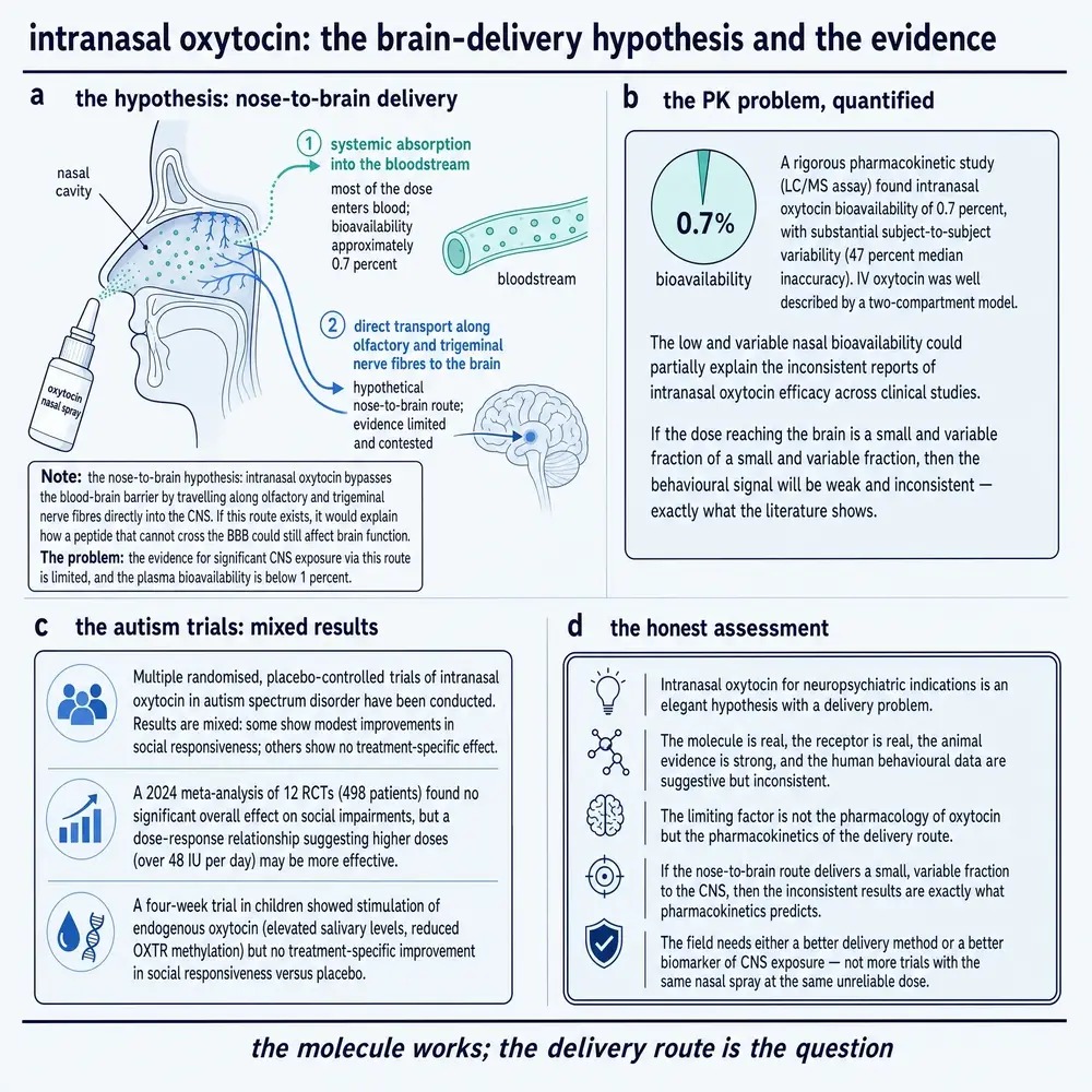
That measurement did not create the argument, it sharpened it. Leng and Ludwig had already set out the sceptical case in 2016, and their charge sheet was blunt: very little of an applied dose reaches cerebrospinal fluid; peripheral concentrations rise to supraphysiological levels with likely effects on gut, heart and reproductive tract; many published oxytocin measurements used methodology that does not survive scrutiny; and the inference from a peripheral measurement to a central state is questionable at best (Leng & Ludwig, 2016). Their prescription — preregistration, declared primary outcomes, prespecified analysis, open data, real dose–response work, and peripheral-administration control arms — is the standard the subsequent literature should be read against.
The defence is not empty. A 2021 review argues that converging human and animal evidence supports functionally relevant effects of the intranasal route and direct nose-to-brain transport, and documents genuine methodological improvement in the field since 2016 (Quintana et al., 2021). A third position exists as well: circulating oxytocin may be actively carried into the brain by the receptor for advanced glycation end-products, with release governed by CD38 and CD157, which would link peripheral and central concentrations through a saturable transporter rather than through the nose at all. That proposal rests chiefly on the authors' own animal in vivo work, reviewed by them (Higashida et al., 2022).
Somebody then tried to watch it happen. A first-in-human study gave radiolabelled oxytocin intranasally to six human volunteers in vivo and imaged them with combined positron emission tomography and magnetic resonance (Winterdahl et al., 2025). Uptake in the nasal cavity was high within five minutes and then declined as the tracer was absorbed systemically. Signal in the trigeminal ganglion and the brain varied between individuals with no clear dependence on dose. Time–activity curves suggested tracer was present in brain regions between 25 and 45 minutes, but the nasal cavity was the dose-limiting organ and spillover from it compromised the co-registration. The authors' conclusion is that the tracer is not presently well suited to central receptor imaging by this route.
This document is not going to resolve the dispute, because nothing read for it can. The defensible position is narrower and still says a good deal: a measured bioavailability of 0.7 per cent with nearly 50 per cent intersubject variability is, by itself, sufficient to produce a literature of small, inconsistent, non-replicating findings without requiring that any individual finding be false. And no method currently in this corpus can establish the central concentration achieved by an intranasal dose in a human being. A field that cannot measure its own exposure variable is a field that will argue for a long time.
19Trust, 2005 to 2026
In 2005 Nature published an experiment that did more than any other to make oxytocin famous. Volunteers given intranasal oxytocin played an economic trust game, in which one player hands money to another and can only get more back if the other chooses to return it. The oxytocin group handed over substantially more, and — the detail that made the finding feel mechanistic rather than incidental — the effect was specific to social risk rather than to risk in general. This was human in vivo work (Kosfeld et al., 2005). The trust molecule was born in that paper, and the popular framing has outlived every subsequent test of it.
In 2020 a registered replication attempted the experiment at greater than 95 per cent power, implementing both a condition matching the original's minimal social contact and a no-contact condition. In the condition that matched the original, oxytocin had no effect on trusting behaviour in these human volunteers (Declerck et al., 2020). An exploratory post-hoc analysis suggested a possible increase among low-trust individuals in the no-contact condition, which the authors themselves flagged as needing confirmation rather than presenting as a rescue.
In 2026 a registered report ran the test again with 211 participants and pooled its data with the 2020 study for a combined 532. Again no effect. This time the analysis went further and applied equivalence testing to the pooled data, which bounded the effect within a range too small to matter. This too was human in vivo (Kroll et al., 2026). There was no moderation by baseline trust, reward sensitivity or punishment sensitivity.
A conventional statistical test can only fail to find an effect. It cannot distinguish “there is nothing here” from “we did not look hard enough”, which is why a null result is usually a weak statement and why a literature of nulls can be argued with indefinitely. Equivalence testing inverts the question: it asks whether the data are inconsistent with any effect larger than a specified size. When a pooled sample of 532 returns equivalence against a smallest-effect-of-interest, the conclusion is not that the effect was missed. It is that if an effect exists, it is smaller than a laboratory experiment of this kind could usefully detect.
This is the cleanest case in the whole compound file for saying that recency supersedes, and the reason is not the date. It is the design. The later studies were larger, they were preregistered so the analysis could not follow the data, and the last of them tested for equivalence rather than merely for difference. Where a newer null supersedes an older positive, it does so on the strength of its method; a 2026 study with the 2005 study's design and sample size would settle nothing.
Two clarifications, because this section is easy to overread. Nothing here implies the 2005 result was fabricated or that its authors did anything wrong; small early-positive results followed by null replications are the expected signature of an underpowered literature exploring a real question, and that is a statement about the field's methods in 2005, not about anyone's integrity. And the trust game is one paradigm. Its collapse bounds what can be claimed about that paradigm, not about every effect the peptide might have.
20Autism, and the largest trial
If intranasal oxytocin promotes social behaviour, the clinical application that follows is obvious, and for fifteen years it was pursued hard. The definitive test is SOARS-B: a multicentre, randomised, placebo-controlled trial in children and adolescents aged 3 to 17 with autism spectrum disorder, running 24 weeks, with a target dose of 48 international units daily by nasal spray, randomised 1:1 and stratified by verbal fluency and age. It is the largest human in vivo test the compound has had (Sikich et al., 2021). Those are trial parameters reported as such. Of 355 screened, 290 were randomised and 277 entered the modified intention-to-treat analysis. The trial had 90 per cent power to detect a five-point difference on its primary outcome.
The primary outcome was the least-squares mean change on the modified social withdrawal subscale of the Aberrant Behavior Checklist. It improved by 3.7 points on oxytocin and 3.5 points on placebo: a difference of 0.2 points, with a confidence interval from −1.5 to 1.0, and a P value of 0.61. All three secondary outcomes agreed — social motivation, sociability, and abbreviated IQ all showed differences indistinguishable from zero.
Two details deserve reporting because they speak to how much weight the result can bear. The first is a piece of authorial scrupulousness: the investigators state that there was no prespecified plan to adjust confidence intervals for multiplicity in the secondary, exploratory and sensitivity analyses, and that those results therefore cannot be used to infer definitive treatment effects. A trial that says this about its own secondary analyses is a trial to believe on its primary. The second is safety, reported here rather than quarantined into a later section: there were three serious adverse events, of which one was judged related to oxytocin — sedation while driving, leading to a motor-vehicle accident. Four participants on oxytocin and three on placebo stopped for adverse events, most of the oxytocin discontinuations for irritability or aggression.
The wider picture is consistent with the trial. A 2026 meta-analysis of 42 double-blind randomised trials of intranasal oxytocin against placebo across autism, schizophrenia spectrum disorders, substance use disorders and other mental disorders, pooling 1,922 participants, found an overall effect of Hedges' g = 0.17 with a confidence interval from −0.05 to 0.38 and heterogeneity of 77 per cent — small, non-significant, and wildly inconsistent between trials (Bonnieux et al., 2026).
Then the diagnostic step. Removing two outlier substance-use trials with very large effects dropped the pooled estimate to g = 0.05, with a confidence interval from −0.03 to 0.12 — and took heterogeneity from 77 per cent to zero. Heterogeneity collapsing to nothing when two studies leave is not a robustness check that failed; it is a finding. It says the apparent inconsistency of this literature was substantially two trials, and that everything else in it is a consistent, tiny, indistinguishable-from-zero effect. The one subgroup that survives is schizophrenia spectrum disorders, at g = 0.12 with a lower bound of 0.01, which is real, positive, and about as small as an effect can be while remaining detectable. Earlier syntheses in that population point the same way: high-level social cognition may improve where lower-level social cognition and neurocognition do not (Bürkner et al., 2017), and a dose–response meta-analysis for negative symptoms of schizophrenia reaches similarly modest conclusions (Sabe et al., 2021).
21What survives
A document that stopped at Section 20 would be making its own kind of error. Not everything failed, and the pattern of what did not fail is informative.
Start with the trial that worked. FOXY was a phase 2a/2b adaptive crossover trial of intranasal oxytocin for apathy in frontotemporal dementia, across eleven clinics, with 94 participants of mean age 65.9 and 40 per cent women; the regimen selected in the adaptive stage was 72 international units twice daily given every third day, which is a trial parameter and not a recommendation. This is human in vivo evidence (Coleman et al., 2025). The Neuropsychiatric Inventory apathy score improved by an estimated 1.32 points against placebo, with a confidence interval from 0.21 to 2.43 and a one-sided P value of 0.010. No adverse events were attributed to oxytocin. The authors describe this as a small reduction in apathy, and it is; it is also a positive primary outcome in a well-conducted trial, which this literature has produced few of.
Second, a methodological result that points the other way from Section 20 and deserves to be reported as conflict rather than smoothed over. A multiverse meta-analysis took 530 effect sizes from 185 oxytocin administration studies and systematically varied the inclusion criteria, the synthesis model and the publication-bias correction, generating 256 defensible meta-analyses of the same literature (Kang et al., 2025). The summary estimates ranged from d = −0.16 to d = 1.45 depending on those choices, which is a devastating comment on any single pooled number including the one in Section 20. But more than nine in ten of the 256 exceeded the range produced by bootstrapped analyses assuming no effect at all.
The clinical meta-analysis and the multiverse analysis are not asking the same question, and once that is seen they stop contradicting each other. The first asks whether intranasal oxytocin reduces clinical symptoms, and finds a pooled effect indistinguishable from zero once two outliers leave. The second asks whether oxytocin administration produces any measurable effect across all outcome domains, and finds that most defensible analytic paths exceed a null benchmark — while also showing that the size of the answer is largely a function of the analyst's choices.
Both can be true at once, and the position this document takes is that they are: a real but small and heavily context-dependent pharmacological effect, and no demonstrated clinical benefit. That is not splitting the difference. It is what the two records say when their questions are kept apart.
Third, and strongest, is a clinical case that has barely been tested. Oxytocin deficiency has only recently been recognised as a possible clinical entity, chiefly in people with hypothalamic–pituitary damage who also lack vasopressin (Atila et al., 2026). Diagnosis is unresolved: basal concentrations are unreliable, stimulation testing is limited, and both an MDMA challenge and neurophysin I measurement are under investigation as alternatives. Preliminary work suggests intranasal oxytocin may improve socioemotional outcomes in these patients, and robust randomised evidence does not exist. An uncontrolled case series of thirteen patients with hypothalamic syndrome given 16 to 24 international units daily for six months reported reductions in body-mass index z-score, hyperphagia, behavioural questionnaire scores and daytime sleepiness, with some measures reversing by six months and no severe adverse effects — human in vivo, but uncontrolled (Wang et al., 2025). A case series is a case series, and thirteen patients with no control arm can establish nothing.
Note the structure of that argument, because it is the opposite of the one in Section 20. Everywhere else in this Part, oxytocin is being given to people who have plenty of it, in the hope of an effect on behaviour. Here it would be given to people who lack it, as replacement, which is how every other pituitary hormone is used and the only setting in which the pharmacology has an obvious rationale. The evidence is thin to the point of absence. The rationale is the best in the field.
Fourth, an honest complication from a small crossover trial in behavioural-variant frontotemporal dementia — human in vivo (Colonnello et al., 2026). The primary outcomes — facial-emotion and intention-recognition tasks — did not improve. Accuracy for recognising harmful intentions actually decreased. Caregiver-rated social engagement improved, and exploratory analyses showed reduced misclassification of fearful and sad expressions as angry, and reduced first-person pronoun use. One trial, several outcome measures, and answers that depend on which measure is read. That is a fair summary of the field, and it is also, as Section 23 notes, the only observation in this corpus of a behavioural change from oxytocin that nobody would want.
22Everywhere else
The public image of oxytocin is a brain hormone with an obstetric day job. The receptor's actual distribution says something closer to the opposite: this is a general-purpose signalling peptide with one famous application. The densest review in the material read for this document makes a related argument at length, holding that the peptide's effects are context-dependent, sexually dimorphic and altered by experience, that many of them depend on its interaction with vasopressin and vasopressin receptors, and that the chemical properties which make the molecule interesting are the same ones that make it hard to measure (Carter et al., 2020). What follows is a tour of tissues in which the receptor has been studied, each entry labelled by the kind of study that produced it, and none of them an indication, a use or a recommendation.
One caution before the tour, because a list of tissues is the easiest thing in this literature to over-read. That a receptor is expressed somewhere is a fact about transcription, or about what a binding assay detected; it is not by itself a fact about what the tissue does, and it is a long way from a fact about what would happen if the peptide were given to a person. Most of the entries below rest on animal work or on human tissue in a dish, and the few human in vivo observations among them are small. The order is anatomical convenience and ranks nothing, neither by importance nor by weight of evidence. Where the evidence is thin, the entry says so instead of filling the space.
Bone. A review of oxytocin and bone sets out the physiological argument (Breuil et al., 2021), and a 2026 mouse study reports directly, in animals, that oxytocin affects bone and body composition (Ryu et al., 2026). The case is that the elevated oxytocin of pregnancy and lactation mobilises calcium from the maternal skeleton to mineralise the fetal one — a hormone of birth doing an accounting job nobody advertises.
Pancreatic islets. One paper carries three tiers, and they are kept apart here (Gu et al., 2021). In human observation, blood oxytocin was lower in gestational diabetes than in healthy pregnancy and was associated with impaired beta-cell function. In animal in vivo work, acute oxytocin increased insulin secretion in gestating and non-gestating mice, three weeks of it increased beta-cell proliferation and mass in gestating mice only, and blocking the receptor with atosiban impaired insulin secretion and produced a gestational-diabetes phenotype — again in gestating mice only. In animal in vitro work on an insulinoma cell line, oxytocin enhanced glucose-stimulated insulin secretion. The pattern across all three is the interesting part: the system appears to matter for the pancreatic adaptation of pregnancy and to be largely idle outside it.
Prostate smooth muscle. In human in vitro work on primary tissue, the receptor is widely expressed in the human prostate and co-localises with the contractile cells of the stroma; adding oxytocin increased the frequency of spontaneous contractions and atosiban reduced them, both significantly, and tissue responsiveness correlated with donor age (Lee et al., 2021).
Gastric emptying. A three-way crossover in ten healthy volunteers, human in vivo, produced a result whose asymmetry is the point: atosiban delayed gastric emptying by 37 per cent against saline, while an oxytocin infusion at 40 milliunits per minute did nothing at all (Ohlsson et al., 2006). Blocking the endogenous system had an effect where adding more peptide did not, which is a signature of a system already operating at the top of its useful range. It also demonstrates, in one small experiment, why “oxytocin does nothing here” and “oxytocin signalling does nothing here” are different claims.
The heart, in the wrong direction. After experimental myocardial infarction in rats — animal in vivo — oxytocin neurones projecting to a brainstem cardiovascular centre became active and drove up cardiac sympathetic nerve activity; blocking the receptor centrally or intravenously prevented the rise and reduced both ventricular arrhythmias and mortality (Roy et al., 2018). Here oxytocin signalling is harmful, and blocking it helps. That is a rodent result and should not be read as a human one, but it is a necessary corrective to any framing of this molecule as inherently protective.
Migraine, and a clean negative. A review notes oxytocin receptor expression in the trigeminovascular system and observes that oxytocin falls at menstruation, which makes a plausible hypothesis about menstrual migraine (Warfvinge et al., 2026). A randomised, double-blind, placebo-controlled provocation study then tested it in humans in vivo: 20 women with episodic migraine, 20 healthy women and 20 men with migraine received atosiban or placebo, and migraine-like attacks in women with migraine did not differ between them, 6 of 20 against 4 of 20 (Fitzek et al., 2026). Healthy women showed measurable vascular changes on atosiban without attacks. A plausible mechanism, tested properly, and not supported.
Spinal locomotor networks. In isolated neonatal rat spinal cord — animal in vitro — oxytocin between 1 nanomolar and 1 micromolar depolarised motoneurones in a dose-dependent way and produced sporadic bursting; tetrodotoxin abolished the response, showing it arises from the network rather than from the recorded cell, and atosiban blocked it (Dose et al., 2014).
Embryonic diapause. The newest entry, and the best argument that this biology is not closed. In mice — animal in vivo and in vitro — nursing, or optogenetic stimulation of oxytocin neurones in nursing patterns, triggered gestational delay; blastocysts turn out to express oxytocin receptors; oxytocin induced a delayed-implantation-like state in cultured embryos; and receptor-knockout embryos transferred to normal surrogates survived poorly during diapause (Minder et al., 2025). A previously unknown physiological role for a hormone described in 1906 and synthesised in the 1950s, found in 2025.
Human studies relating circulating oxytocin to obesity and metabolic syndrome exist and point in opposite directions: one 2025 cohort found plasma oxytocin higher in people with metabolic syndrome and positively correlated with body-mass index, another found it lower in metabolically unhealthy obesity. Both used immunoassay. Neither is cited for a value in this document, because with a measurement problem of the size described in Section 18 there is no principled way to choose between them, and averaging two contradictory immunoassay results produces a number with no referent. The disagreement is reported; the values are not.
Taken together, these are not side effects of a social hormone. They are the ordinary business of a receptor that appears in bone, gut, pancreas, prostate, spinal cord, blastocyst and brainstem, and whose most studied job happens to be the one visible from outside the body. Two of the entries above — the cardiac one and the migraine one — point away from the molecule's reputation, and they are here for that reason.
23Safety, in two separate literatures
Oxytocin's safety record splits cleanly in two, and conflating the halves produces opposite errors in both directions: treating a well-tolerated nasal spray as evidence that intravenous infusion is benign, or treating obstetric haemodynamic warnings as if they applied to a research dose given into a nose.
Parenteral, in obstetrics
This is a large exposure over a long time, with a recognised set of dose-related harms. The clearest guideline-level acknowledgement is the World Health Organization's own: observational studies in caesarean section suggest that rapid intravenous injection produces harmful haemodynamic effects, which is why the Guideline Development Group prefers dilution and slow administration of the recommended quantity. That is stated in the guideline, not derived from a trial comparing the two.
Three recent human in vivo studies fill in the picture. A randomised trial in 22 elective caesareans compared intramyometrial injection with continuous intravenous infusion after a small intravenous bolus: total blood loss did not differ, but the intramyometrial route produced smaller falls in blood pressure and required less vasopressor support (Naruse et al., 2026). A randomised, double-blind trial in 200 caesareans found significantly greater blood-pressure reductions and heart-rate increases with oxytocin than with carbetocin (Samimi et al., 2026). And a study of 70 elective caesareans found subjective adverse symptoms — headache, chest pain, burning — significantly more frequent with oxytocin than with carbetocin (Zagrodnik et al., 2025). That last one allocated patients by even and odd calendar days and collected unblinded self-report, which makes it a signal rather than a finding, and it is reported with that weakness attached.
A pharmacovigilance analysis of the World Health Organization's global adverse-reaction database compared 11,258 oxytocin reports with 374 carbetocin reports, finding higher reporting odds for carbetocin on hypertension, hypotension and tachycardia (Stämpfli et al., 2023). Spontaneous reports have no denominator: they cannot establish incidence, they cannot establish causation, and here the two report counts differ by a factor of thirty. This is a signal to be checked, not a comparison of risk.
The dose–harm cohorts described in Section 13 belong to this literature too, and so does their shared confounding.
Intranasal, in behavioural and psychiatric research
The systematic evidence here is consistent and thin at the same time. A PRISMA-guided systematic review of nine randomised trials in 331 participants aged 60 and over found adverse effects predominantly mild and inconsistent between studies, with no significant association with severe adverse outcomes at 24 to 72 international units, given as single or repeated doses, in the short term (Sawares et al., 2025). The same review summarises two earlier bodies of work: 38 controlled trials concluding that acute doses of 18 to 40 international units are generally well tolerated in mostly young male samples, and eleven studies in 261 children and adolescents reporting mostly mild events — dizziness, drowsiness, dry throat, nasal irritation, headache. A randomised trial of chronic intranasal oxytocin in older men reaches similar conclusions (Rung et al., 2021). All doses in this paragraph are trial parameters.
Inside the efficacy trials the picture holds. In the largest and longest paediatric exposure in this corpus — 290 children and adolescents randomised for 24 weeks — there were three serious adverse events, of which one was attributed to oxytocin, and it was sedation (Sikich et al., 2021). In the frontotemporal dementia trial, two adverse events reached 5 per cent of participants, upper respiratory tract infection and headache, and none was attributed to treatment (Coleman et al., 2025).
The crossover trial in behavioural-variant frontotemporal dementia described oxytocin as safe and well tolerated, and in the sense the safety framework uses, it was. It also found that participants became less accurate at recognising harmful intentions — human in vivo (Colonnello et al., 2026). Nobody was looking for that. It is not an adverse event of the kind a trial records, it would not appear in any tolerability table, and it is exactly the sort of effect a drug given to change social cognition might be expected to produce.
No study in this corpus was designed to detect behavioural harm from intranasal oxytocin. The one observation of it was incidental. “Well tolerated in short trials” is the accurate summary of the intranasal safety record; “safe” is a stronger word than the evidence supports.
The vasopressin overlap
Return to the two amino acids. Oxytocin has intrinsic antidiuretic activity because it resembles vasopressin closely enough to act weakly at its receptor, and a high-dose infusion given with a large volume of hypotonic fluid is the classical route to water intoxication. That mechanism follows from the structural similarity described in Section 01 and from the neurohypophysial physiology reviewed in the endocrine literature (Atila et al., 2026). What this document cannot do is quantify it: nothing read here reports an incidence of oxytocin-associated hyponatraemia, and no figure is asserted. Related verified records address the neighbouring questions — urinary oxytocin secretion after pituitary surgery in relation to early vasopressin deficiency, human in vivo (Constanthin et al., 2025) — without supplying the missing number.
Two things that are safety issues without being pharmacology
The first is supply. Cost-effectiveness modelling for a low-resource setting states plainly that oxytocin's real-world effectiveness is compromised by its cold-chain requirement and by manufacturing quality, and models heat-stable carbetocin as averting tens of thousands of haemorrhages and over a hundred maternal deaths a year in one country while being cost-saving (Ononge et al., 2025). That is a model rather than a trial, and it is labelled as one. But a drug that degrades in the heat is less effective than its trials say, and that is a property of the product rather than of the molecule.
The second is the one the midwives described in Section 13: titration under time pressure without a clear protocol. Between the guideline's admission that no evidence compares infusion regimens and the qualitative account of how infusions are actually managed, there is a coherent picture of where the risk in this drug really sits, and it is not in the chemistry.
One further signal is worth naming because of how it was resolved. The apparent effect of intranasal oxytocin in substance use disorder was, in the 2026 clinical meta-analysis, entirely a matter of two outlier trials (Bonnieux et al., 2026). The correct response to a signal built from small single-site studies is a multisite randomised trial, and one has now been run in alcohol use disorder (Tiouririne et al., 2026). That is how a question of this kind is meant to be closed.
24What is not known, and the standing constraint
A monograph that ends by summarising what it has established would be less useful than one that ends by naming what it could not. Before the list, it is worth seeing what kind of evidence this document is built on, because the shape of the answer explains several of the gaps that follow.
The classification behind the figure is per record rather than per claim, and it is deliberately blunt. A study is counted once, by the kind of subject it actually observed: a person, an animal, human tissue or cells, animal tissue or cells. A trial in people is human in vivo whether it succeeded or failed. A recording from a rat is animal in vivo whether it was elegant or crude. A receptor structure solved from protein produced in cultured cells is human in vitro even though its subject is a human molecule, because nothing about a person was observed. Reviews, meta-analyses and historical papers are counted in their own category rather than inheriting the design of the studies underneath them, since a synthesis is a different kind of object from an experiment and merging the two would inflate the primary categories exactly where a reader is most likely to be counting. Every cited record falls into one category and one only, and the categories are required to add up to the reference list.
Human evidence and reviews together account for the majority of what is cited here, and the animal in vivo literature is the third-largest block — which is a fair reflection of a field whose most influential mechanistic results come from rodents and whose most consequential clinical results come from very large trials in women. The gaps below are stated without hedging, and each is an absence in the material actually read for this document rather than a rhetorical gesture.
There is no validated measurement of central oxytocin concentration after an intranasal dose in a human being. The attempt to make one with positron emission tomography did not succeed, and its authors said so (Winterdahl et al., 2025). Every behavioural inference in the intranasal literature therefore rests on an exposure nobody has measured at the site of action.
There is no randomised comparison of intravenous regimens for the recommended prophylactic quantity. Injection speeds from one minute to forty and dilutions from one millilitre to a litre all appear in the underlying trials, and the guideline states that no direct comparative evidence exists.
There is no long-duration intranasal exposure record. Nothing in this corpus runs longer than about six months, and the largest trial that reached 24 weeks was in children.
No study has been designed to detect behavioural harm. The single observation of it was incidental to a trial looking for benefit (Colonnello et al., 2026).
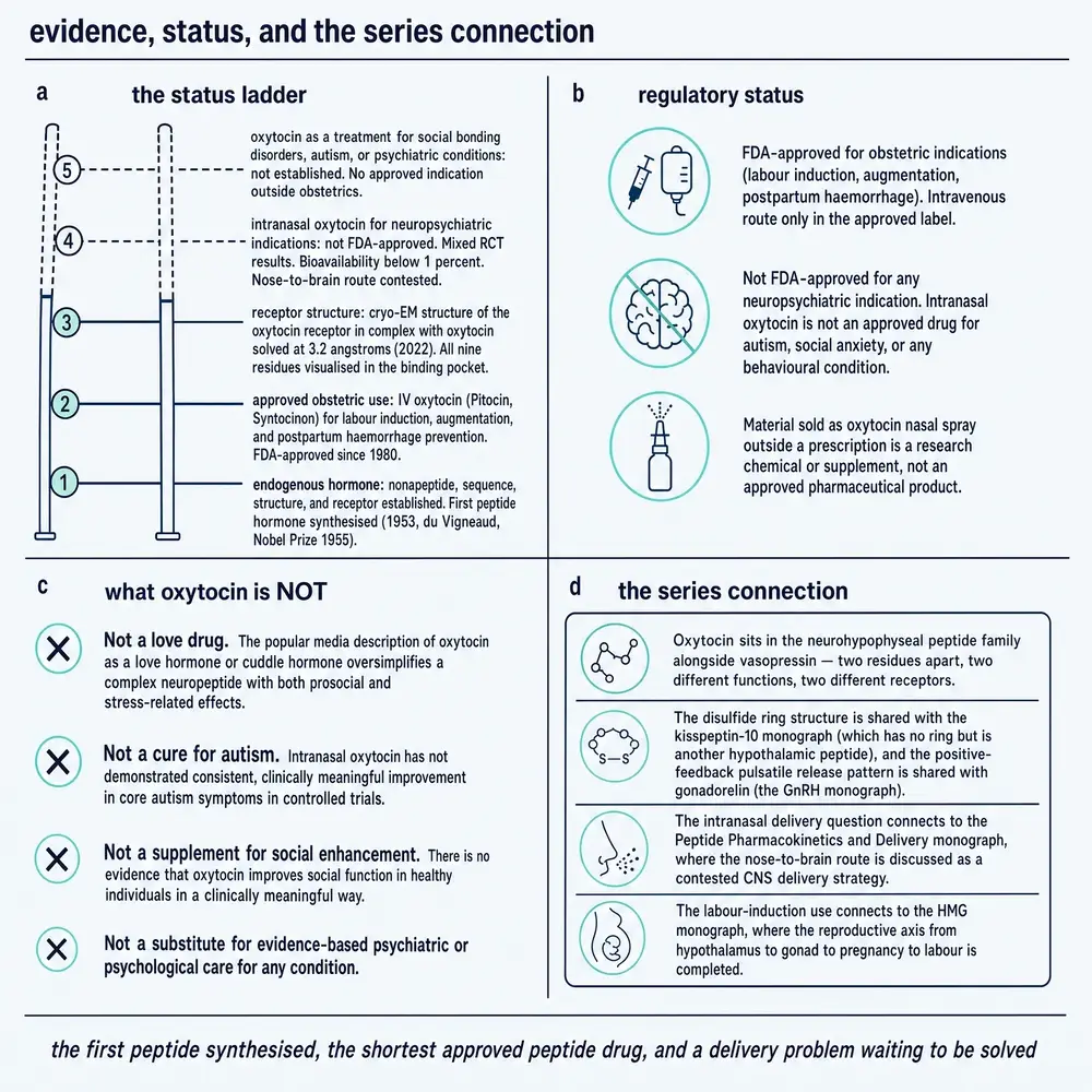
Cumulative intrapartum dose has not been causally separated from labour difficulty. Three cohorts agree on the direction and none can rule out confounding by indication, and only a randomised comparison of regimens would settle it.
There is no quantified incidence of oxytocin-associated hyponatraemia in this corpus, although the mechanism is well understood.
There is no small-molecule full agonist of the oxytocin receptor. The active-state structural paper says so, and until one exists the field cannot separate the peptide's delivery problem from its pharmacology (Meyerowitz et al., 2022).
Set against that list, what the molecule is remains remarkable. Nine amino acids, two of them the only difference from a hormone with an unrelated job. The first hormone whose structure was proved by making it, and therefore the origin of every peptide drug since. A reference-standard obstetric medicine whose regimen is unresolved after a century of use. An animal literature of real depth, a human behavioural literature that has largely failed to reproduce, and a receptor whose atomic structure — magnesium, cholesterol and all — was only solved in the last five years. Whether the last of those eventually rescues the second is the open question, and nothing in this corpus answers it.
The corpus in question is described in full in Section 26. Its shape is worth one last look, because the number that matters is not how much was collected but how much survived being read.
This monograph describes published research. It does not recommend human use of oxytocin or of any related compound, and it specifies no dose, route or schedule for any person. The quantities reported in it are parameters of registered trials, exposures recorded in observational studies, or the content of a public health guideline, and each is given with its population and duration attached. Nothing in this document is medical advice, and decisions about the clinical use of an approved medicine belong to a patient and their clinician.
Section 25References
Every entry below was resolved against the National Library of Medicine's records during this build and read back against its author, journal, year and title line. None was written from recall. The build refuses to run if any identifier fails to resolve, and a separate gate parses every author–year citation out of the prose above and asserts it against these records, so a citation that is correctly formatted and attached to the wrong paper is caught rather than printed.
Of the 71 records, 60 were read as open-access full text and 11 have no PubMed Central identifier and were read at abstract level with their metadata verified. The second group is named in Section 27.
- Atila C, Refardt J, Christ-Crain M. Arginine Vasopressin Deficiency and Oxytocin Deficiency in the Endocrine Clinic. J Clin Endocrinol Metab. 2026;111(4):922-937.
PMID 41363127 · doi:10.1210/clinem/dgaf651 · PMC13017940 - Barrett CE, Arambula SE, Young LJ. The oxytocin system promotes resilience to the effects of neonatal isolation on adult social attachment in female prairie voles. Transl Psychiatry. 2015;5(7):e606.
PMID 26196439 · doi:10.1038/tp.2015.73 · PMC5068726 - Bonnieux J, Gumuchian ST, Harboun A, Trespalacios F, Bélisle KA, Haddad A, et al.. Does intranasal oxytocin reduce symptoms of mental disorders? A meta-analysis of clinical trials. Neurosci Biobehav Rev. 2026;187:106749.
PMID 42134427 · doi:10.1016/j.neubiorev.2026.106749 - Breuil V, Trojani MC, Ez-Zoubir A. Oxytocin and Bone: Review and Perspectives. Int J Mol Sci. 2021;22(16).
PMID 34445256 · doi:10.3390/ijms22168551 · PMC8395200 - Busłowicz JT, Brüggmann D, Al Naimi A, Hoock SC, Deuster E, Schaarschmidt W, et al.. Short-term maternal outcomes after intraoperative administration of prophylactic oxytocin during cesarean sections: a retrospective cohort study with a comparison of different administration protocols. Arch Gynecol Obstet. 2026;313(1).
PMID 41902958 · doi:10.1007/s00404-026-08391-6 · PMC13032929 - Bürkner PC, Williams DR, Simmons TC, Woolley JD. Intranasal Oxytocin May Improve High-Level Social Cognition in Schizophrenia, But Not Social Cognition or Neurocognition in General: A Multilevel Bayesian Meta-analysis. Schizophr Bull. 2017;43(6):1291-1303.
PMID 28586471 · doi:10.1093/schbul/sbx053 · PMC5737621 - Camerino C. The Long Way of Oxytocin from the Uterus to the Heart in 70 Years from Its Discovery. Int J Mol Sci. 2023;24(3).
PMID 36768879 · doi:10.3390/ijms24032556 · PMC9916674 - Carter CS, Kingsbury MA. Oxytocin and oxygen: the evolution of a solution to the 'stress of life'. Philos Trans R Soc Lond B Biol Sci. 2022;377(1858):20210054.
PMID 35856299 · doi:10.1098/rstb.2021.0054 · PMC9272143 - Carter CS, Kenkel WM, MacLean EL, Wilson SR, Perkeybile AM, Yee JR, et al.. Is Oxytocin "Nature's Medicine"?. Pharmacol Rev. 2020;72(4):829-861.
PMID 32912963 · doi:10.1124/pr.120.019398 · PMC7495339 - Chen K, Ye Y, Troje NF, Zhou W. Oxytocin modulates human chemosensory decoding of sex in a dose-dependent manner. Elife. 2021;10.
PMID 33439831 · doi:10.7554/eLife.59376 · PMC7806258 - Chrisman AN, Sugimoto C, Butler-Struben H, Minie VA, Eagle AL, Anderson D, et al.. Oxytocin neurons in the anterior and posterior paraventricular nucleus have distinct behavioral functions and electrophysiological profiles. Neuropsychopharmacology. 2026;51(5):946-955.
PMID 41548026 · doi:10.1038/s41386-026-02352-y · PMC13003577 - Coleman KKL, Berry S, Cummings J, Hsiung GR, Laforce R, Huey E, et al.. Intranasal oxytocin for apathy in people with frontotemporal dementia (FOXY): a multicentre, randomised, double-blind, placebo-controlled, adaptive, crossover, phase 2a/2b superiority trial. Lancet Neurol. 2025;24(2):128-139.
PMID 39862881 · doi:10.1016/S1474-4422(24)00456-3 - Colonnello V, Capellari S, Guarino M, Parchi P, Sambati L, De Matteis M, et al.. Oxytocin reduces anger bias, harm-intention recognition, and self-focus in behavioral variant frontotemporal dementia: a randomized double-blind placebo-controlled crossover trial. Front Aging Neurosci. 2026;18:1735220.
PMID 41658013 · doi:10.3389/fnagi.2026.1735220 · PMC12872833 - Constanthin PE, Isidor N, De Seigneux S, Momjian S. Urinary oxytocin secretion after pituitary surgery, early arginine vasopressin deficiency and syndrome of inappropriate antidiuresis. Endocrine. 2025;88(1):262-272.
PMID 39681826 · doi:10.1007/s12020-024-04131-5 · PMC11933140 - Declerck CH, Boone C, Pauwels L, Vogt B, Fehr E. A registered replication study on oxytocin and trust. Nat Hum Behav. 2020;4(6):646-655.
PMID 32514040 · doi:10.1038/s41562-020-0878-x - Dose F, Zanon P, Coslovich T, Taccola G. Nanomolar oxytocin synergizes with weak electrical afferent stimulation to activate the locomotor CpG of the rat spinal cord in vitro. PLoS One. 2014;9(3):e92967.
PMID 24658101 · doi:10.1371/journal.pone.0092967 · PMC3962494 - DU VIGNEAUD V. Oxytocin, the principal oxytocic hormone of the posterior pituitary gland: its isolation, structure, and synthesis. Experientia. 1955:9-26.
PMID 13305558 - DU VIGNEAUD V. Trail of sulfur research: from insulin to oxytocin. Science. 1956;123(3205):967-74.
PMID 13324123 · doi:10.1126/science.123.3205.967 - Dölen G, Darvishzadeh A, Huang KW, Malenka RC. Social reward requires coordinated activity of nucleus accumbens oxytocin and serotonin. Nature. 2013;501(7466):179-84.
PMID 24025838 · doi:10.1038/nature12518 · PMC4091761 - Fitzek MP, Kleist P, Handtmann C, Overeem LH, Hoehne CL, Lange KS, et al.. Oxytocin receptor antagonism in migraine: a randomized, double-blind, placebo-controlled provocation study. J Headache Pain. 2026;27(1).
PMID 41699456 · doi:10.1186/s10194-026-02297-z · PMC12927216 - Gallos ID, Yunas I, Devall AJ, Podesek M, Tobias A, Price MJ, et al.. Uterotonic agents for preventing postpartum haemorrhage: a network meta-analysis. Cochrane Database Syst Rev. 2025;4(4):CD011689.
PMID 40237648 · doi:10.1002/14651858.CD011689.pub4 · PMC12002006 - Grinevich V, Neumann ID. Brain oxytocin: how puzzle stones from animal studies translate into psychiatry. Mol Psychiatry. 2021;26(1):265-279.
PMID 32514104 · doi:10.1038/s41380-020-0802-9 · PMC7278240 - Gu P, Lin Y, Wan Q, Su D, Shu Q. Oxytocin signal contributes to the adaptative growth of islets during gestation. Endocr Connect. 2021;10(7):694-706.
PMID 34077390 · doi:10.1530/EC-21-0043 · PMC8240721 - Higashida H, Furuhara K, Lopatina O, Gerasimenko M, Hori O, Hattori T, et al.. Oxytocin Dynamics in the Body and Brain Regulated by the Receptor for Advanced Glycation End-Products, CD38, CD157, and Nicotinamide Riboside. Front Neurosci. 2022;16:858070.
PMID 35873827 · doi:10.3389/fnins.2022.858070 · PMC9301327 - Horie K, Blumenthal SA, Inoue K, Yada S, Nishimori K, Young LJ. Male, but not female, oxytocin receptor knockout prairie voles (Microtus ochrogaster) show impaired consolation behavior. Horm Behav. 2025;169:105708.
PMID 39965529 · doi:10.1016/j.yhbeh.2025.105708 - Insel TR, Shapiro LE. Oxytocin receptor distribution reflects social organization in monogamous and polygamous voles. Proc Natl Acad Sci U S A. 1992;89(13):5981-5.
PMID 1321430 · doi:10.1073/pnas.89.13.5981 · PMC402122 - Jung D, Han HB, Kim J, Kim JH, Froemke RC, Choi JH. Oxytocin selectively biases sensory-prefrontal communication through network-level suppression and theta coupling. Proc Natl Acad Sci U S A. 2026;123(24):e2603966123.
PMID 42263121 · doi:10.1073/pnas.2603966123 · PMC13273337 - Kang H, Deilhaug E, Walle KM, Sartorius AI, Quintana DS. A multiverse meta-analysis of oxytocin administration studies. Biol Psychol. 2025;201:109112.
PMID 40848964 · doi:10.1016/j.biopsycho.2025.109112 - Kosfeld M, Heinrichs M, Zak PJ, Fischbacher U, Fehr E. Oxytocin increases trust in humans. Nature. 2005;435(7042):673-6.
PMID 15931222 · doi:10.1038/nature03701 - Kroll CF, Schruers KRJ, Viechtbauer W, Vingerhoets C, Seidel L, Riedl A, et al.. Absence of a meaningful effect of intranasal oxytocin on trusting behavior: a registered report with pooled equivalence testing. Cortex. 2026;198:208-233.
PMID 41880977 · doi:10.1016/j.cortex.2026.03.006 - Lee SN, Kraska J, Papargiris M, Teng L, Niranjan B, Hammar J, et al.. Oxytocin receptor antagonists as a novel pharmacological agent for reducing smooth muscle tone in the human prostate. Sci Rep. 2021;11(1):6352.
PMID 33737570 · doi:10.1038/s41598-021-85439-4 · PMC7973579 - Leng G, Russell JA. The Peptide Oxytocin Antagonist F-792, When Given Systemically, Does Not Act Centrally in Lactating Rats. J Neuroendocrinol. 2016;28(4):n/a.
PMID 26497634 · doi:10.1111/jne.12331 · PMC4982133 - Leng G, Ludwig M. Intranasal Oxytocin: Myths and Delusions. Biol Psychiatry. 2016;79(3):243-50.
PMID 26049207 · doi:10.1016/j.biopsych.2015.05.003 - Liang Y, Liang Z, Zhong J, Yue R, Jiang X. Dose-response relationship between oxytocin exposure during labor induction and neonatal hyperbilirubinemia. Front Pediatr. 2025;13:1714769.
PMID 41357803 · doi:10.3389/fped.2025.1714769 · PMC12678289 - Marlin BJ, Mitre M, D'amour JA, Chao MV, Froemke RC. Oxytocin enables maternal behaviour by balancing cortical inhibition. Nature. 2015;520(7548):499-504.
PMID 25874674 · doi:10.1038/nature14402 · PMC4409554 - Meyerowitz JG, Robertson MJ, Barros-Álvarez X, Panova O, Nwokonko RM, Gao Y, et al.. The oxytocin signaling complex reveals a molecular switch for cation dependence. Nat Struct Mol Biol. 2022;29(3):274-281.
PMID 35241813 · doi:10.1038/s41594-022-00728-4 · PMC12101619 - Minder JL, Winokur SB, Stephens J, Tong J, Cassel NL, Schuster L, et al.. Oxytocin induces embryonic diapause. Sci Adv. 2025;11(10):eadt1763.
PMID 40043121 · doi:10.1126/sciadv.adt1763 · PMC11881891 - Nagasawa M, Mitsui S, En S, Ohtani N, Ohta M, Sakuma Y, et al.. Social evolution. Oxytocin-gaze positive loop and the coevolution of human-dog bonds. Science. 2015;348(6232):333-6.
PMID 25883356 · doi:10.1126/science.1261022 - Naruse S, Akinaga C, Mazda Y, Nakajima Y. Intramyometrial injection versus intravenous infusion of oxytocin for maintaining uterine contractility during elective caesarean delivery in a randomised controlled trial. Sci Rep. 2026;16(1).
PMID 41917148 · doi:10.1038/s41598-026-46727-z · PMC13186971 - Ocampo Daza D, Bergqvist CA, Larhammar D. The Evolution of Oxytocin and Vasotocin Receptor Genes in Jawed Vertebrates: A Clear Case for Gene Duplications Through Ancestral Whole-Genome Duplications. Front Endocrinol (Lausanne). 2021;12:792644.
PMID 35185783 · doi:10.3389/fendo.2021.792644 · PMC8851675 - Ohlsson B, Björgell O, Ekberg O, Darwiche G. The oxytocin/vasopressin receptor antagonist atosiban delays the gastric emptying of a semisolid meal compared to saline in human. BMC Gastroenterol. 2006;6:11.
PMID 16542457 · doi:10.1186/1471-230X-6-11 · PMC1434749 - Ononge S, Kakaire O, Mwembezi J, Nakatudde H, Mutumba R, Mugahi R. Cost-effectiveness results comparing heat-stable carbetocin & other uterotonics in postpartum heamorrhage prevention in Uganda. PLOS Glob Public Health. 2025;5(4):e0003562.
PMID 40261858 · doi:10.1371/journal.pgph.0003562 · PMC12013952 - Passoni I, Leonzino M, Gigliucci V, Chini B, Busnelli M. Carbetocin is a Functional Selective Gq Agonist That Does Not Promote Oxytocin Receptor Recycling After Inducing β-Arrestin-Independent Internalisation. J Neuroendocrinol. 2016;28(4):n/a.
PMID 26751410 · doi:10.1111/jne.12363 · PMC5021139 - Payrastre C, Debost-Legrand A, Roussel L, Accoceberry M, Rouzaire M, Gallot D. Association between total oxytocin dose and maternal and neonatal morbidity during labour induction: a retrospective study. BMC Pregnancy Childbirth. 2025;25(1):857.
PMID 40817207 · doi:10.1186/s12884-025-08003-2 · PMC12355890 - Perkeybile AM, Kenkel WM, Yee JR, Waugh RE, Lillard TS, Ferris CF, et al.. Oxytocin treatment at birth accelerates an epigenetic shift in the oxytocin receptor gene in the maternal brain. BMC Pregnancy Childbirth. 2025;25(1):777.
PMID 40684112 · doi:10.1186/s12884-025-07868-7 · PMC12275451 - Prifti KK, Dave RM, Mitchum KT, Rich JL, Gill RM, Mbadhi MN, et al.. Energy substrate preference and metabolic flexibility in quiescent and oxytocin-stimulated human myometrial cells. iScience. 2026;29(6):116248.
PMID 42317739 · doi:10.1016/j.isci.2026.116248 · PMC13273498 - Quintana DS, Rokicki J, van der Meer D, Alnæs D, Kaufmann T, Córdova-Palomera A, et al.. Oxytocin pathway gene networks in the human brain. Nat Commun. 2019;10(1):668.
PMID 30737392 · doi:10.1038/s41467-019-08503-8 · PMC6368605 - Quintana DS, Lischke A, Grace S, Scheele D, Ma Y, Becker B. Advances in the field of intranasal oxytocin research: lessons learned and future directions for clinical research. Mol Psychiatry. 2021;26(1):80-91.
PMID 32807845 · doi:10.1038/s41380-020-00864-7 · PMC7815514 - Roy RK, Augustine RA, Brown CH, Schwenke DO. Activation of oxytocin neurons in the paraventricular nucleus drives cardiac sympathetic nerve activation following myocardial infarction in rats. Commun Biol. 2018;1:160.
PMID 30320228 · doi:10.1038/s42003-018-0169-5 · PMC6172223 - Rung JM, Horta M, Tammi EM, Perez E, Ojeda MC, Lin T, et al.. Safety and tolerability of chronic intranasal oxytocin in older men: results from a randomized controlled trial. Psychopharmacology (Berl). 2021;238(9):2405-2418.
PMID 33982141 · doi:10.1007/s00213-021-05862-3 · PMC8115997 - Ryu V, Gumerova AA, Pevnev G, Korkmaz F, Kannangara H, Cullen L, et al.. Single transcript level atlas of oxytocin and the oxytocin receptor in the mouse brain. Elife. 2026;15.
PMID 41729202 · doi:10.7554/eLife.95215 · PMC13008351 - Sabe M, Zhao N, Crippa A, Strauss GP, Kaiser S. Intranasal Oxytocin for Negative Symptoms of Schizophrenia: Systematic Review, Meta-Analysis, and Dose-Response Meta-Analysis of Randomized Controlled Trials. Int J Neuropsychopharmacol. 2021;24(8):601-614.
PMID 33890987 · doi:10.1093/ijnp/pyab020 · PMC8378078 - Samimi M, Hosseiniara R, Akbari H, Beyrami D. Carbetocin versus oxytocin with or without tranexamic acid for preventing postpartum hemorrhage in cesarean delivery: a randomized controlled trial. BMC Pregnancy Childbirth. 2026;26(1):183.
PMID 41593544 · doi:10.1186/s12884-026-08674-5 · PMC12918010 - Sawares A, Olver J, Morcos M, Norman T. Oxytocin in old age psychiatry: A systematic review of the safety of using intranasal oxytocin in older adults. Australas Psychiatry. 2025;33(1):33-44.
PMID 39474844 · doi:10.1177/10398562241291335 · PMC11804147 - Shafer SL, Ririe DG, Miller S, Curry RS, Hsu DT, Sullivan GM, et al.. Plasma pharmacokinetics of intravenous and intranasal oxytocin in nonpregnant adults. Br J Anaesth. 2025;134(5):1513-1522.
PMID 40121179 · doi:10.1016/j.bja.2024.12.046 · PMC12106900 - Sikich L, Kolevzon A, King BH, McDougle CJ, Sanders KB, Kim SJ, et al.. Intranasal Oxytocin in Children and Adolescents with Autism Spectrum Disorder. N Engl J Med. 2021;385(16):1462-1473.
PMID 34644471 · doi:10.1056/NEJMoa2103583 · PMC9701092 - Stämpfli D, Dommrich R, Orbach-Zinger S, Burden AM, Heesen M. An explorative analysis of pharmacovigilance data of oxytocin and its analogue carbetocin, with a focus on haemodynamic adverse effects. Int J Clin Pharm. 2023;45(4):922-928.
PMID 37162656 · doi:10.1007/s11096-023-01587-9 · PMC10366022 - Theofanopoulou C, Andirkó A, Boeckx C, Jarvis ED. Oxytocin and vasotocin receptor variation and the evolution of human prosociality. Compr Psychoneuroendocrinol. 2022;11:100139.
PMID 35757177 · doi:10.1016/j.cpnec.2022.100139 · PMC9227999 - Tiouririne NA, Legan JA, Devine E, Lee K, Weerts EM, Ellis JD, et al.. Intranasal Oxytocin for Alcohol Use Disorder: A Randomized, Double-Blind, Placebo-Controlled Multisite Trial Assessing Efficacy and Safety. Alcohol Clin Exp Res (Hoboken). 2026;50(7):e70326.
PMID 42458234 · doi:10.1111/acer.70326 · PMC13373018 - Uvnäs-Moberg K, Ekström-Bergström A, Berg M, Buckley S, Pajalic Z, Hadjigeorgiou E, et al.. Maternal plasma levels of oxytocin during physiological childbirth - a systematic review with implications for uterine contractions and central actions of oxytocin. BMC Pregnancy Childbirth. 2019;19(1):285.
PMID 31399062 · doi:10.1186/s12884-019-2365-9 · PMC6688382 - Van-Quynh Duong T, Yaw AM, Zhou G, Sinha N, Cherukuri AS, Nguyen D, et al.. Interaction between time-of-day and oxytocin efficacy in mice and humans with and without gestational diabetes. Mol Metab. 2026;103:102269.
PMID 41109427 · doi:10.1016/j.molmet.2025.102269 · PMC12902303 - Waltenspühl Y, Schöppe J, Ehrenmann J, Kummer L, Plückthun A. Crystal structure of the human oxytocin receptor. Sci Adv. 2020;6(29):eabb5419.
PMID 32832646 · doi:10.1126/sciadv.abb5419 · PMC7439316 - Wang Y, Wang Y, Cao B, Li X, Ding Y, Meng X, et al.. Long-term intranasal oxytocin therapy in patients with hypothalamic syndrome: case series and literature review. Endocr Connect. 2025;14(10).
PMID 41091101 · doi:10.1530/EC-25-0092 · PMC12583497 - Warfvinge K, Edvinsson JCA, Maddahi A, Edvinsson L. Hypothalamic and sex-related hormones in migraine. J Headache Pain. 2026;27(1).
PMID 41680623 · doi:10.1186/s10194-026-02289-z · PMC12927229 - Winterdahl M, Nielsen EN, Hansen SB, Dias AH, Vendelbo MH, Jakobsen S, et al.. First-in-human intranasal [13N]oxytocin PET: evaluation of feasibility, biodistribution, and radiation dosimetry. EJNMMI Res. 2025;15(1):137.
PMID 41251983 · doi:10.1186/s13550-025-01329-0 · PMC12627303 - Xiao Y, Shen Q, Lin Y, Wen Z, Gao Y. Midwives' experiences in the clinical management of oxytocin infusion for labor induction and augmentation: a qualitative study. BMC Nurs. 2025;24(1):909.
PMID 40646480 · doi:10.1186/s12912-025-03556-1 · PMC12255094 - Xu H, Li Y, Cui X, Zhang Y, Wu X, Zhuo SS, et al.. Oxytocin attenuates fear learning via enhancing somatostatin interneuron-mediated local GABAergic inhibition in the prelimbic cortex. Sci Adv. 2026;12(24):eaef8400.
PMID 42284412 · doi:10.1126/sciadv.aef8400 · PMC13262635 - Yamaguchi T, Yan R, Khan M, Kuno S, Tewatia K, Osakada T, et al.. The neural mechanisms supporting the rise and fall of maternal aggression. Nature. 2026;654(8120):1012-1022.
PMID 41986710 · doi:10.1038/s41586-026-10354-5 · PMC13135462 - Yamamuro K, Ikehara M, Noriyama Y, Okuda M, Okumura K, Matsuoka K, et al.. Oxytocin receptor neurons in the paraventricular thalamus as a nexus for social behaviour and fear. Brain. 2026;149(6):2151-2165.
PMID 41861062 · doi:10.1093/brain/awaf421 · PMC13233147 - Zagrodnik E, Szczuko M, Kordek A, Surówka A, Szydłowska I, Rzewuska B, et al.. Adverse Effects of Oxytocin Are More Prevalent than Those Associated with Carbetocin Administration During Cesarean Section. J Clin Med. 2025;14(20).
PMID 41156081 · doi:10.3390/jcm14207211 · PMC12565578 - Özdemir M, Çeber Turfan E, Keskin HL. Oxytocin administration during labor does not affect maternal serum oxytocin levels, early postpartum breastfeeding attitude and maternal attachment. PLoS One. 2025;20(12):e0337762.
PMID 41348825 · doi:10.1371/journal.pone.0337762 · PMC12680139
Sources with no PubMed record. Guideline, database and structural-archive material is listed separately so that the numbered list above stays wholly machine-verified.
- World Health Organization. WHO recommendations on maternal and newborn care for a positive postnatal experience, and the consolidated recommendations on the prevention and treatment of postpartum haemorrhage. Guidelines Review Committee. The 10 IU intramuscular or intravenous prophylactic recommendation quoted in Section 12, the stated preference for slow intravenous over intramuscular administration where intravenous access already exists, the caesarean-section recommendation, and the Guideline Development Group's own remarks on injection speed, dilution volume and the absence of direct comparative evidence, were read from the guideline text held in this project's corpus rather than from a summary of it.
https://www.who.int/publications - UniProt Consortium. P01178 (OXT_HUMAN), oxytocin-neurophysin 1 preprohormone; and P30559 (OXYR_HUMAN), oxytocin receptor. The gene symbols OXT and OXTR, the prepropeptide architecture and the receptor's accession are taken from these records. Sequence, mass and disulfide connectivity quoted in Sections 01 and 04 come from the cited literature, not from the database.
https://www.uniprot.org/uniprotkb/P01178/entry - Protein Data Bank. Coordinate depositions accompanying the two oxytocin receptor structures cited in this document. The retosiban-bound inactive-state crystal structure and the oxytocin-bound active-state cryo-EM complex were deposited with the papers cited as Waltenspuhl et al. 2020 and Meyerowitz et al. 2022. Resolutions, residue contacts, mutagenesis costs and the cholesterol site quoted in Sections 05 and 06 are as published in those papers; no coordinate file was opened and no accession code is asserted here, because none was verified against the archive during this build.
https://www.rcsb.org/
Section 26How this document was assembled
The corpus was built against project 05, the Therapeutic Peptide Research Library, and against PubMed and PubMed Central. As with every compound in this series the interesting arithmetic is not what was collected but what had to be refused — and this molecule presents the identity problem in an unusually mild form and the volume problem in the most severe form the series has met.
The identity problem, and why it is small here
“Oxytocin” is not a word with another meaning, not a gene symbol belonging to something else, and not an abbreviation. It is an unambiguous designation, and the disqualify lists that have done the heavy lifting in this pipeline since No. 22 are almost empty for it. Three things still needed configuring.
Superstring analogues. Demoxytocin, desamino-oxytocin and carba-oxytocin all contain the subject's name as a substring. A naive matcher admits a paper about any of them as a paper about oxytocin. They are stripped before matching rather than counted.
The siblings, and why they refuse only by a margin. Carbetocin and atosiban are the analogue and the antagonist, and both have literatures of their own. Strict dominance — a named relative mentioned at least as often as the subject takes the document back — would have been too aggressive here, because a paper genuinely about both names both. The rule used is a threefold margin, and it refused 47 documents: 35 dominated by atosiban and 12 by carbetocin. 0 were refused for any other reason. That is 47 refusals out of 1,041 string matches, which is a low rate and the correct one for a name this clean.
Vasopressin is context, not a rival. This is the consequential setting. Oxytocin and vasopressin differ at two of nine positions, are released from the same gland, are measured by assays that cross-react, and are discussed together in a large fraction of the honest literature about either. Wiring vasopressin in as a competing compound would have refused a great many genuine documents on the strength of a comparison the papers themselves are making. It is counted and reported, and it never refuses a document. Desmopressin is treated the same way.
The matcher was break-tested before the first sweep rather than after, and the identity gate ran ahead of every other screen, per A9.
The local store
Every file with a document extension in the project's stores was opened — 45,975 of them — and its extracted text searched. 1,041 contained a designation, 47 were refused as described above, and 994 were admitted as being about oxytocin.
Those were then classified by what kind of document they are, because a corpus figure that silently includes vendor pages is a claim about coverage the document does not have. The result is 677 peer-reviewed full texts against 317 commercial and derived documents. That ratio is unusual for this series and it has an obvious cause: oxytocin is a century-old licensed medicine on the World Health Organization's essential medicines list, not a research-use compound sold on the open market, so its footprint is overwhelmingly scientific rather than commercial.
The external harvest, and the volume problem
The PubMed query returned 29,748 records, of which 28,611 passed record-level relevance. That is the largest external surface any compound in this series has produced, by a wide margin, and it is a fact about the molecule rather than about the query: oxytocin has been studied continuously since 1895 and is used in obstetrics worldwide.
A separate full-text sweep of PubMed Central returned 11,580 body-text matches. A17 moves the substantive-use screen in front of the fetch where a surface is too large to retrieve, and this surface is fourteen times the 800-document threshold, so the front screen was applied: a title-and-abstract-scoped query reduced 11,580 body-text matches to 4,514 plausible targets, and 8,773 documents were counted as surface and never read. Counting them as corpus would have been the easy overstatement, and the distinction between a surface and a corpus is the reason this section exists.
The two routes were then merged by key rather than added. Of the indexed records, 7,498 carry a PubMed Central identifier and are therefore retrievable; the body-text route contributed 4,514. Their naive sum is 12,012 and their keyed union is 7,657, so 4,355 documents would have been counted twice by an addition that looks arithmetically innocent. From that union, 817 full texts were retrieved under a per-route budget and screened on the far side, where nothing is counted without being read.
| Far-side screen | Documents | Enters the corpus |
|---|---|---|
| Substantive discussion of the compound | 224 | yes |
| Passing the substantive-mention threshold | 66 | yes |
| Named as a tool or comparator only | 447 | no |
| Not about this compound | 10 | no |
| Refused by the identity gate | 11 | no |
| No retrievable body text | 59 | no |
The largest cell in that table is the one that does not enter the corpus. 447 fetched documents name oxytocin only as a laboratory tool or a comparator — atosiban used to block a receptor in an experiment about something else, oxytocin used to contract a tissue in an assay about something else. Admitting them would have doubled the corpus figure and improved nothing.
The reading corpus is 1,256 unique scientific full texts, about 30,657 printed-page equivalents. That figure is a keyed union of the local store and the external harvest, not a sum: 89 documents were reachable by both routes and are counted once.
Retractions
Every indexed record in the harvest — 28,613 of them — was queried against the National Library of Medicine for retraction status and for expressions of concern. 43 are retracted and 12 carry an expression of concern. That is a rate of roughly two in a thousand, and it is what a surface this size should be expected to contain; the clustering is informative, because most of the retracted work is obstetric trial literature comparing uterotonic agents, which is a field with a documented integrity problem of its own.
Six of the flagged records were among the 817 documents this build actually fetched and read: five retracted trials of misoprostol, carboprost or tranexamic acid against oxytocin, and one paper under an expression of concern. None of the 71 cited records is retracted or flagged, and no claim in this document rests on a withdrawn paper. The six are counted in the corpus figure because they were genuinely read, and they are named here rather than quietly removed, which would change a number without changing what happened.
The gap this document cannot close
11 of the 71 cited records have no PubMed Central identifier and were not read as open-access full text by this pipeline: du Vigneaud's 1955 and 1956 papers, Kosfeld's 2005 trust experiment, Nagasawa's 2015 dog-gaze study, Leng and Ludwig's 2016 critique, Declerck's 2020 replication, Coleman's 2025 dementia trial, Horie's 2025 knockout voles, Kang's 2025 multiverse analysis, Bonnieux's 2026 meta-analysis and Kroll's 2026 registered report. Their identifiers, authors, journals, years and titles are verified against the National Library of Medicine and their abstracts were read. Where a number from one of them appears in this document — the apathy estimate, the pooled effect sizes, the multiverse range — it is a number the abstract itself states, and it is labelled here rather than allowed to hide behind the corpus figure.
Two of those eleven are load-bearing for Part Four's argument, and that should be said plainly: the trust replications and the clinical meta-analysis are the records on which the recency judgement in Section 19 and the pooled estimate in Section 20 rest. Both are recent, neither is open access, and both are cited from verified indexed records with their abstracts read in full.
| Stage | What it did | Result |
|---|---|---|
| 00b | break-test the identity matcher before the first sweep | siblings by margin, vasopressin as context |
| 01b – 01g | sweep the local stores, gate, classify by source kind | 994 admitted, 677 peer-reviewed |
| 02 – 02b | PubMed and PubMed Central surfaces, front-screened under the retrieval ceiling | 29,748 and 11,580 |
| 02c – 02d | select targets and merge by key, not by sum | keyed union 7,657, 4,355 double-counts avoided |
| 02e | query every indexed record for retraction and concern status | 43 retracted, 12 flagged, none cited |
| 03 – 03c | fetch and screen on the far side | 817 fetched, 290 kept |
| 05 – 05c | resolve every reference against NCBI; inject figures and captions | 71 references, 19 figures |
| 05b | assert every author–year citation in the prose against the resolved records | run before assembly |
| 06 – 07c | assemble, number figures, render both editions, stamp running furniture | 19 figures |
| 11 – 15 | density, margins, artwork, contrast and format gates | run against both editions |
What this build found in its own pipeline
This series records, repeatedly, that a fix written into its standard is not the same as a fix present in the code. Three defects were found and repaired during this build, and all three would have produced a plausible document rather than an error.
The reference stage reused the harvest record wherever one existed. The harvest stores a journal's full title, because that is what the recency screen needs; house style section 5 requires the ISO abbreviation, and the reference formatter reads that field. Every one of the 71 references would have printed with an expanded journal name, and no gate could have seen it, because both strings are true. The stage now re-fetches every cited record from the National Library of Medicine rather than preferring a cached copy.
An authored figure carried an invented confidence interval. The uterotonic comparison in Section 11 originally drew carbetocin with a plotted interval, because every other row had one and the drawing looked wrong without it. The review does not publish an interval for that comparison; it publishes a verdict. The bar was removed and the verdict printed as words, which is section 4's rule that a caption must say what a figure is not.
The evidence-tier figure hand-typed its own total. It now derives every bar from a per-record classification and asserts the sum against the number of references the build actually resolved, so adding a citation without classifying it breaks the figure instead of quietly producing a chart that no longer adds up.
Section 27Evidence handling
Study type is named in the sentence that reports the finding. A result in a vole is called a result in a vole. For this compound that labelling carries more weight than usual, because the popular understanding of oxytocin rests substantially on rodent work and the human work that followed has not reproduced all of it. Where an animal result is quoted, the species is named; where a human result is quoted, the design, the size and the duration are given.
A peripheral measurement is not a central one. This is the standing rule for this compound and it is applied throughout. A plasma, saliva or urine oxytocin concentration is a statement about the periphery. The experiment in Section 14 shows the two compartments can be dissociated pharmacologically, and the pharmacokinetic study in Section 18 shows how little of an intranasal dose reaches the circulation at all. Any inference from one compartment to the other is flagged at the point it is made.
The assay is part of the finding. Nineteen of the twenty studies in the childbirth systematic review used radioimmunoassay, and a 2025 mass-spectrometry study found immunoassay reading systematically lower on the same samples. This document therefore reports the shape of the physiological curve, which is consistent across studies, and declines to print absolute concentrations from immunoassay literature. Two human studies relating circulating oxytocin to metabolic disease, pointing in opposite directions and both using immunoassay, are discussed in Section 22 as an instance of the problem rather than resolved by choosing one.
Conflicting evidence is presented as conflict, and recency is not a tiebreak on its own. Three cases are load-bearing, and they resolve differently. In the trust literature, a newer null does supersede an older positive, and Section 19 says why: the later studies were larger, preregistered, and tested for equivalence rather than only for difference, so the case rests on design and not on date. In the prairie-vole literature, a newer null does not supersede, because constitutive receptor deletion and acute pharmacological blockade answer different questions, and Section 17 states the dissociation rather than declaring a winner. In the clinical meta-analysis literature, two recent syntheses disagree, and Section 21 shows that they are asking different questions and that both answers can hold.
Adverse and null findings sit beside the efficacy figures. The one drug-related serious adverse event in the largest autism trial is reported in the same section as the trial's primary outcome. The dose–harm cohorts appear in Part Three beside the evidence that the drug prevents haemorrhage, not in a later section. The null on intrapartum oxytocin and maternal bonding is reported beside the animal mechanism that motivates the worry.
Confounding by indication is named where it applies. Three observational cohorts agree that higher cumulative intrapartum exposure travels with worse outcomes. In every one of them a large dose is also a marker of a difficult labour, and the authors of the largest say so. The association is reported; the causal claim is not made.
Guideline text is attributed to the guideline and read from it. The recommended quantity, the stated route preference, the caesarean recommendation and the Guideline Development Group's own remarks on injection speed, dilution volume and the absence of comparative evidence were read from the guideline held in this project's corpus rather than from a summary of it. All are reported as guideline parameters and none as a direction for use.
Two records were deliberately not used, and the reason is on the record. An ophthalmology study reporting plasma oxytocin in glaucoma prints two group means that are internally inconsistent with the direction the paper states; the discrepancy could not be resolved from the read text, so nothing in this document comes from it. Two metabolic-disease studies reporting opposite directions by immunoassay are discussed as a disagreement and cited for no value.
Absence is reported as a finding. Section 24 lists seven things this corpus does not contain, including a quantified incidence of oxytocin-associated hyponatraemia and any validated measurement of central oxytocin concentration after an intranasal dose. Each is an absence in what was actually read, not a rhetorical gesture, and each is stated rather than smoothed over.
Continue exploring