
LL-37 The only cathelicidin a human being has, and the trouble with an antibiotic that also talks to the immune system
Every mammal makes its own antibiotics, and most make several: the pig, the cow and the sheep each carry a small arsenal of chemically unrelated ones. Human beings make one. It is thirty-seven amino acids long, it lives coiled up inside a larger protein in the granules of white blood cells, and it is released by being cut in half in the open air. It has never been approved as a medicine anywhere, and after thirty years of work the reason is not that it fails to kill bacteria. It is that killing bacteria may be the least interesting thing it does. The same thirty-seven residues summon blood vessels, package loose DNA, recruit the cells of the immune system, and — in a paper published this year — determine whether an animal with a lung infection dies of a blood clot or of the infection the clot was built to trap. A molecule with that many jobs has no side effects, because it has no single main effect to be beside.
Findings are labelled by the kind of study that produced them, in the sentence that reports them. A result in a mouse is called a result in a mouse. This matters more here than in most monographs in this series, because the mouse version of this peptide is a different molecule with a different sequence, cut by a different enzyme, under the control of a promoter the mouse does not share with us. Where a finding belongs to mouse CRAMP rather than to human LL-37, it says so.
Two further rules apply throughout. Results obtained with an engineered derivative — OP-145, SAAP-148, KR-12 — belong to that derivative and are attributed to it. And a null result obtained with scrambled LL-37, which is this field's standard negative control, is a null about a scrambled peptide and not about this one. No dose, route or schedule for any person is recommended anywhere in this document; where a study's parameters are reported, they are reported as that study's, with its population and duration attached.
Section 01A pig probe, and the wrong clone
In the early 1990s a laboratory at Stockholm University was trying to finish an argument its head had started twelve years earlier. Hans Boman had spent his career on a question that sounds naive and is not: how does an animal without antibodies survive in a world of bacteria? Insects have no adaptive immune system, no memory, nothing to make an antiserum from — and yet a moth that survives an infection is measurably harder to infect afterwards. In 1981 Boman's group isolated the molecules responsible from the cecropia moth and called them cecropins (Steiner et al., 1981). They were small, positively charged peptides that punched holes in bacterial membranes, and they founded a field. The paper that announced them already named the criterion this whole document turns on: cecropin A, the authors noted, is specific for bacteria — in contrast to melittin, the lytic component of bee venom, which destroys bacterial and animal cells alike.
By 1994 that field had a shopping list. Antibacterial peptides had been pulled out of frogs, pigs, cows and rabbits, and the obvious next question was what the human versions were. One candidate looked especially promising. PR-39 is a proline- and arginine-rich peptide purified from pig small intestine and later traced to pig bone marrow. If pigs had it, the reasoning went, we probably had something like it. So Birgitta Agerberth and colleagues built a PCR probe from the PR-39 gene and used it to screen a human bone-marrow cDNA library.
The probe caught eight clones, and not one of them was a human PR-39. They carried the information for a peptide with no proline-arginine repeats at all — a 39-residue sequence, as predicted from the reading frame, that contained no cysteine and no tryptophan, which made it unlike every human antibacterial peptide then known. All the human ones described up to that point were defensins, and every defensin is stapled together by three disulfide bridges. This thing had nothing to staple. They named it after the first four residues of the predicted product: FALL-39 (Agerberth et al., 1995).
Two details from that first paper are worth holding onto, because the rest of this document keeps returning to them. The first is that residues 13 to 34 were predicted to form what the authors called a perfect amphipathic helix — a spiral with all the greasy side chains on one face and all the charged ones on the other. The second is that circular dichroism showed the synthetic peptide was not helical in plain buffer. It only became helical when the medium changed. This molecule does not have a shape so much as it has a shape it adopts when it arrives somewhere.
Section 02The same gene, from the other end, in another country
Six months later, and by a completely different route, a group at Rigshospitalet in Copenhagen arrived at the same molecule. Jack Cowland, Anders Johnsen and Niels Borregaard were not looking for a peptide at all. They were cataloguing the contents of neutrophil granules — the small packets of antimicrobial machinery that a white blood cell carries around and dumps onto whatever it is attacking. Neutrophils have two main kinds. Azurophil granules hold the aggressive enzymes; specific granules hold a different inventory. In the specific granules there was an unassigned protein band at about 19 kilodaltons.
They cut it up, sequenced the fragments, pulled a full-length cDNA from a leukaemia library, and found that their clone included the sequence that Stockholm had published as FALL-39. They named the protein hCAP-18, for human cationic antimicrobial protein of 18 kDa — a name that has been slightly wrong from the day it was coined, since the sequence gives 19,301 daltons and their own gel said 19 (Cowland et al., 1995).
What Copenhagen added was the crucial structural observation. The protein had two halves that behaved completely differently. The N-terminal region was conserved — it matched sequences already known from pig and cow proteins — and the C-terminal region was wildly variable between species. In October of the same year, Margherita Zanetti, Renato Gennaro and Domenico Romeo in Trieste gave that arrangement a name. A protein with a conserved pro-region resembling the cystatin family of protease inhibitors, attached to a variable antimicrobial tail, was a cathelicidin (Zanetti et al., 1995).
So within a single year, one human gene acquired three names from three cities: FALL-39 from the DNA, hCAP-18 from the protein, and membership of the cathelicidins from comparative anatomy. None of those names is the one we use.
Section 03The correction that became the name
The 1995 paper had made a prediction. Reading the open frame, and assuming the processing enzyme would cut at a pair of basic residues in the usual way, the mature peptide should be 39 residues long and should start phenylalanine-alanine. That was a reasonable guess and it was wrong.
In 1996 the Stockholm group went and looked. They raised antibodies, located the peptide in granulocytes, degranulated the cells, and isolated whatever the cells actually released. Then they sequenced it. The real product was two residues shorter and began leucine-leucine. Following the convention they had already established — first residues, then length — they called it LL-37 (Gudmundsson et al., 1996).
This is worth pausing on, because it is the only compound in this monograph series whose name commemorates the fixing of a mistake. FALL-39 and LL-37 are not two molecules and not even two hypotheses; they are the same C-terminus with two extra residues on the front. The reference database still carries both as separate features of the same protein — FALL-39 as residues 132 to 170, LL-37 as residues 134 to 170 — which is a small permanent monument to a laboratory that published a prediction, tested it, found it two residues off, and said so.
The same 1996 paper reported something with much larger consequences. Having sequenced the whole gene — a compact thing, 1,963 base pairs, four exons, with the signal sequence and the conserved cathelin region on exons one to three and the entire mature peptide on exon four — the authors went looking for relatives. There were none. Their conclusion, stated plainly, was that this gene is the only member of the cathelin family present in the human genome.
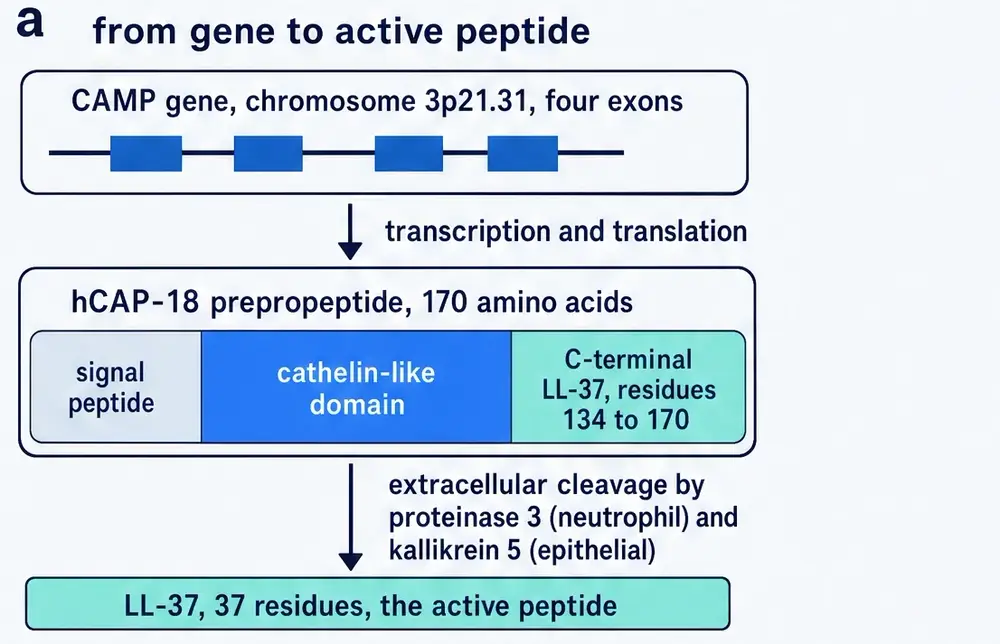
That sentence is easy to read past. It should not be. Every other mammal that has been looked at carefully carries several cathelicidins, and they are not variations on a theme — they are structurally unrelated molecules that happen to share a delivery system. The pig has the proline-rich PR-39 and the disulfide-looped protegrins and the helical PMAPs. Cattle have the helical BMAPs, the proline-rich bactenecins, and indolicidin, which is thirteen residues long and a quarter tryptophan. Sheep have their own set. Each species carries a small arsenal of chemically different weapons behind one conserved pro-region.
What follows presses on that only-member claim until the comparative zoology is visible: one human cathelicidin against families of them in the mammals we usually compare ourselves to.
Section 04What “only one” means
We carry one, and it is helical. There is no proline-rich human cathelicidin and no disulfide-looped one. If a bacterium evolves a way to resist a cationic amphipathic helix — and several have, by remodelling the charge on their membrane surface or by simply secreting a protease — there is no second cathelicidin to fall back on.
This is the fact that makes LL-37 interesting to a physiologist and frustrating to a pharmacologist. It has no redundancy, so evolution has had to load everything onto it: the organism cannot afford a single-copy defence gene that only does one thing. And that is exactly why it is difficult to turn into a drug. Everything in Part Three follows from the compression of many functions into one small molecule, and everything in Part Four follows from the fact that those functions cannot be separated.
Section 05The molecule, counted
LL-37 is LLGDFFRKSKEKIGKEFKRIVQRIKDFLRNLVPRTES. Every number in the table below was derived from that string during the preparation of this document rather than copied from a review, because a sequence is one of the few things in pharmacology that can be checked arithmetically in a few seconds and it is worth doing.
| Property | Value | Consequence |
|---|---|---|
| Length | 37 residues | short enough to synthesise, long enough to span a membrane |
| Molecular formula | C205H340N60O53 | no sulfur — there is no cysteine and no methionine |
| Average mass | 4,493.3 Da | — |
| Basic residues | 11 (6 Lys, 5 Arg) | the charge that finds a bacterial surface |
| Acidic residues | 5 (2 Asp, 3 Glu) | — |
| Net side-chain charge | +6 at neutral pH | strongly cationic; binds anionic lipid and DNA alike |
| Cysteine | none | no disulfide bridge, unlike every human defensin |
| Tryptophan | none | — |
| Proline | one, at position 34 | in the disordered tail, not the helix |
The 1995 paper's title claimed the molecule was cysteine-free; its abstract added that it lacked tryptophan. Both claims are confirmed by counting the letters, which is the cheapest form of peer review available and one this series makes a habit of.
What the composition buys is a molecule that is conditionally structured. In water LL-37 is largely disordered. Put it in contact with an anionic surface — the outer leaflet of a bacterial membrane, a micelle, a stretch of DNA — and the eleven positive charges anchor it while the hydrophobic face folds into a helix and inserts. Nuclear magnetic resonance in lipid micelles resolves this properly: the structure is not a single straight helix but a curved helix–bend–helix running from residue 2 to residue 31, with the bend located between glycine 14 and glutamate 16, followed by a disordered C-terminal tail (Wang, 2008). All four of the peptide's phenylalanines — positions 5, 6, 17 and 27 — make contact with the lipid, along with the arginines, which is direct evidence that the entire amphipathic stretch lies against the membrane rather than dangling from it.
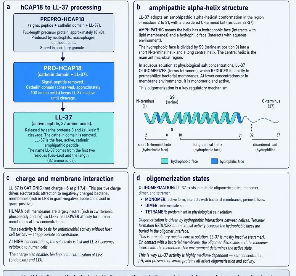
The same study identified the smallest fragment of LL-37 that still kills bacteria: residues 18 to 29, twelve residues beginning lysine-arginine, hence KR-12. That fragment matters more than its size suggests, and Part Five returns to it, because KR-12 was reported to be toxic to bacteria and not to human cells — a separation the parent molecule does not achieve.
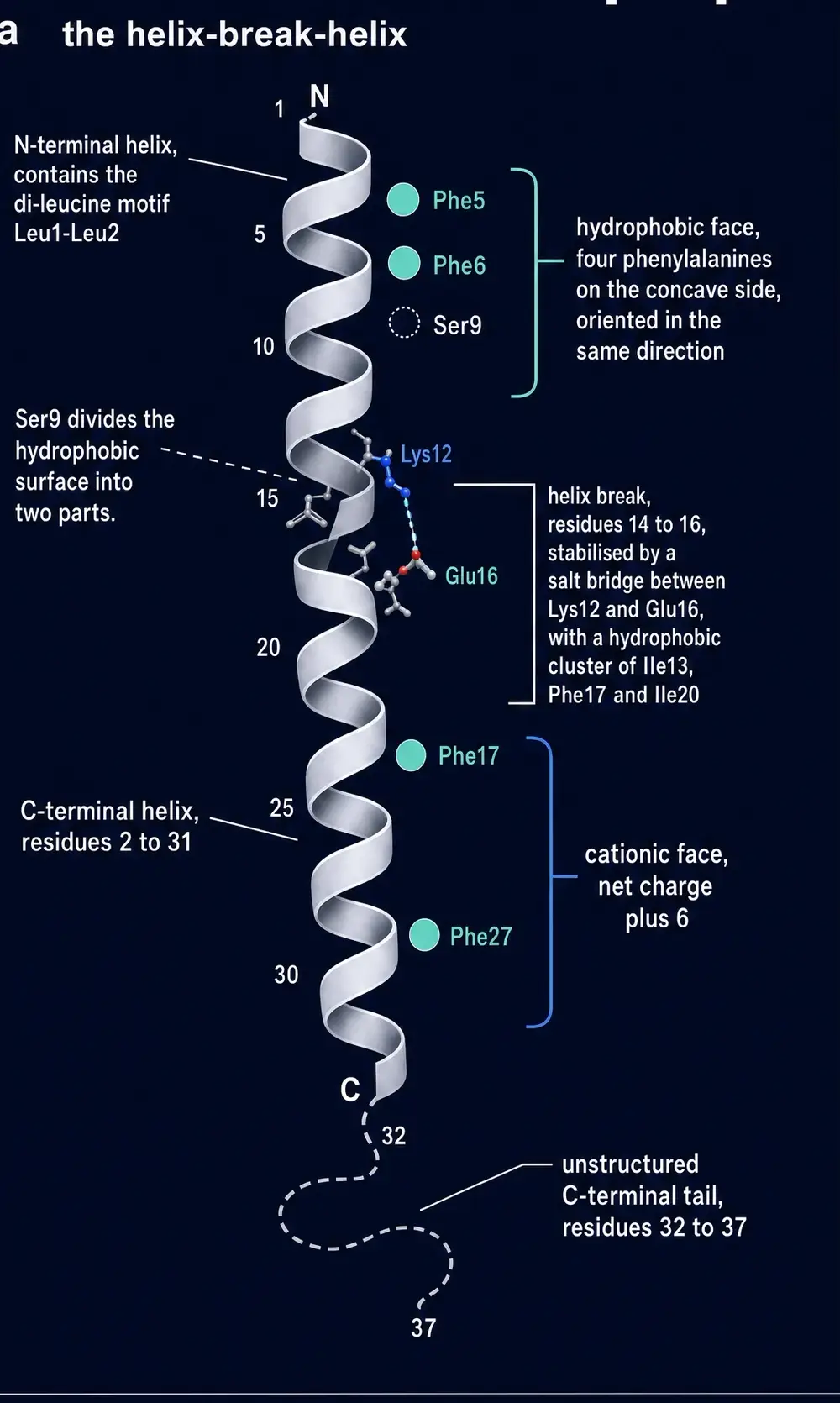
The peptide is the last fifth of a 170-residue protein. To understand when a human being has any of it, you have to look not at the peptide but at the promoter of the gene that encodes it — and there the story stops being about immunology and becomes, briefly, about genome parasites.
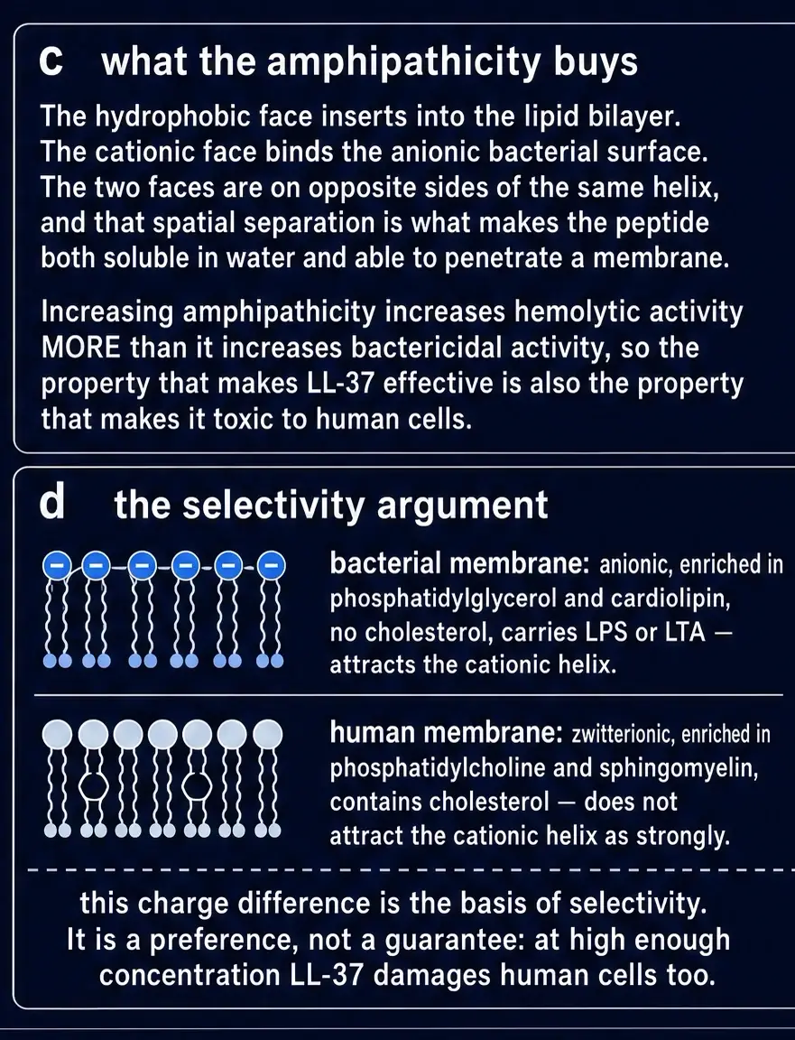
That amphipathic segregation is not a metaphor borrowed from detergent chemistry. It is the geometric reason the same thirty-seven residues can bind an anionic bacterial outer leaflet, insert, and — above a threshold surface density — destroy the barrier that keeps the bacterium alive. The wheel below is the shortest picture of that fact.
Section 06The parasite that installed a vitamin D switch
Roughly half the human genome consists of mobile elements: sequences whose principal activity, over evolutionary time, has been copying themselves into new locations. Alu elements are the most numerous family in primates, about 300 base pairs each, present in something over a million copies. They are usually described as junk, and usually that is accurate.
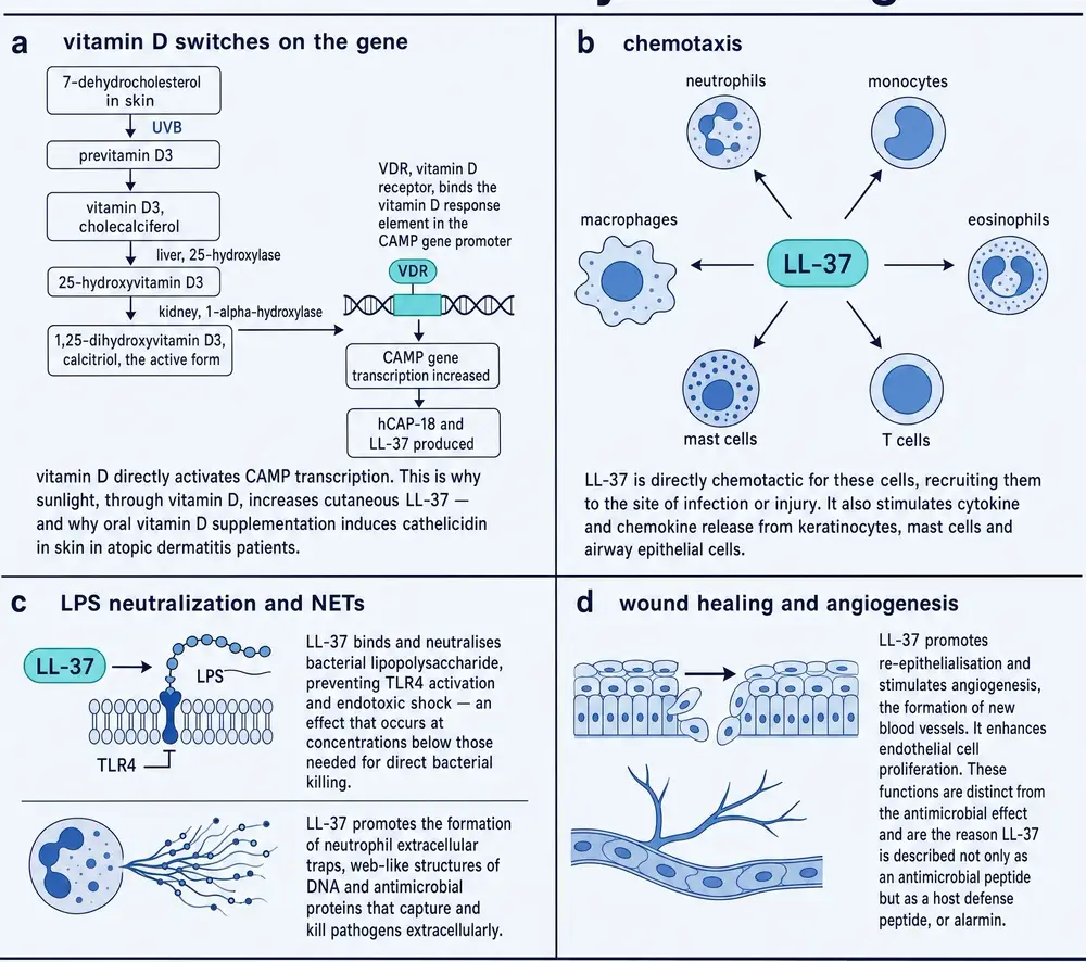
In 2005 Adrian Gombart, working with Niels Borregaard and Phillip Koeffler, reported that the human CAMP gene — the gene for this peptide — carries a consensus vitamin D response element in its proximal promoter, that the vitamin D receptor binds it, and that 1,25-dihydroxyvitamin D3 strongly induces cathelicidin in human myeloid cells, keratinocytes and colon lines (Gombart et al., 2005). That alone would be a useful piece of regulatory biology. The part that makes it remarkable is what they found when they looked at other species. The induction did not happen in murine cells. Cathelicidin messenger RNA in the bone marrow of vitamin-D-receptor-deficient mice was much the same as in wild-type animals. And when they compared genomes, the response element turned out to sit inside a short interspersed element that is present in primates and absent from the mouse, rat and dog genomes altogether.
A follow-up study four years later established the lineage in detail. The element is an AluSx, inserted in the ancestor of humans, apes, Old World monkeys and New World monkeys, and structurally conserved in every primate examined — conserved, on the authors' estimate, under purifying selection for 55 to 60 million years (Gombart et al., 2009). New World monkeys kept additional hormone-response sites in the same element that the human lineage let decay; the human gene responds to neither retinoic acid nor thyroid hormone.
It is worth being precise about what that establishes and what it does not. It establishes a mechanism: a transcription factor activated by a vitamin binds a specific site in a specific promoter and raises the amount of a specific antimicrobial peptide, in human cells, reproducibly. It does not establish that giving a person vitamin D makes them better at fighting infection, which is a different claim requiring a different kind of evidence — and Section 23 returns to how that evidence has actually turned out. What the exaptation does explain is a puzzle that predates it: why an element of innate immunity should depend on sunlight at all. A defence system wired to a molecule the skin manufactures under ultraviolet light is a defence system with a season and a latitude, and the observation that tuberculosis has both is far older than any of this molecular detail.
The clinical payoff arrived in 2006. Robert Modlin's group at UCLA showed that when a human macrophage's Toll-like receptors detect a mycobacterial lipopeptide, the cell responds by upregulating the vitamin D receptor and the enzyme that converts circulating vitamin D into its active form. The macrophage, in other words, builds its own local vitamin D system on demand, and the output of that system is cathelicidin, and the cathelicidin kills intracellular Mycobacterium tuberculosis. In mouse monocytes the equivalent job is done principally by nitric oxide instead. The same paper reported that serum from African-American donors, a group with both lower 25-hydroxyvitamin D and higher tuberculosis susceptibility, supported that induction inefficiently (Liu et al., 2006).
What makes the finding hard to dismiss is not that vitamin D raises a transcript. Plenty of genes do that. It is that the response element sits in DNA the mouse never received, so every knockout and infection model that treats murine CRAMP as a stand-in for LL-37 is missing the principal human control on the gene. The map below places that element.
Take these findings together and something uncomfortable follows. The single most important regulatory input to this gene in human beings is an input that mice do not have, carried on a piece of DNA that arrived in our ancestors after the rodent lineage had already split off.
Almost every mechanistic experiment in this field — the knockouts, the infection models, the wound-healing studies — has been done in mice. Those experiments are not worthless; they are experiments about a gene wired to a different switchboard, and they should be read that way. It is the reason this document labels the species in every sentence that reports an animal result.
Section 07Armed outside the cell, and only outside
A neutrophil does not store LL-37. It stores hCAP18, which is inert, and it stores it in the specific granules, physically separated from the aggressive proteases in the azurophil granules. The peptide only exists once the holoprotein has been cut.
Where that cut happens turns out to be a decision, and different species have made different ones. In 2001 Ole Sørensen and colleagues in Copenhagen worked out the human answer, and it is counter-intuitive. Using immunoelectron microscopy they confirmed that hCAP18 and the azurophil enzymes both end up in the phagolysosome — the compartment where a swallowed bacterium is digested. Then they looked for cleaved peptide there and found none. There was no detectable processing inside the cell after phagocytosis. Cleavage happened in exocytosed material, outside the neutrophil, and of the three azurophil serine proteases only proteinase 3 did it (Sørensen et al., 2001).
Pig and cow cathelicidins are cleaved by elastase, intracellularly. As the authors observed, the generation of an active antimicrobial peptide from a common pro-protein happens differently in closely related species.
The consequence is not a technicality. Cutting inside the phagolysosome would deliver a membrane-active peptide precisely where the bacterium is and nowhere else — a targeted release. Cutting outside puts it into the extracellular space, at a wound margin or an inflamed airway, where it meets host cells, host membranes, host DNA and host plasma proteins on its way to anything bacterial. Every activity described in Part Three is a consequence of that address. So is every problem described in Part Four.
Section 08Where it is, and how much of it there is
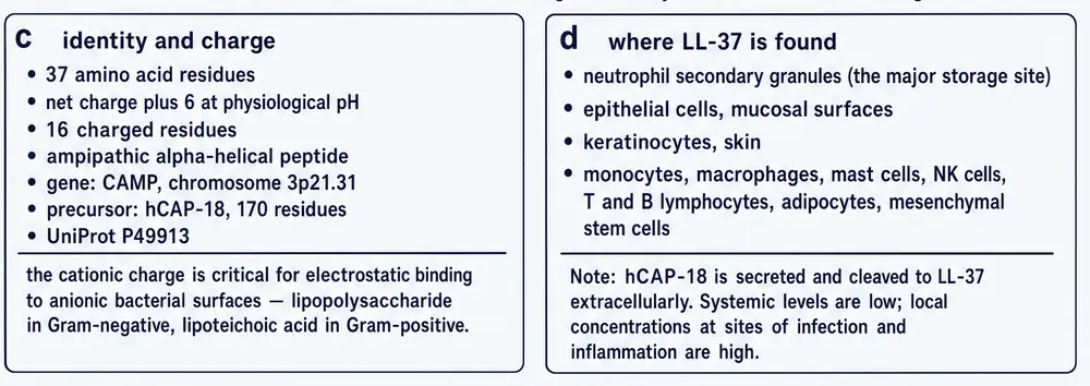
hCAP18 is not confined to neutrophils, though that is where it is most concentrated. It has been reported in keratinocytes, in airway epithelium, in the gut, in saliva, in sweat, in seminal plasma and in breast milk. In the skin its expression is low in healthy tissue and rises sharply on injury or inflammation, which is the observation that made it interesting to the wound literature.
Those concentrations are not interchangeable units on a single scale. A nanomolar reading in plasma and a micromolar reading in a neutrophil granule describe different biological situations, and the log plot below exists to stop them being averaged into a false middle.
The quantitative anchors come from an ELISA developed in Copenhagen in 1997. A neutrophil contains about 0.627 micrograms of hCAP18 per million cells. Plasma contains about 1.18 micrograms per millilitre — which the authors noted was several-fold higher than other neutrophil specific-granule proteins — circulating as high-molecular-weight complexes rather than free protein, and barely excreted in urine. The bone marrow appears to be the major source of the plasma pool (Sørensen et al., 1997).
That plasma figure repays a moment's arithmetic. At 19,301 daltons, 1.18 µg/mL is about 61 nanomolar. The concentrations at which LL-37 kills bacteria in a test tube are typically quoted between one and thirty-two micromolar — that is, between sixteen and roughly five hundred times higher than the amount of holoprotein circulating in blood. Some of that gap is real biology: the peptide is not meant to work in bulk plasma, it is meant to work in the small volume between a degranulating neutrophil and its target, where local concentrations are much higher than anything a blood sample can measure. But the gap is also the reason to be careful with any sentence of the form “LL-37 kills X”. It kills X at concentrations, in buffers, that were chosen to make the experiment work.
And there is a further complication, which Section 15 takes up properly: the plasma in which that 61 nanomolar is dissolved does not merely dilute the peptide. It actively inhibits it.
Before the killing chemistry, the inventory: where the peptide is made, what charge it carries at physiological pH, and how those facts constrain every later claim about systemic use. The identity plate collects the numbers that the rest of the monograph keeps referring back to.
Section 09Killing
The founding activity, and the one the molecule is named for, is direct antibacterial killing. LL-37 has reported activity against Gram-negative and Gram-positive organisms, against some fungi, and against enveloped viruses. Its mechanism against bacteria is physical rather than enzymatic: the cationic face binds the anionic outer surface of a bacterial membrane, the hydrophobic face inserts, and above a threshold surface density the membrane loses integrity.
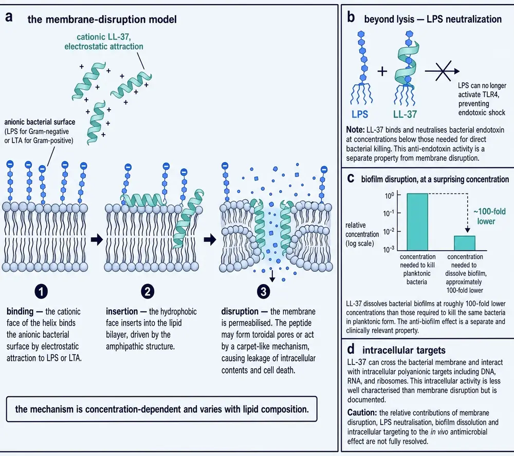
How exactly it loses integrity is genuinely unsettled and this document reports it as unsettled. The two long-standing models are the carpet mechanism, in which peptide accumulates parallel to the surface until the bilayer disintegrates detergent-like, and the toroidal pore, in which peptide and lipid head-groups together line a transient channel. Different model membranes and different techniques favour different answers, and the honest summary is that LL-37 does not appear to have one mechanism so much as a concentration-dependent series of them.
Two properties follow from a physical mechanism and both matter clinically. The first is that resistance is harder to acquire than it is against an antibiotic with a protein target, because there is no single enzyme to mutate — though bacteria have found routes, principally by reducing the negative charge of their surface or by secreting proteases that digest the peptide. The second is that a mechanism which depends on charge is exquisitely sensitive to the ionic strength of its surroundings, which is why so much of the older literature was run in low-salt buffers.
Section 10Neutralising
Killing a Gram-negative bacterium releases its lipopolysaccharide, and lipopolysaccharide is the most potent inflammatory signal the innate immune system recognises. An antibiotic that lyses bacteria without dealing with the debris can make a patient acutely worse.
LL-37 binds lipopolysaccharide directly and neutralises its ability to activate cells. This is not incidental to the design: the same positive charge that finds a bacterial membrane finds the phosphate groups of lipid A. The family was recognised for this early — rabbit CAP18, the protein our own is named after, was characterised as an LPS-binding protein before anyone was interested in its antibacterial activity. It is one of the few places where LL-37's multifunctionality is unambiguously an advantage: the molecule cleans up after itself.
It is worth attaching the usual caution. Endotoxin neutralisation is measured by mixing peptide and lipopolysaccharide and then asking whether cells still respond, and the concentrations at which that works are the concentrations of Section 08 — micromolar, in buffer. Whole blood contains lipopolysaccharide-binding protein, soluble CD14 and the lipoproteins of Section 15, all of which compete for the same molecule. That the peptide binds endotoxin is not in doubt; that it is the thing binding endotoxin in a septic patient is a much larger claim, and it is not one the tube experiment makes.
Section 11Calling, and building
By the early 2000s it was clear that LL-37 did things to human cells that had nothing to do with bacteria. The receptor that accounts for most of them is formyl peptide receptor 2, a G-protein-coupled receptor also known in the older literature as FPRL1. Through it, LL-37 is chemotactic for neutrophils, monocytes and T cells — the peptide dumped at a site of infection recruits more of the cells that make it.
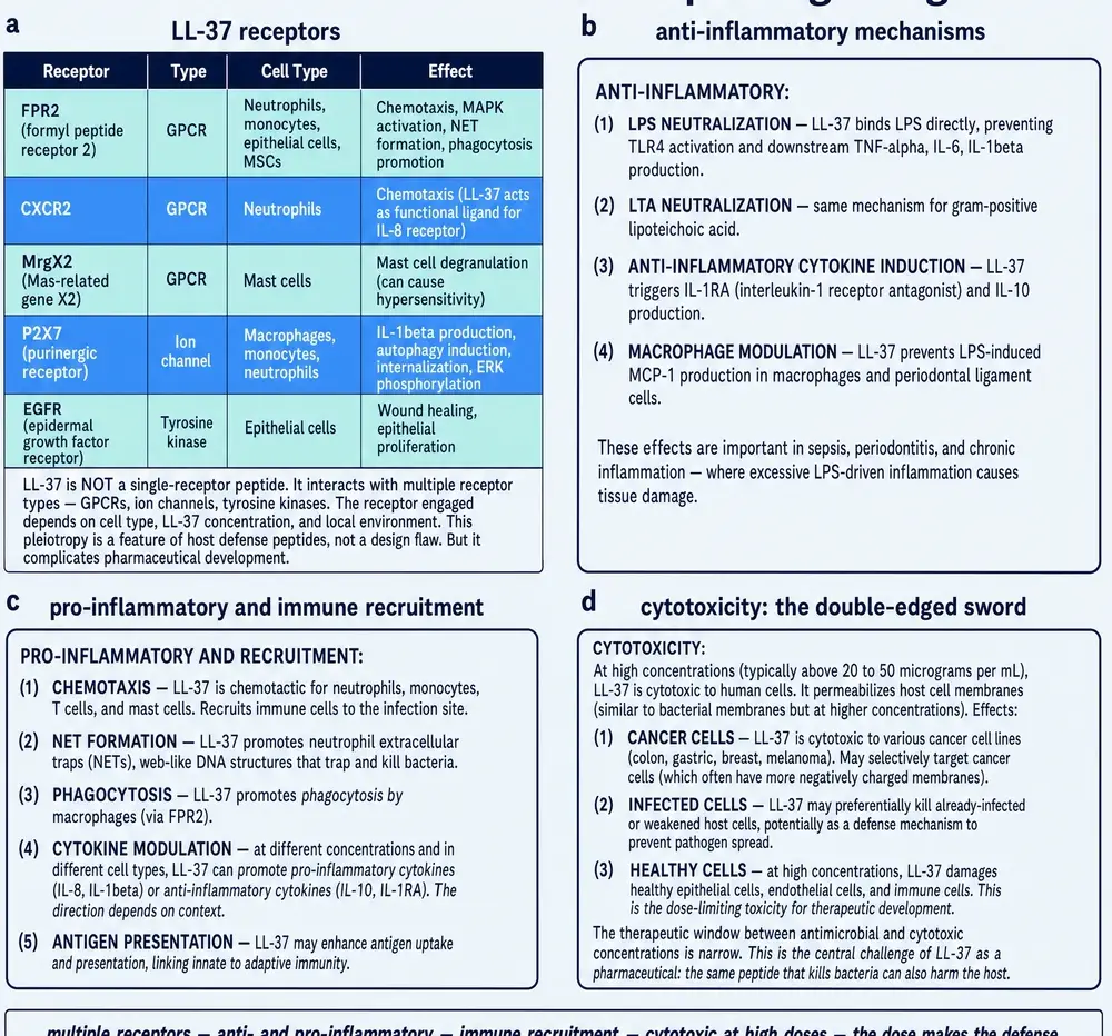
The more surprising finding came in 2003, from a collaboration between Marburg, Munich and San Diego. LL-37 induces angiogenesis. Applied to the chick chorioallantoic membrane it produced new vessels; in a rabbit hind-limb ischaemia model it produced neovascularisation; on cultured endothelial cells it drove proliferation and the formation of vessel-like structures, and the effect was blocked by interfering with FPRL1 (Koczulla et al., 2003). The same paper showed reduced vascularisation during wound repair in CRAMP-deficient mice — a mouse result, about the mouse peptide, and labelled as such here.
This is the point at which LL-37 stopped being an antibiotic in the literature's imagination and became something harder to name. A molecule that kills bacteria, recruits leukocytes and grows blood vessels is not a drug candidate with side effects. It is a wound-healing programme in a single sequence.
None of those non-killing jobs requires a second active site. They are the same amphipathic helix read by a different partner — DNA, a G-protein-coupled receptor, a growth-factor pathway — and the practical consequence is that you cannot delete the host-facing work without deleting the antimicrobial work. The panel that follows makes that indivisibility visible.
Of everything LL-37 does, the property with the largest consequences is the one that follows most directly from its chemistry and was noticed last. A peptide with eleven positive charges binds polyanions. DNA is a polyanion.
Section 12Condensing DNA — the function that cuts both ways
A study published this year characterises the interaction carefully. LL-37 binds double-stranded DNA non-specifically along the phosphodiester backbone, condenses it, and then assembles progressively larger complexes from smaller domains until complete complexation is reached at a DNA-to-peptide ratio of about 1 to 1.7 by weight. The morphology depends on how much peptide is present: relatively little gives loose aggregates, more gives well-defined disc-like complexes around 150 nanometres across. The condensed DNA loses its characteristic B-form signature. Crucially, the peptide's amphipathic helical structure is required — this is not generic polycation behaviour. And when the same experiment is done on neutrophil extracellular traps, high LL-37 concentrations significantly reduce the projected area of the trap (Zielke et al., 2026).
Read one way, that is housekeeping: a neutrophil that has just exploded its own chromatin into a web to catch bacteria has an obvious interest in keeping that web compact and in coating it with something antimicrobial.
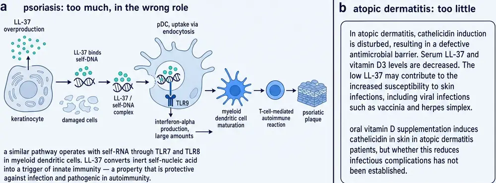
Read the other way, it is the mechanism of an autoimmune disease. In 2007 Roberto Lande and Michel Gilliet's group asked why plasmacytoid dendritic cells — which are built to detect microbial DNA through endosomal Toll-like receptor 9 and which normally ignore the body's own DNA entirely — stop ignoring it in psoriasis. The answer was LL-37. The peptide converts inert self-DNA into a potent trigger by binding it into aggregated, condensed structures that are delivered to and retained within the early endocytic compartment where TLR9 sits, and the cells respond with type I interferon (Lande et al., 2007).
There is no second mechanism here and no separable domain. The chemistry that packages a bacterium's DNA in a neutrophil trap packages the host's DNA in inflamed skin. Whether the result is defence or disease is a question about whose DNA is lying around, in what quantity, and where — not a question about the peptide.
This has a consequence that runs straight into Part Five. If you wanted a version of LL-37 that killed bacteria without ever complexing self-DNA, you would have to remove the property that does both, and that property is the charge. Eleven basic residues are what find the anionic surface of a bacterial membrane; they are also what find the phosphate backbone of a chromosome. The two activities are not separate functions housed in separate parts of the molecule, to be engineered apart. They are one electrostatic fact, used twice. Every derivative in Section 22 is an attempt to trade some of that charge away and keep enough of it, and the reason none of them is simply “LL-37, improved” is that the trade is real.
DNA binding is also the hinge on which the molecule’s reputation turns. The same property that stabilises a neutrophil extracellular trap can deliver self-DNA into an endosome and light an interferon programme. The schematic that follows separates those two readings without pretending they use different chemistry.
Section 13The newest job, and the sharpest statement of the problem
In May 2026 a group at the University of Calgary published a study in Science that, more than any other single experiment in this literature, explains why LL-37 has not become a medicine. They imaged the lung microcirculation of septic mice directly.
What they saw was a cascade. Cathelicidin localised neutrophils to the vessel wall and initiated immunothrombi — clots built of platelets and neutrophils — acting through formyl peptide receptors, the same receptor family that mediates chemotaxis and angiogenesis in Section 11. Those immunothrombi captured bacteria. And the cathelicidin enabled antimicrobial activity in the platelets themselves. The clot was not a complication of the infection; it was an organ of defence (Brown et al., 2026).
Then they blocked it. Blocking cathelicidin prevented immunothrombosis and attenuated early death from sepsis — precisely the result a drug developer would want, and precisely the result that a paper written five years earlier might have stopped at. The animals then died later, of uncontrolled infection.
This is a mouse study and the peptide in it is CRAMP. What generalises is not the number but the shape: the benefit and the harm were the same event, observed at different times. The authors' own conclusion points at a different target — inhibiting leukotriene B4, which amplifies the cascade, relieved the vascular compromise while preserving host defence — and that is the useful lesson. Where a molecule's harm and benefit are inseparable, the tractable target is usually not that molecule.
Section 14The newest functions, weighted as new
The list is still growing, and this section reports the recent additions with the caution that recency deserves. Each is a single primary report or a small number of them, and none has yet been independently replicated in the way the older findings have.
A group at the Kunming Institute of Zoology has reported that LL-37 binds low-density lipoprotein through its apolipoprotein B-100 domains, increases ApoB-100 solubility, inhibits LDL modification and aggregation, and promotes LDL uptake through the LDL receptor in both hepatocytes and macrophages. In Apoe-deficient mice with hypercholesterolaemia the murine homologue Cramp accelerated cholesterol clearance and reduced hepatic lipid accumulation (Fang et al., 2026). If it holds up, an antimicrobial peptide is also a lipoprotein chaperone.
In January 2026 a group in Chongqing reported the darker version of the same kind of finding. In mouse models of breast cancer lung metastasis, a neutrophil subset recruited during macrometastasis forms extracellular traps, and cathelicidin embedded in those traps binds adenine nucleotide translocator 1 in CD8 T cells, opens the mitochondrial permeability transition pore, and kills them (Wang et al., 2026). The peptide as an immunosuppressive agent in the metastatic niche is a long way from a natural antibiotic, and it is again a mouse result about Cramp.
The cancer literature more broadly runs in both directions and has done for years: LL-37 is reported as pro-tumour in ovarian, lung and breast models and anti-tumour in gastric and colon models, with the direction appearing to depend on tissue, receptor repertoire and concentration. A review published this year frames the same duality for the gut, arguing that the peptide's effect “varies with peptide concentration, receptor binding status, disease stage, and local microenvironment” (Liu et al., 2026). That is the honest position, and it is also a description of a molecule that is very hard to prescribe.
Section 15The plasma problem
In 1998 a group at the Karolinska Institutet — including Birgitta Agerberth, who had found the molecule — published a short paper with an uncomfortable title: Apolipoprotein A-I binds and inhibits the human antibacterial/cytotoxic peptide LL-37. Two things in that title are worth noticing. The first is that by 1998 the people who discovered the peptide were already calling it cytotoxic in the same breath as antibacterial. The second is the finding itself.
Human plasma inhibits LL-37's antibacterial and cytotoxic activity. It does not do so by degrading the peptide — LL-37 is fairly stable in plasma. The group ran plasma proteins over an LL-37 affinity column and pulled off a single 26 kDa band, which sequenced as apolipoprotein A-I, the principal protein of high-density lipoprotein. Surface plasmon resonance put the interaction in the low micromolar range. Fifty micromolar apoA-I inhibited the antibacterial activity of fifty micromolar LL-37 by about half of what whole plasma achieves, and an antibody against apoA-I completely blocked plasma's inhibition up to 25 µM peptide (Wang et al., 1998).
Set that beside Section 08. The peptide's precursor circulates in plasma at about 61 nanomolar, and the plasma it circulates in contains a scavenger that binds it. This is not a defect in the organism — it is almost certainly the point, since a freely active membrane-lytic peptide in the bloodstream would be a catastrophe. But it means that any therapeutic strategy involving systemic LL-37 has to contend with a buffering system evolved specifically to prevent systemic LL-37 from doing anything. It is one of the clearest reasons the only human trials of the molecule have been topical.
That constraint is worth stating as a constraint rather than an accident. A topical route confines the peptide to a compartment where the scavenger is scarce, the concentration can be held high, and systemic exposure is negligible — which is exactly the setting the wound and middle-ear studies of Part Five occupy. It also forecloses most of what the molecule is interesting for. Nothing in the tuberculosis work, the sepsis work or the lipoprotein work of Part Three describes an effect a cream could produce, and no route yet demonstrated in a person reaches those compartments.
Section 16The selectivity problem
The mechanism that makes LL-37 hard for bacteria to resist is the mechanism that makes it dangerous to us. Membranes are membranes. What distinguishes a bacterial membrane from a human one is a matter of degree — more anionic lipid on the outer leaflet, no cholesterol — and a peptide that discriminates on charge discriminates imperfectly.
In practice this shows up as a therapeutic window that is narrow and, worse, assay-dependent. Reported haemolytic and cytotoxic thresholds for LL-37 sit in the same order of magnitude as the bactericidal concentrations rather than comfortably above them, and where in that band a given experiment lands depends on the cell type, the serum content of the medium, the ionic strength and the incubation time. This document does not quote a single therapeutic index for LL-37 because the literature does not support one; what it supports is the statement that the killing range and the toxicity range overlap.
The engineering programme in Section 22 exists almost entirely because of this section.
Section 17The control problem, and why it belongs in a monograph
This section is unusual for this series in that it is about the literature rather than the molecule, but it changes how several of the numbers above should be read.
LL-37 research uses two standard comparators. One is D-LL-37, the all-D-amino-acid enantiomer, used to test whether an effect requires a specific chiral interaction such as receptor binding — a D-peptide still forms an amphipathic helix and still disrupts membranes, but will not fit a stereospecific pocket. The other is scrambled LL-37, a peptide with the same composition and charge in a randomised order, which is the negative control for anything that depends on the sequence rather than on bulk cationicity.
Both of those designations contain the subject's designation in full. Every sentence in the literature about scrambled LL-37 is, by design, a null result — that is what a negative control is for. A reader skimming, or an automated extraction system, that misses the word “scrambled” will read the field's own controls as evidence that the peptide does nothing.
The enantiomer carries the opposite risk. Because a D-peptide still folds into an amphipathic helix and still disrupts a membrane, an effect that survives in D-LL-37 is usually evidence that the effect is physical rather than receptor-mediated — and an effect that disappears is evidence that a specific protein is involved. That is a genuinely informative comparison, and it is also one that is easy to report backwards. A sentence beginning “D-LL-37 was inactive” may be the strongest evidence in a paper that the activity is real and receptor-dependent.
Those two control designations are why an uncritical literature search over-counts papers about LL-37: many hits are really about D-LL-37 or a scrambled sequence used as a negative arm. The note below records how this document removed them.
The retrieval pipeline behind this monograph strips both designations from every document before it counts anything, so that a paper whose control arm is named forty times cannot be admitted as a paper about the peptide. The same treatment is applied to species-qualified forms of the holoprotein — “rabbit CAP-18” and its relatives — because a rabbit paper introduces the protein once with its species and then writes the bare name for the rest of the article. The Apparatus reports how many documents each rule removed.
Section 18When it turns on the host
Four human conditions illustrate the two directions in which this molecule fails.
Psoriasis. Section 12 gave the mechanism: LL-37 complexed with self-DNA activates plasmacytoid dendritic cells through TLR9. The peptide is also, separately, an autoantigen: two-thirds of patients with moderate-to-severe plaque psoriasis carry CD4- or CD8-positive T cells specific for LL-37, those cells produce interferon-γ and Th17 cytokines, and their presence in blood correlates with disease activity (Lande et al., 2014). So the same molecule drives both the innate and the adaptive arm of the same disease. Here the problem is excess.
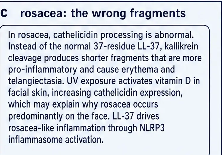
Rosacea. The 2007 study from Richard Gallo's group is the most instructive of the four, because the abnormality is not simply quantity. People with rosacea express abnormally high cathelicidin in facial skin and the processed forms they carry are different from those in unaffected people. The cause is increased stratum corneum tryptic enzyme, a kallikrein-family protease, cutting the same holoprotein in different places. Injecting the rosacea-specific peptide forms into mouse skin produced inflammation; adding the enzyme did; increasing protease activity genetically did; and deleting Camp abolished the effect (Yamasaki et al., 2007). The condition affects around 3 per cent of the US population over 30.
What that paper demonstrates is subtler than “too much cathelicidin causes rosacea”. It demonstrates that the identity of the fragment matters — that a change in the protease environment turns a defensive molecule into an inflammatory one without changing how much of the parent protein is present. Section 07's observation that human processing happens outside the cell, in whatever protease environment happens to obtain, is what makes this possible.
That is the distinction the commissioned plate is there to hold: rosacea as a processing disorder, not an abundance disorder. The same holoprotein, cut differently, yields a different clinical story — which is why counting cathelicidin in skin without asking which fragment is present answers the wrong question.
Chronic wounds. Here the problem runs the other way. Cathelicidin is reported low or absent at the edge of chronic ulcers, and that observation is the entire rationale for the only randomised trial of the peptide in Section 21.
Tuberculosis. Also a deficiency direction, and the one with the clearest mechanism, via Section 06.
Two further associations should be mentioned and weighted carefully. Citrullination — the enzymatic conversion of arginine to citrulline, of which LL-37 has five candidate residues — alters the peptide's charge and its immunological behaviour, and citrullinated LL-37 has been studied as an autoantigen in rheumatoid arthritis. And a body of work relates LL-37 to amyloid: the peptide interacts with amyloid-β, modulates its aggregation, and can itself self-assemble, with reviews proposing a shared role for both as host-defence molecules whose chronic activation contributes to neuroinflammation (Asti, 2026). That last is a hypothesis under construction rather than an established finding, and this document reports it as one.
None of the four is a story about the molecule being present or absent. Each is a story about how much of it, which fragment, and next to what — which is an uncomfortable shape for any therapeutic hypothesis, because it means there is no direction of intervention that is right in general.
So the clinical pattern is not “too little peptide” or “too much peptide” as a single rule. It is the wrong fragment, at the wrong concentration, next to the wrong cargo. That is why the same literature can look like a deficiency disease in one tissue and an autoinflammatory disease in another. The summary figure compresses that bidirectional map.
Section 19The species problem, stated once, plainly
Three separate findings from three separate laboratories, already reported above, converge on a single conclusion, and it is worth collecting them because no one of them alone carries the weight.
| Level | Human | Mouse |
|---|---|---|
| Peptide | LL-37, 37 residues | CRAMP, a different sequence |
| Gene | CAMP | Cnlp |
| Activating protease | proteinase 3, extracellular | elastase-family, as in other non-primates |
| Vitamin D response element | present, inside an AluSx | absent from the genome |
| Induction by 1,25(OH)2D3 | strong | not observed |
| Principal TLR-driven killing mechanism in macrophages | vitamin D → cathelicidin | nitric oxide |
A knockout mouse tells you what happens to a mouse when you remove the mouse's peptide. That is a real and useful experiment — the Group A Streptococcus work that first proved a cathelicidin matters in vivo was exactly this, and it remains the field's foundation (Nizet et al., 2001). But the inference from it to human physiology has to cross four differences, and the one in the promoter is the largest, because it means the human gene's most powerful natural stimulus does not exist in the model organism.
This is not an argument that the animal literature is wrong. It is an argument for a habit: whenever this document reports a finding, it names the system it came from, and the reader can decide how far it travels.
Section 20What has actually been given to people
Thirty years of laboratory work on a human molecule with obvious therapeutic appeal ought to have produced a substantial clinical record. It has not, and the shape of what exists is more informative than its size.
Searching the United States clinical trials registry for LL-37, cathelicidin or ropocamptide as an intervention returns 31 studies. Read one at a time, the great majority turn out not to be studies of the peptide at all. The largest single class administers vitamin D and measures cathelicidin as an output — a rational design given Section 06, but a trial of a vitamin, not of a peptide. The next largest class administers nothing and measures LL-37 in saliva, gingival crevicular fluid or serum as a biomarker of periodontal disease, caries, smoking status or COVID-19 severity.
In two registered studies the peptide itself was given to human beings.
The first is NCT02225366, run at MD Anderson Cancer Center with the National Cancer Institute: intratumoral injection of LL-37 into cutaneous melanoma deposits, once weekly for eight weeks, as a dose-finding study with up to four dose levels and two participants per level. The registry record states the intended size in its own words: “Up to 36 participants will be treated in this study.” The enrolment field reads 4. The study opened on 8 July 2015 and completed on 24 November 2020 — five years and four patients. The same record notes plainly that “LL37 is not FDA approved or commercially available. It is currently being used for research purposes only.”
The second is NCT04098562, a phase 2 study of an LL-37 cream in 40 patients with diabetic foot ulcers, sponsored by a university faculty in Indonesia, with a listed primary completion date of December 2019. Its registry status is UNKNOWN and no report of its results was located during the preparation of this document.
That is the entire registered human experience of administering this molecule. It is worth stating explicitly what that does and does not mean. It does not mean the peptide is dangerous or that it failed; a trial that never enrolled has produced no evidence of any kind. It means that the human evidence base for LL-37 is, as of this compilation, a single completed randomised trial — and that trial is not in this registry.
Section 21The one randomised trial, reported as it reads
Between 2017 and 2020 a Swedish company, Promore Pharma, ran a placebo-controlled phase IIb study of topical LL-37 in hard-to-heal venous leg ulcers, mostly at sites in Poland. It was called HEAL LL-37, and it is the only randomised controlled trial of this molecule in existence. The results were published in Wound Repair and Regeneration in 2021 (Mahlapuu et al., 2021).
The design was sound. One hundred and forty-eight patients, three arms — LL-37 at 0.5 mg/mL, LL-37 at 1.6 mg/mL, and placebo — applied twice weekly at dressing changes on top of compression therapy, which is standard care. The population was the right one: mean age 67.6 years, median ulcer duration 20.3 months, mean wound area 11.6 cm2 at randomisation. These were not easy wounds.
The result, in the paper's own words, is that the efficacy analysis performed on the full study population did not identify any significant improvement in healing in patients treated with LL-37 as compared with placebo.
A post-hoc analysis then found statistically significant improvement in several interrelated healing parameters in the subgroup of patients whose wounds were at least 10 cm2 at randomisation — a group with a known poor prognosis, and therefore the group with the most room to improve. The authors are careful about this and call, correctly, for a further study adequately powered to assess that population. The peptide was safe and well tolerated at both concentrations, which is a genuine finding in its own right and the strongest piece of human safety data the molecule has.
The sponsor's own announcement of these results describes them as positive and gives the number of patients treated as 144. The peer-reviewed report gives 148 and states the null result for the full population in its abstract. Where a company statement and the primary paper disagree, this document reports the paper. A post-hoc subgroup finding on interrelated endpoints, without pre-specification or multiplicity control, is a hypothesis worth testing. It is not a demonstration of efficacy, and describing it as one is how a molecule acquires a reputation its evidence does not support.
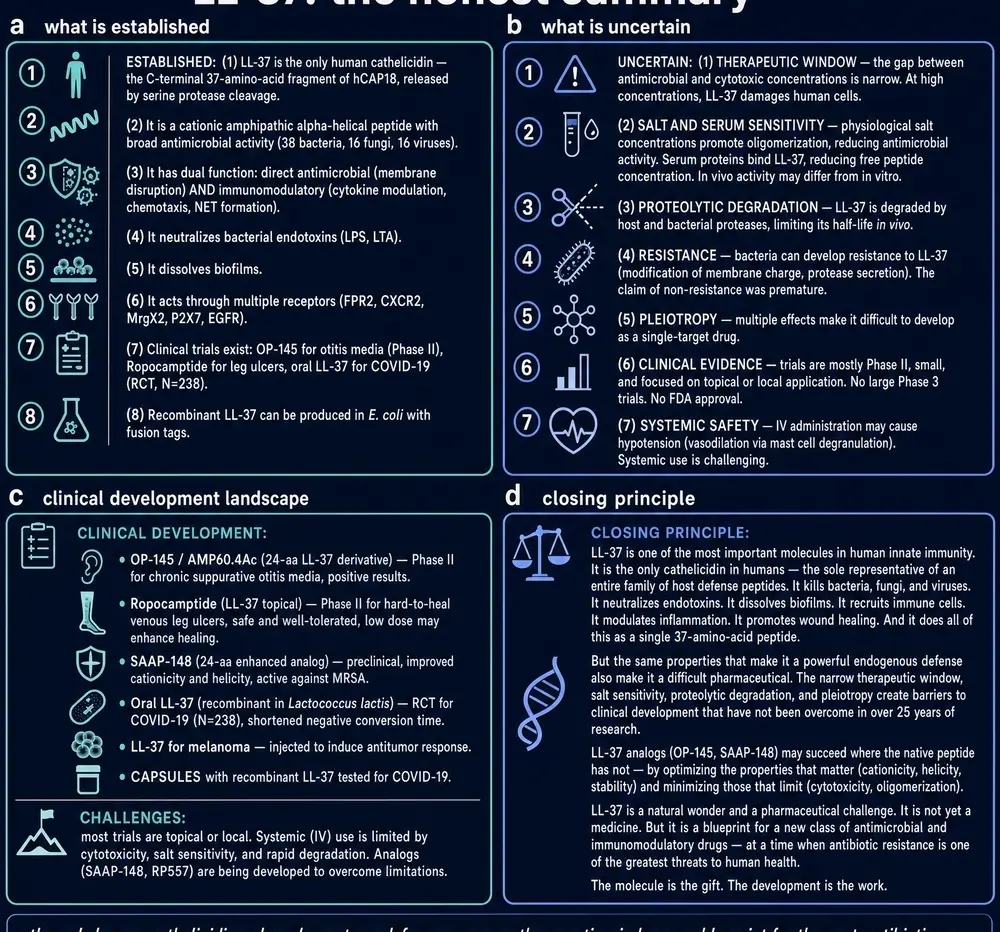
If the parent peptide’s completed randomised trial did not meet its primary endpoint, the natural next move in this literature has been to change the molecule. That move is real engineering; it is also a change of subject.
Section 22The derivatives carry most of the rest
Faced with Sections 15 and 16 — a peptide inhibited by plasma, toxic to host cells near its working concentration, and rapidly degraded — the field did the sensible thing and started modifying it.
The truncations came first, because LL-37 turned out to contain smaller active units. KR-12, residues 18 to 29, is the shortest fragment retaining antibacterial activity, and when it was characterised structurally it showed a property the parent lacks: a selective toxic effect on bacteria but not on human cells (Wang, 2008). Others — GF-17, FK-13, KS-30, LL-23 — map different parts of the same helix onto different balances of potency and toxicity.
Then came deliberate engineering. OP-145, also written P60.4Ac, is an LL-37-derived variant developed in the Netherlands and taken into clinical work for chronic suppurative otitis media — and its record is instructive, because it is better than the parent’s. A dose-finding study in 16 subjects was followed by a randomised, double-blind, placebo-controlled phase IIa study in 34 adults; ototopical drops at the selected 0.5 mg/mL were safe and well tolerated, and produced treatment success in 47 per cent of cases against 6 per cent on placebo (Peek et al., 2020). That is a derivative result and belongs to the derivative. SAAP-148 is a later derivative with strong anti-biofilm activity. D-LL-37 resists proteolysis by construction. And the ceragenins, of which CSA-13 is the best known, are not peptides at all — they are steroid-based small molecules designed to present the same pattern of charge and hydrophobicity, which makes them cheap to make, stable to proteases and insensitive to ionic strength (Łuckiewicz et al., 2026).
The pattern across that list is consistent and slightly awkward: the further a molecule is from the natural sequence, the better it behaves as a drug. That is a strong argument for the engineering programme. It is also the reason none of its results transfer back. A clinical finding for OP-145 is a clinical finding about OP-145.
If the peptide is hard to give, the obvious alternative is to make people produce more of it, and Section 06 supplies an unusually well-understood lever for doing so. That reasoning is why cathelicidin appears as an endpoint in so many vitamin D trials — in tuberculosis, in chronic obstructive pulmonary disease, in respiratory infection, in critical illness, in pregnancy.
Section 23The vitamin D detour
Raising the biomarker is straightforward. What has not followed reliably is clinical benefit. The vitamin D supplementation literature as a whole is one of the most extensively studied and most equivocal bodies of evidence in modern medicine, and the cathelicidin-endpoint trials sit inside it: they can generally show that the intervention did what it was supposed to do at the molecular level, and they have not established that doing so changes outcomes in an unselected population.
This is a pattern that recurs across this monograph series, and it is worth naming plainly because it is the single most common way a mechanism becomes overstated: moving a marker is not the same as helping anybody. The vitamin D–cathelicidin axis is real, evolutionarily conserved, mechanistically detailed and, on current evidence, not by itself a therapy.
Section 24Why it is hard to drug, and what would have to be true
Collect the obstacles and they fall into two classes, and only one of them is an engineering problem.
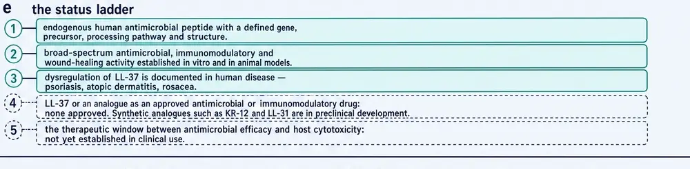
Those engineering constraints are not a prelude to a rescue that has already arrived. They are the reason the derivatives exist, and the reason evidence gathered on a derivative does not transfer back to the parent sequence. The status board that follows is drawn to that rule rather than to the more flattering summary the field sometimes prefers.
The tractable ones are familiar to anyone who has worked on peptides. LL-37 is degraded by proteases, including bacterial ones. It is inhibited by plasma apolipoprotein A-I. Its activity falls with rising ionic strength. Thirty-seven residues of synthetic peptide is expensive at scale. Each of these has a known engineering answer — D-amino acids, cyclisation, lipidation, truncation, non-peptide mimicry — and Section 22 is the field applying them.
The untractable one is the subject of this whole document. LL-37's value to the organism is its context-dependence. It kills bacteria at a wound edge, recruits the cells that will clear them, builds the vessels that will feed the repair, packages the DNA those cells release, and stops. A drug cannot be context-dependent in that way, because a drug arrives at a dose chosen in advance by someone who cannot see the context. What a developer needs is a molecule that does one thing, whose dose can be titrated against one endpoint, and whose therapeutic index can be measured. The 2026 sepsis experiment is the cleanest demonstration available that LL-37 is not that molecule: the harm and the benefit were not two effects to be separated, they were one event observed at two times.
The field's implicit answer has been to engineer the context-dependence away. It is a reasonable answer and it works, and it ends with a compound that is no longer LL-37 — which is why a monograph on this molecule ends up being, in large part, a monograph about why its descendants exist.
Section 25What would change the picture
Four things, each specific enough to be falsified.
An adequately powered trial in the population the post-hoc analysis identified. HEAL LL-37 generated a real hypothesis: that topical LL-37 helps large, long-standing venous ulcers. That hypothesis has been available since 2021 and has not been tested. A pre-specified trial restricted to wounds of at least 10 cm2, with a single primary endpoint, would settle it either way. Its absence is itself a finding about the molecule's commercial position.
A human study in which the peptide, rather than vitamin D, is the variable. Almost everything called a cathelicidin trial is a vitamin trial. The mechanism deserves a direct test.
A model organism that has the Alu element. The vitamin D–cathelicidin axis cannot be studied in mice, and the exaptation work establishes that it is intact in Old and New World monkeys. Any claim about this pathway's importance in whole-organism defence currently rests on human correlation and cell culture.
A derivative with a measured therapeutic index in humans. KR-12's reported separation of antibacterial from cytotoxic activity, obtained in vitro, is the most promising single observation in the engineering literature and has not been converted into a human number.
None of those requires a new idea. All of them require someone to fund a trial of a molecule that cannot be patented, in an indication where the standard of care is a compression bandage.
It is worth ending on what the evidence for this molecule actually consists of, because the volume of publication is easy to mistake for the weight of proof. Thirty years of work have produced several thousand papers, and almost all of them are about buffers, cultured cells, or a mouse whose version of this gene lacks the switch that matters most in ours.
That is not a reason to dismiss LL-37. It is the reason this document has been so insistent about naming the system behind every sentence. The molecule is genuinely remarkable: a single-copy human defence gene, handed a vitamin D switch by a transposon sixty million years ago, armed outside the cell by one specific protease, doing eight jobs that cannot be separated from one another. Whether it is also a medicine is a question that one completed randomised trial has begun to answer, and it answered no.
What remains, after the registry, the one completed randomised trial, and the derivative programme, is a molecule whose mechanistic literature is deep and whose clinical literature is thin. The last figure states that imbalance without converting it into a development narrative the evidence does not support.
Nothing in this document recommends the use of LL-37, any fragment of it, or any derivative of it, in any person. No dose, route, schedule or duration is proposed for human use. Where the parameters of a study are reported, they are that study's parameters, reported with its population and its duration, and their appearance here is a description of what was done and not a suggestion that it be repeated. LL-37 holds no marketing authorisation in any jurisdiction, and the United States registry record for the only completed oncology study of the molecule states in its own words that it is not approved and not commercially available.
Section 26References
Generated from verified NCBI records rather than from recall. Author lists, journal names, volumes, pages and identifiers are taken from the PubMed record for each citation, and the build refuses to run if any identifier fails to resolve. This is not a formality. During the preparation of this monograph, three identifiers recalled from memory were checked against PubMed and two of them resolved to real but entirely unrelated papers on apoptosis. That is the seventh consecutive monograph in this series in which recalled identifiers were wrong, and it is why nothing in the list below was typed.
- Agerberth B, Gunne H, Odeberg J, Kogner P, Boman HG, Gudmundsson GH. FALL-39, a putative human peptide antibiotic, is cysteine-free and expressed in bone marrow and testis. Proc Natl Acad Sci U S A. 1995;92(1):195-9.
PMID 7529412 · doi:10.1073/pnas.92.1.195 · PMC42844 - Asti AL. β-Amyloid (Aβ) and Human Cathelicidin LL-37: Two Sides of the Same Coin?. Int J Mol Sci. 2026;27(12).
PMID 42353177 · doi:10.3390/ijms27125460 · PMC13300153 - Brown L, Schlechte J, Rashid M, Lou Y, Nguyen AP, Hassanabad MF, et al.. Visualizing a lung neutrophil-platelet immunothrombosis cascade during sepsis in mice. Science. 2026;392(6801):eadv0377.
PMID 42207885 · doi:10.1126/science.adv0377 - Cowland JB, Johnsen AH, Borregaard N. hCAP-18, a cathelin/pro-bactenecin-like protein of human neutrophil specific granules. FEBS Lett. 1995;368(1):173-6.
PMID 7615076 · doi:10.1016/0014-5793(95)00634-l - Fang Y, Zhang Z, Cao Q, Wang G, Duan Z, Meng P, et al.. Cathelicidin LL-37-ApoB-100 interaction promotes LDL clearance and attenuates cholesterol accumulation in the liver. Sci China Life Sci. 2026;69(2):492-505.
PMID 40971038 · doi:10.1007/s11427-025-3006-2 - Gombart AF, Borregaard N, Koeffler HP. Human cathelicidin antimicrobial peptide (CAMP) gene is a direct target of the vitamin D receptor and is strongly up-regulated in myeloid cells by 1,25-dihydroxyvitamin D3. FASEB J. 2005;19(9):1067-77.
PMID 15985530 · doi:10.1096/fj.04-3284com - Gombart AF, Saito T, Koeffler HP. Exaptation of an ancient Alu short interspersed element provides a highly conserved vitamin D-mediated innate immune response in humans and primates. BMC Genomics. 2009;10:321.
PMID 19607716 · doi:10.1186/1471-2164-10-321 · PMC2716374 - Gudmundsson GH, Agerberth B, Odeberg J, Bergman T, Olsson B, Salcedo R. The human gene FALL39 and processing of the cathelin precursor to the antibacterial peptide LL-37 in granulocytes. Eur J Biochem. 1996;238(2):325-32.
PMID 8681941 · doi:10.1111/j.1432-1033.1996.0325z.x - Koczulla R, von Degenfeld G, Kupatt C, Krötz F, Zahler S, Gloe T, et al.. An angiogenic role for the human peptide antibiotic LL-37/hCAP-18. J Clin Invest. 2003;111(11):1665-72.
PMID 12782669 · doi:10.1172/JCI17545 · PMC156109 - Lande R, Gregorio J, Facchinetti V, Chatterjee B, Wang YH, Homey B, et al.. Plasmacytoid dendritic cells sense self-DNA coupled with antimicrobial peptide. Nature. 2007;449(7162):564-9.
PMID 17873860 · doi:10.1038/nature06116 - Lande R, Botti E, Jandus C, Dojcinovic D, Fanelli G, Conrad C, et al.. The antimicrobial peptide LL37 is a T-cell autoantigen in psoriasis. Nat Commun. 2014;5:5621.
PMID 25470744 · doi:10.1038/ncomms6621 - Liu PT, Stenger S, Li H, Wenzel L, Tan BH, Krutzik SR, et al.. Toll-like receptor triggering of a vitamin D-mediated human antimicrobial response. Science. 2006;311(5768):1770-3.
PMID 16497887 · doi:10.1126/science.1123933 - Liu Q, Xu P, Zhang C. LL-37: Biological Mechanisms and Emerging Therapeutic Applications in Intestinal Disease. Immun Inflamm Dis. 2026;14(5):e70451.
PMID 42095322 · doi:10.1002/iid3.70451 · PMC13150692 - Mahlapuu M, Sidorowicz A, Mikosinski J, Krzyżanowski M, Orleanski J, Twardowska-Saucha K, et al.. Evaluation of LL-37 in healing of hard-to-heal venous leg ulcers: A multicentric prospective randomized placebo-controlled clinical trial. Wound Repair Regen. 2021;29(6):938-950.
PMID 34687253 · doi:10.1111/wrr.12977 · PMC9298190 - Nizet V, Ohtake T, Lauth X, Trowbridge J, Rudisill J, Dorschner RA, et al.. Innate antimicrobial peptide protects the skin from invasive bacterial infection. Nature. 2001;414(6862):454-7.
PMID 11719807 · doi:10.1038/35106587 - Ong PY, Ohtake T, Brandt C, Strickland I, Boguniewicz M, Ganz T, et al.. Endogenous antimicrobial peptides and skin infections in atopic dermatitis. N Engl J Med. 2002;347(15):1151-60.
PMID 12374875 · doi:10.1056/NEJMoa021481 - Overhage J, Campisano A, Bains M, Torfs EC, Rehm BH, Hancock RE. Human host defense peptide LL-37 prevents bacterial biofilm formation. Infect Immun. 2008;76(9):4176-82.
PMID 18591225 · doi:10.1128/IAI.00318-08 · PMC2519444 - Peek NFAW, Nell MJ, Brand R, Jansen-Werkhoven T, van Hoogdalem EJ, Verrijk R, et al.. Ototopical drops containing a novel antibacterial synthetic peptide: Safety and efficacy in adults with chronic suppurative otitis media. PLoS One. 2020;15(4):e0231573.
PMID 32287316 · doi:10.1371/journal.pone.0231573 · PMC7156094 - Steiner H, Hultmark D, Engström A, Bennich H, Boman HG. Sequence and specificity of two antibacterial proteins involved in insect immunity. Nature. 1981;292(5820):246-8.
PMID 7019715 · doi:10.1038/292246a0 - Sørensen O, Cowland JB, Askaa J, Borregaard N. An ELISA for hCAP-18, the cathelicidin present in human neutrophils and plasma. J Immunol Methods. 1997;206(1-2):53-9.
PMID 9328568 · doi:10.1016/s0022-1759(97)00084-7 - Sørensen OE, Follin P, Johnsen AH, Calafat J, Tjabringa GS, Hiemstra PS, et al.. Human cathelicidin, hCAP-18, is processed to the antimicrobial peptide LL-37 by extracellular cleavage with proteinase 3. Blood. 2001;97(12):3951-9.
PMID 11389039 · doi:10.1182/blood.v97.12.3951 - Wang G. Structures of human host defense cathelicidin LL-37 and its smallest antimicrobial peptide KR-12 in lipid micelles. J Biol Chem. 2008;283(47):32637-43.
PMID 18818205 · doi:10.1074/jbc.M805533200 - Wang R, Liu X, Hou Y, Chen S, Liu Y, Lu Z, et al.. The Ly6ghigh Neutrophil Subset Dictates Breast Cancer Lung Metastasis via CD8+ T Cell Death. Cancer Commun (Lond). 2026;46:0003.
PMID 41625479 · doi:10.34133/cancomm.0003 · PMC12857760 - Wang Y, Agerberth B, Löthgren A, Almstedt A, Johansson J. Apolipoprotein A-I binds and inhibits the human antibacterial/cytotoxic peptide LL-37. J Biol Chem. 1998;273(50):33115-8.
PMID 9837875 · doi:10.1074/jbc.273.50.33115 - Yamasaki K, Di Nardo A, Bardan A, Murakami M, Ohtake T, Coda A, et al.. Increased serine protease activity and cathelicidin promotes skin inflammation in rosacea. Nat Med. 2007;13(8):975-80.
PMID 17676051 · doi:10.1038/nm1616 - Zanetti M, Gennaro R, Romeo D. Cathelicidins: a novel protein family with a common proregion and a variable C-terminal antimicrobial domain. FEBS Lett. 1995;374(1):1-5.
PMID 7589491 · doi:10.1016/0014-5793(95)01050-o - Zielke C, Rad B, Nielsen JE, Li J, Pimcharoen S, Sawant M, et al.. Human cathelicidin peptide LL-37 compacts nucleic acids and alters neutrophil extracellular trap structure. Sci Rep. 2026;16(1).
PMID 42156793 · doi:10.1038/s41598-026-48091-4 · PMC13379582 - Łuckiewicz M, Wnorowska U, Zakrzewska M, Błażejczyk I, Daniluk T, Kondziołka W, et al.. Exploring the role of cathelicidin LL-37 and ceragenins in wound healing processes. Eur J Pharmacol. 2026;1019:178727.
PMID 41791569 · doi:10.1016/j.ejphar.2026.178727
Sources without a PubMed record
Registry records, sequence-database entries and company announcements have no PubMed record and are listed separately, so that the generated list above remains wholly machine-verified.
- UniProt Consortium. Cathelicidin antimicrobial peptide (CAMP_HUMAN), accession P49913. UniProtKB entry. The sequence, feature boundaries, chain length and mass quoted in Sections 05 and 19 were retrieved from this record during the preparation of this document, and the formula, average mass and net charge were recomputed from the sequence rather than quoted.
https://www.uniprot.org/uniprotkb/P49913 - United States National Library of Medicine. Induction of Antitumor Response in Melanoma Patients Using the Antimicrobial Peptide LL37 — ClinicalTrials.gov NCT02225366. Phase 1/2, MD Anderson Cancer Center with the National Cancer Institute. Protocol text: “Up to 36 participants will be treated in this study.” Enrolment field: 4. Opened 8 July 2015, completed 24 November 2020. The record states that LL37 “is not FDA approved or commercially available”. Read directly from the registry, not from a summary.
https://clinicaltrials.gov/study/NCT02225366 - United States National Library of Medicine. The Efficacy of LL-37 Cream on Aerobic Bacteria Colonization Pattern, Inflammation Response, and Healing Rate of Diabetic Foot Ulcers — ClinicalTrials.gov NCT04098562. Phase 2, n = 40. Registry status UNKNOWN; listed primary completion December 2019; no results report located during the preparation of this document.
https://clinicaltrials.gov/study/NCT04098562 - United States National Library of Medicine. ClinicalTrials.gov — searched for LL-37, cathelicidin and ropocamptide as interventions. Searched 3 August 2026; 31 studies returned and read individually. The randomised venous-leg-ulcer trial reported in Section 21 is NOT among them, having been run and registered in Europe. That is a fact about the registry rather than about the trial.
https://clinicaltrials.gov/ - Promore Pharma AB. Promore Pharma announces positive results from Phase IIb study of ropocamptide in the treatment of venous leg ulcers. Company announcement, November 2020. Cited here only as the source of a discrepancy: it describes the trial as positive and gives the number treated as 144, where the peer-reviewed report gives 148 and states that the full-population efficacy analysis found no significant improvement over placebo.
Section 27How this document was assembled
The corpus was built against project 05, the Therapeutic Peptide Research Library, and against PubMed and PubMed Central. As with every compound in this series the interesting arithmetic is not what was collected but what had to be refused, and for this molecule the refusals were unusually large and unusually varied.
The identity problem, in numbers
LL-37 carries more distinct name collisions than any subject in this series so far, and they pull in different directions. They are listed here because the size of each one is a fact about the literature, not merely about the pipeline.
| Colliding string | What it also means | Measured surface |
|---|---|---|
| CAMP | the gene symbol — and cyclic adenosine monophosphate, and catabolite activator protein | 112,067 PubMed records against 4,309 for the whole of cathelicidin |
| IL-37 | interleukin-37, an unrelated anti-inflammatory cytokine one character away | 919 records |
| CRAMP | the mouse orthologue — and the ordinary English word for a muscle spasm | 4,481 records, more than the entire cathelicidin literature |
| CAP-18 | the human holoprotein — and the rabbit protein it was named after | — |
| LL-23, LL-29, LL-31 | the subject's own truncation fragments, sharing its exact prefix | in the reference database as separate features |
| D-LL-37, scrambled LL-37 | the enantiomer and the standard negative control — both contain the subject's designation in full | — |
| non-human cathelicidins | PR-39, protegrins, indolicidin, BMAPs, SMAP-29 — different molecules sharing every corroboration term | 982 records |
Two of those deserve comment because they were found by measurement rather than anticipated. The gene symbol CAMP returns roughly twenty-six times as many PubMed records as the entire cathelicidin literature, essentially all of them about cyclic AMP; the matcher therefore admits the gene symbol only case-sensitively, only beside a cathelicidin term, and never when a cyclic-nucleotide term sits in the same window. And CRAMP — which ought to be a safe, specific abbreviation — returns more records than the family it belongs to, because it is also the word for a muscle spasm. That collision was dangerous in a way a rejection count would never have shown: it does not admit junk, it moves hits into a sibling's counter, where they silently refuse genuine documents by out-voting them. Making that arm case-sensitive was verified to be load-bearing by breaking it on purpose and watching a genuine cathelicidin document be refused.
The matcher was break-tested before the first sweep rather than after, on twenty-seven constructed traps — each collision above in isolation, the subject named beside interleukin-37, a cyclic-AMP paper, a rabbit CAP-18 paper, a paper whose only mention is the scrambled control, and a plate-well designation LL-370. Twenty-six passed on the first run. The one that failed was the rabbit holoprotein, where bare CAP-18 out-counted the species-qualified form and admitted a rabbit paper as a human one; it was fixed by stripping the species-prefixed form before counting, and the suite then passed twenty-seven of twenty-seven.
The local store
Every file with a document extension in the project's stores was opened — 45,975 of them — and its extracted text searched. 704 contained a designation and 18 were refused: 12 because a relative dominated the subject, 6 on a known non-subject expansion, and 0 for naming the class without corroboration. That leaves 686 admitted.
Those were then classified by what kind of document they are, because a corpus figure that silently includes vendor pages is a claim about coverage the document does not have. This compound produces an unusual result by the standards of this series: 497 peer-reviewed full texts against 187 commercial and derived documents. GHK-Cu measured twenty to one the other way, sermorelin fifty to one and DSIP sixty-three to one. LL-37 is the first subject in this series whose local footprint is predominantly scientific rather than commercial, which is itself a finding: this is a molecule with a large research literature and a small grey market, and most of the compounds in this series are the reverse.
The external harvest, and two ceilings
The PubMed query ran in five arms — the peptide by unambiguous designation, the 1995–97 founding literature that never uses the name “LL-37” at all, the gene and its vitamin D regulation, the engineered derivatives that carry the clinical record, and the non-human orthologues harvested deliberately as context. It returned 3,682 records, of which 3,476 passed the relevance screen. The query was partitioned by publication date and every partition reconciled against the unpartitioned total, with a shortfall of zero.
The PubMed Central body-text sweep hit a harder problem. PubMed indexes titles, abstracts and MeSH terms only, and LL-37 is the reference host-defence peptide — a standard positive control across membrane biophysics, immunology and materials science — so a great deal of work that measures it never names it outside the Methods. The body-text surface is 18,576 documents. That is far too many to retrieve in order to discard most, and NCBI's search interface caps result paging at 9,999 without returning an error — it returns malformed JSON, so the failure arrives as a parsing exception several frames from its cause. The query was therefore partitioned by date, each partition asserted under the ceiling, and the union keyed by identifier: 18,576 recovered against 18,576 reported, shortfall zero.
The screen then moved in front of the fetch. The only evidence available without retrieving a document is whether an indexer put the molecule in its title or abstract — the indexer's judgement that the paper is about the peptide rather than merely using it. That subset is 2,373 documents, and it was fetched. 16,204 documents were counted and not read, and are reported here as a separate number rather than folded into a corpus figure. A corpus of 18,576 would be a claim about coverage this document does not have.
| Stage | What it did | Result |
|---|---|---|
| 00b | break-test the identity matcher against constructed traps, before any sweep | 27 / 27 |
| 01b, 01c, 01g | sweep the local stores; gate on identity; classify by source kind | 686 admitted, 497 peer-reviewed |
| 02, 02a | measure the retrieval surface arm by arm; harvest PubMed, partitioned | 3,476 kept |
| 02b | PubMed Central body-text sweep, partitioned, screened in front of the fetch | 2,373 targeted, 16,204 counted and not read |
| 02d | merge the two routes keyed on identifier, not summed | overstatement of 1,781 avoided |
| 03, 03c | fetch full text; strip derivative designations; screen by substantive use | 2,340 unique full texts |
| 05, 05b | resolve every reference against NCBI; check every author–year in the prose against the resolved record | 28 references |
| 06 – 07c | assemble, number figures, render both editions, stamp running furniture | 27 figures |
| 11, 11b, 11c, 11d, 12 | page density, figure placement, margins, artwork integrity, contrast | gated |
| 14 | release manifest — every work product, every location | reported |
What was removed by the control-and-derivative rule
Section 17 explains why this matters. Before any counting, every document had D-LL-37, scrambled LL-37 and species-prefixed forms of the holoprotein substituted out, so that a paper whose negative control is named repeatedly cannot clear a substantive-use threshold on mentions of a molecule that is not this one, and so that its nulls are not read as the subject's. The same stripping is what allows the rabbit and mouse literatures to be counted as the context they are.
The commissioned artwork, and the three panels that were withheld
Five commissioned plates were delivered for this compound after both editions had been gated and filed. Nine crops of them are admitted and appear in the figure series above; three panels were withheld. Supplied art is treated here as evidence to be checked rather than as decoration, and the full audit — every printed value, verified, qualified or refused — travels with the delivery bundle.
A 37-tile rendering of the sequence was withheld because it draws the wrong residues at positions 35 and 36, putting an arginine where the sequence has a threonine and a threonine where it has a glutamate. The plate refutes itself: the sequence string printed directly beneath the tiles is correct, and the tiles as drawn imply a net charge of +8 where the same plate’s adjacent panel prints +6. The count of charged residues is 16 either way, which is why that number does not catch it.
A helical wheel was withheld because it draws both of the acidic residues in its window — Asp4 and Glu11 — as cationic arginines, and carries no acidic category in its legend at all. The panel’s whole payload is the separation of a cationic face from a hydrophobic one, and deleting the acidic residues manufactures a cleaner separation than the molecule has. Note the direction: the error makes the argument look stronger than the evidence supports, which is the kind most likely to survive review.
A concentration window was withheld because it places antimicrobial activity at 0.1–1 µM. The minimum inhibitory concentration verified from the primary source is 64 µg/mL, or 14.2 µM — which that panel places inside its own cytotoxic band, and which would have contradicted this document’s own Figure 12 two pages away.
Two further panels were admitted with corrections stated in their captions rather than withheld, because in each case the error is an annotation on a panel whose payload is sound: a helix segment given the span of the whole motif, and a skin measurement described as a serum one. And one verification ran the other way — chasing a plate’s biofilm ratio into its primary source produced a measurement this document did not previously carry, and it is that same measurement which refuted the concentration window.
Section 28Evidence handling
Study type is named in the sentence that reports the finding. A result in a mouse is called a result in a mouse; a result in a buffer is called a result in a buffer. This document does not use the construction “LL-37 has been shown to” without saying in what.
The species rule. Findings obtained in mice are findings about CRAMP. The two peptides differ in sequence, in activating protease, and — most consequentially — in the presence of the vitamin D response element that is the human gene's most powerful natural stimulus. Section 19 sets the differences out in full. Where an animal finding is reported here, the species is named in the same sentence, and no mouse result is written as though it were a human one.
The derivative rule. OP-145, P60.4Ac, SAAP-148, KR-12, GF-17, 17BIPHE2 and the ceragenins are engineered molecules, several of which carry more human data than LL-37 does. Their results are attributed to them.
The control rule. A null obtained with scrambled LL-37 is a null about a scrambled peptide.
Conflicting evidence is presented as conflict. Where a company announcement and a peer-reviewed report of the same trial disagree — as they do for HEAL LL-37, on both the enrolment figure and the verdict — both are reported and the paper is preferred, with the reason stated. Where the cancer literature runs in two directions, it is described as running in two directions rather than resolved to whichever direction suits the narrative.
Registry and regulatory claims are verified against the instrument. The enrolment figures, dates, planned sample size and approval status quoted in Sections 20 and 21 were read from the ClinicalTrials.gov records themselves, not from a summary of them. The sequence, mass, formula, charge and feature boundaries in Sections 05 and 19 were computed from the UniProt entry during this build, and the arithmetic was validated on a peptide with a published formula before being applied to this one.
Absence is reported as a finding. Where a value is quoted widely and cannot be traced — and where a trial exists but has never reported — this document says so rather than repeating the value or passing over the trial.
Continue exploring