
Thymosin Alpha-1 Twenty-eight residues, sixty years, a thousand papers, and the awkward relationship between how much evidence there is and how good it is
Thymosin alpha-1 is approved in more than thirty-five countries and has been given to very large numbers of people. It is also a compound whose apparent effect shrinks, in every indication anyone has examined carefully, in close proportion to how well the study was designed. Both statements are true, and the space between them is the most interesting thing about it.
01A gland in search of a purpose
For most of the history of anatomy the thymus was a puzzle without a solution. It sits behind the breastbone, above the heart. It is largest in infancy, and from puberty onward it shrinks, its working tissue steadily replaced by fat, until in later life little remains but a pale smear in the mediastinal fat. An organ that is biggest in children and nearly gone in adults does not look like an organ that matters.
That view collapsed in 1961, when neonatal thymectomy was shown to have severe and lasting immunological consequences: a marked deficiency of lymphocytes in blood and lymphoid tissue, an inability to mount cell-mediated responses or to make antibodies against certain antigens, and a wasting syndrome in which the animals failed to grow and their lymphoid tissue atrophied (Costantini et al., 2019). The thymus was not a vestige. It was where the immune system learned its trade.
Then came the experiment that created this entire field. The damage done by neonatal thymectomy could be prevented by grafting thymic tissue back — and the graft still worked when it was sealed inside a chamber whose pores were too small to let cells through (Costantini et al., 2019). A tissue that cannot export cells and still repairs the immune system must be exporting something else. Something soluble. Something, in the vocabulary of the period, hormonal.
The search began in 1965 in the laboratory of Abraham White, who set out to find the thymic factors responsible for the organ's physiological function (Hannappel & Huff, 2003). In 1966, White and Allan Goldstein, working with F. D. Slater, reported the preparation, assay and partial purification of a thymic factor that promoted lymphocyte production. They named it thymosin (Goldstein et al., 1966).
What followed was a decade of demonstrations, published mostly in the Proceedings of the National Academy of Sciences, that this extract did things. It prolonged skin-graft survival in mice (Hardy et al., 1968). It accelerated the development of resistance to virus-induced tumours in newborn mice (Zisblatt et al., 1970). Incubated with bone marrow cells, it made them acquire the surface characteristics of T cells (Bach et al., 1971). In a mouse strain that spontaneously develops an autoimmune disease resembling lupus, it corrected an abnormality of thymocyte DNA synthesis, and the correction depended on both dose and duration (Dauphinee et al., 1974). All of these are animal experiments, and not one of them identified a molecule.
The preparation went through versions. An early one, thymosin fraction 3, induced lymphocyte production in mice, prevented wasting disease and restored immune competence in thymectomised animals. It was purified further into a heat-stable, acetone-insoluble preparation called thymosin fraction 5, which could be made in quantity and was clean enough for clinical use (Costantini et al., 2019; Li et al., 2010). The raw material for that process was, as one review puts it with unusual candour, literally tons of fresh frozen calf thymus and acetone (Li et al., 2010).
Fraction 5 was not a compound. It was a mixture of at least forty polypeptides ranging from about 1,000 to 15,000 daltons, with isoelectric points spread from pH 3.5 to 9.5 (Costantini et al., 2019). Eleven years after the naming, one of those forty was pulled out and sequenced.
02Twenty-eight residues
In February 1977, twelve authors reported in PNAS the isolation and complete sequence of the peptide from the acidic end of that mixture. It was heat-stable, highly acidic, and composed of twenty-eight amino acids (Goldstein et al., 1977). Two years later the chemistry was completed: the amino terminus is an acetylated serine, the isoelectric point is 4.2, and — the finding that mattered commercially — a chemically synthesised molecule was as active as the natural one in the group's bioassays (Low & Goldstein, 1979).
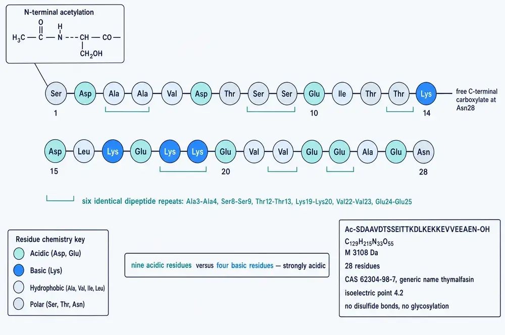
The composition is unusual enough to be worth reading off the diagram. Nine of the twenty-eight residues are acidic and only four are basic, which is what makes the molecule so strongly acidic and gives it the low isoelectric point that put it in the alpha region in the first place. There is no cysteine, so there are no disulfide bridges and no folded core held together by them. There is no proline and no glycine, the two residues that most constrain a peptide backbone. And there is no aromatic residue at all — no phenylalanine, tryptophan or tyrosine — which has a practical consequence familiar to anyone who has handled the peptide: it barely absorbs ultraviolet light at 280 nanometres, the wavelength at which protein concentration is normally measured. In total the molecule is built from ten of the twenty amino acids.
What that adds up to is a peptide with almost no fixed shape, and Section 05 takes up what has followed from that. It is not a defect, and it is not unusual among signalling peptides, but it means the familiar lock-and-key picture of a drug fitting a receptor pocket is the wrong mental image from the outset.
03Where it comes from
Thymosin alpha-1 is not synthesised as itself. It is the first twenty-eight residues of prothymosin alpha, an acidic nuclear protein of about 109 residues that is itself intrinsically disordered and has roles in chromatin remodelling and transcription which have nothing to do with immunity. The short peptide is released from the front of it by legumain, a lysosomal asparaginyl endopeptidase that cuts selectively after asparagine (Tao et al., 2023; Garaci et al., 2024).
The closure claimed in that caption carries more weight than it looks, because for a decade the opposite was seriously argued. In 1984, Haritos, Tsolas and Horecker used a radioimmunoassay against thymosin alpha-1 to survey rat tissues. They found cross-reacting material in thymus, and also in brain, liver, kidney, lung and spleen at fifteen to sixty-five per cent of thymic levels. But when they purified it with a procedure designed to avoid proteolysis, what they recovered was the intact precursor, not the short peptide. Their conclusion was blunt: prothymosin alpha is the endogenous molecule, and thymosin alpha-1 and its sister fragments are formed by proteolytic modification during the preparation of fraction 5. No peptide matching thymosin alpha-1 in size or chromatographic behaviour was detected at all (Haritos et al., 1984).
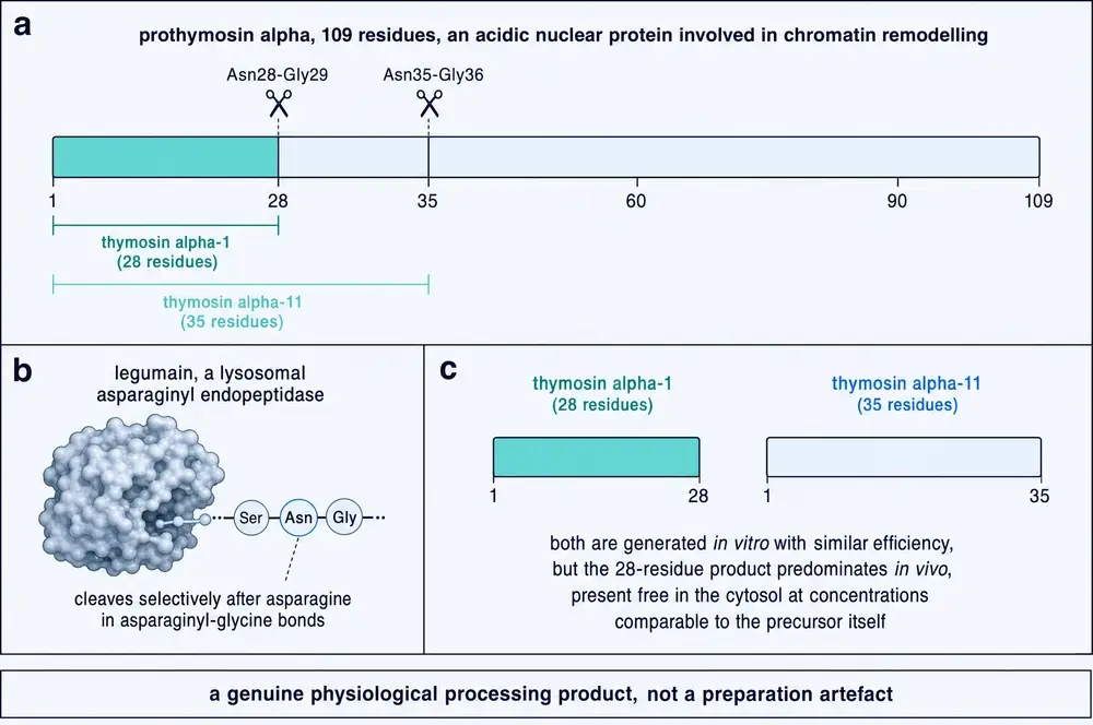
If that had stood, thymosin alpha-1 would not be a hormone that was discovered but a fragment manufactured by the discovery process. It did not stand unopposed for long. Extracting calf thymocytes under isotonic conditions in the presence of protease inhibitors recovered thymosin alpha-1 itself, both inside the cells and in the medium they had been sitting in, while the precursor was not detected in either; those authors concluded that thymocytes make the peptide by natural processing (Freire et al., 1985). Thymoma epithelial cells were separately shown to contain it and to be endocrinologically active (Savino et al., 1985).
The modern position, and the one Figure 2 draws, is that legumain releases the peptide physiologically and the question is therefore settled. It should be said plainly that the corpus assembled for this monograph does not contain that resolution. The processing chemistry is well established. What is not established is the in vivo abundance claim on which the settlement rests: the two tissue surveys that looked for free thymosin alpha-1 in animals did not find it, and the one analysis of human blood puts low-molecular-weight material of the right size at under a tenth of the total cross-reacting immunoreactivity. Forty years on, the honest answer is that the question is open, and this document treats it that way.
There is a third piece, and it comes from inside Goldstein's own laboratory. In 1984 Zatz and colleagues, with Goldstein as senior author, showed that incubating stimulated human lymphocytes with fraction 5 markedly increased production of T-cell growth factor. Thymosin alpha-1 alone did not do it. Their stated conclusion was that the biological activity of thymosin fraction 5 cannot be attributed to thymosin alpha-1, and that some further, undefined component of the mixture was responsible (Zatz et al., 1984). This is a human cell-culture study, and it says that the peptide extracted as the active principle of fraction 5 was not, in that assay, its active principle.
Within the thymus, antibodies raised against the synthetic peptide localise it to the epithelial cells — singly, in groups, and in the whorled structures called Hassall's corpuscles — rather than to the thymocytes those epithelial cells educate (Dalakas et al., 1981). This is a human tissue-staining study, and it remains the clearest anatomical answer to the question of where the peptide is made.
04What "thymosin" means, and what it does not
The word is a persistent source of confusion, and the confusion is built into its history. When the 1977 sequence paper proposed a nomenclature for the peptides of fraction 5, it sorted them by where they stopped in an electric field: below pH 5.0, alpha; between 5.0 and 7.0, beta; above 7.0, gamma (Goldstein et al., 1977; Severa et al., 2019). The numeral records only the order in which peptides were pulled out of a region. Thymosin alpha-1 means, and only means, the first peptide isolated from the acidic end of a calf thymus extract. It is a filing reference that has been mistaken for a taxonomy ever since.
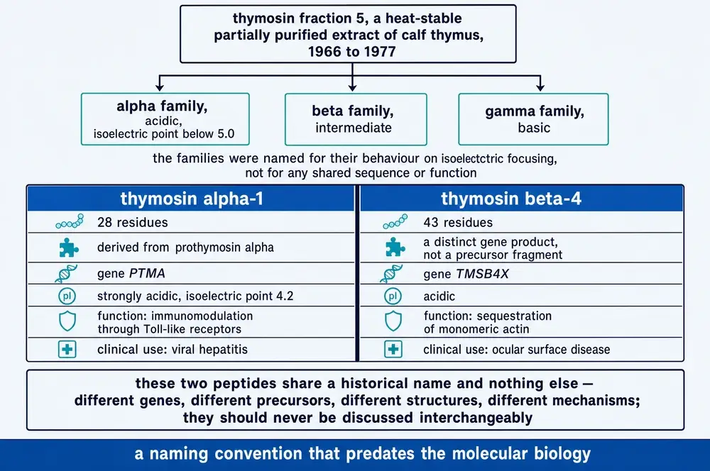
The practical consequence is that a great deal of writing about "thymosin" is about a different molecule. Thymosin beta-4 — sold in the research-chemical market as TB-500 — is 43 residues long, comes from its own gene rather than from a precursor, and works by binding monomeric actin. It has no mechanism in common with the compound described in this document. Any claim that transfers between them on the strength of the shared word is an error, and the error is common enough that the discovery scan behind this monograph was built to exclude it: an asset counted as being about thymosin alpha-1 only if it named the compound explicitly, and generic mentions of thymosin were recorded as context but never treated as a match.
One more member of the family deserves a sentence, because it is the field's earliest and best warning about itself. Polypeptide beta-1 was the most prominent single component of fraction 5. It had no biological activity in the group's assays. When it was sequenced it proved to be 74 residues long and identical to ubiquitin — one of the most abundant and universal proteins in all of eukaryotic biology, present in every cell of every tissue, and not in any sense a thymic hormone (Low & Goldstein, 1979; Severa et al., 2019). The most abundant thing in the thymus extract was the most abundant thing in everything.
Making it
Supply was solved by chemistry rather than by extraction, which is just as well given the tonnage of calf thymus the alternative required. The first total synthesis was published in 1979 (Birr & Stollenwerk, 1979). The following year an automated solid-phase route appeared, co-authored by R. B. Merrifield (Wang et al., 1980) — the inventor of solid-phase peptide synthesis, who would receive the Nobel Prize in Chemistry for it four years later. A twenty-eight-residue peptide with no cysteine and no aromatics is, by the standards of that method, an easy target. From 1980 the molecule was effectively unlimited, and the constraint on the field stopped being chemistry and became evidence.
Recombinant production came later and met a specific obstacle. Bacteria do not perform the N-terminal acetylation, and the unacetylated peptide is not quite the same molecule: the acetyl group on serine 1 is part of what lets the N-terminus insert into a membrane. Working around this has occupied a surprising share of the literature — co-expression of the acetyltransferase RimJ, concatemer constructs cleaved with hydroxylamine, fusions to human serum albumin, to immunoglobulin Fc, and to conformationally disordered PAS polymer chains. Most of those engineering papers exist for a second reason as well, which Section 10 takes up: the natural molecule disappears from the bloodstream in about two hours.
05A molecule with no fixed shape
Most drug mechanisms are told as a shape story: a molecule with a defined structure fits a pocket in a receptor with a complementary structure, and something happens. Thymosin alpha-1 does not have a defined structure. In water it is intrinsically disordered, wandering through a broad ensemble of conformations with no dominant member. What shape it adopts depends entirely on what it is next to.
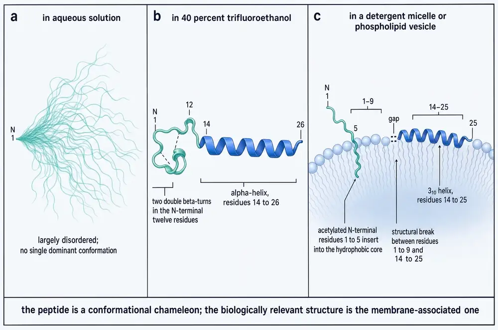
The helical structure in panel b comes from 800 MHz nuclear magnetic resonance in a trifluoroethanol–water mixture (Elizondo-Riojas et al., 2011). It is worth knowing that trifluoroethanol is a helix-inducing co-solvent, and that a group who later tried and failed to reproduce one of this compound's headline biological results pointed out that the structure had been determined under non-standard conditions and might not represent either the aqueous or the membrane-bound state (Armirotti et al., 2019). Panel c is the one that matters mechanistically: in a membrane environment the peptide inserts its first five residues, acetyl group foremost, into the lipid interior (Mandaliti et al., 2017).
06Getting to the membrane
Here is the fact that shapes everything downstream, and it is stated plainly in the field's own reviews: no specific high-affinity receptor for thymosin alpha-1 has ever been identified (Garaci et al., 2024; Simonova et al., 2025). For a compound approved in dozens of countries and studied for half a century, that is a remarkable sentence to have to write.
What has been proposed instead is a route rather than a receptor.
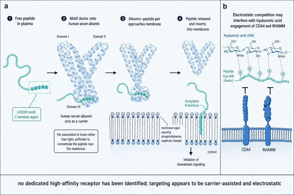
Phosphatidylserine is normally kept on the inner leaflet of the cell membrane and flips to the outside when a cell becomes apoptotic or stressed. A peptide that preferentially inserts into phosphatidylserine-exposing membranes is therefore a peptide that concentrates itself at sites of cell death and tissue damage — which is an elegant targeting story, and one that would explain why a molecule with no receptor might still act selectively. It should be read for what it is: a structural and computational account, supported by model-membrane work, not a demonstration in living tissue (Mandaliti et al., 2017; Garaci et al., 2024). The hyaluronic-acid interaction in panel b was described by its own authors as preliminary, with no functional CD44 or RHAMM data attached (Mandaliti et al., 2017).
07Toll-like receptors and the dendritic cell
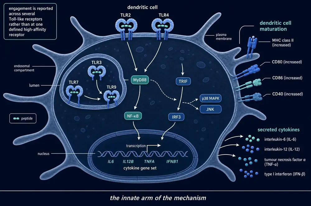
The dominant mechanistic account holds that thymosin alpha-1 acts as an agonist at Toll-like receptors — the sensors innate immune cells use to detect bacterial and viral molecular patterns — and that engaging them matures dendritic cells into effective antigen-presenting cells.
That caveat in the caption deserves expanding, because it is the crux. The evidence for Toll-like receptor involvement is evidence of pathway dependence, not of binding. The strongest single experiment took bone-marrow precursor cells from mice lacking each of TLR2, TLR3, TLR4, TLR9, MyD88 and TRIF in turn, exposed them to the peptide, and found that its promotion of dendritic cell generation was mostly TLR9- and TRIF-dependent (Renga et al., 2020). That is a well-controlled mouse cell experiment and it establishes that the pathway is required. It does not show the peptide touching the receptor. Separately, in transfected human embryonic kidney cells, the peptide accelerated the trafficking of TLR9 into lysosomes where TLR9 signalling occurs — but only in the presence of a conventional TLR9 agonist, and in a cell type that is not an immune cell (Romani et al., 2017).
Two further points cut against the tidy picture in Figure 6. First, the adapter is inconsistent: the knockout study above implicates TRIF, while the widely repeated formula in the review literature is "TLR9/MyD88". Second, and more seriously, the only measured binding affinity anywhere in this literature puts the peptide's interaction with TLR2 at a dissociation constant of about 35 micromolar — roughly a thousand times weaker than the peak concentrations reached in human blood after an injected dose. On that number, direct occupancy of TLR2 at therapeutic exposures is not credible, and the figure comes from a study of a fusion peptide rather than a dedicated investigation of thymosin alpha-1 itself.
Where the signalling has been worked out properly, it has been worked out downstream of the receptor. Using fibroblasts and macrophages from mice lacking TRAF6 or IKKβ, one group showed that both are required for the peptide to activate NF-κB and induce interleukin-6, and that it assembles a signalling complex containing TRAF6, p62 and atypical protein kinase C (Zhang et al., 2005). In human immature dendritic cells the peptide rapidly activates p38 (Yao et al., 2007). These are solid cell experiments. They place the compound in the Toll-like receptor and interleukin-1 receptor signalling family without identifying what it binds.
08What follows downstream
Granting the upstream uncertainty, the cellular consequences reported for this compound are reasonably consistent across laboratories, and they are what the clinical programme has always been built on.
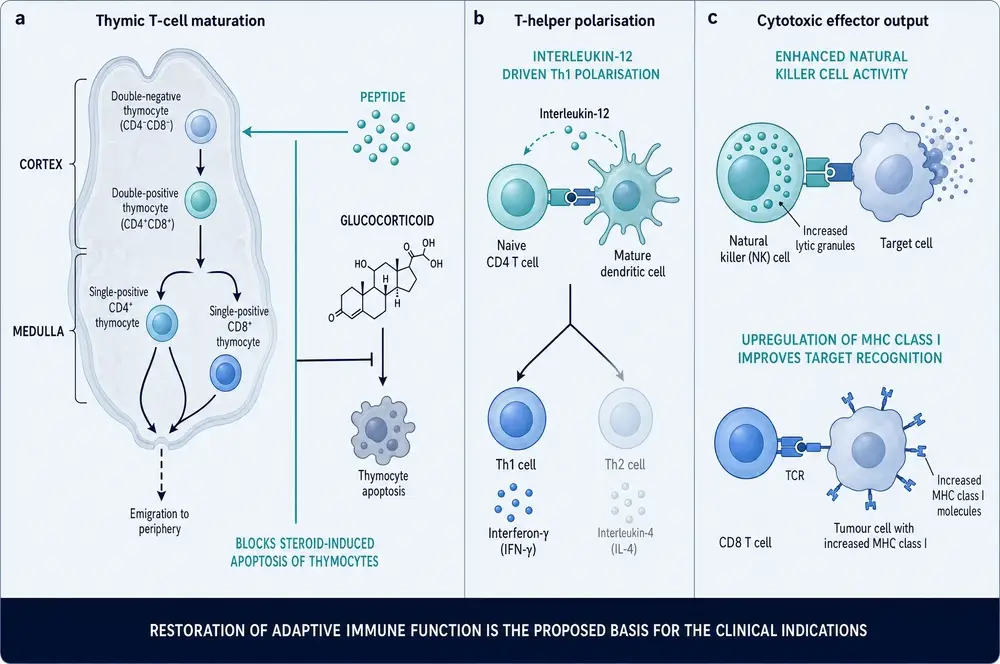
The steroid-apoptosis claim in panel a is worth pausing on, because it would have an obvious clinical corollary if it were established: developing thymocytes are exquisitely sensitive to glucocorticoids, and a molecule that protected them would matter in any illness accompanied by a surge of endogenous cortisol — which is to say, in critical illness. But it is not established. It enters the literature as a hedged sentence in a review — the peptide "may prevent" steroid-induced death of thymocytes (Li et al., 2010) — and travels from there into figures and summaries as though it had been measured. No experiment testing it appears anywhere in this corpus. The interleukin-12 step in panel b has the same character: it is load-bearing for the whole Th1-polarisation account, and no primary measurement of interleukin-12 induction by this peptide could be located. Two of the most-repeated cellular effects of thymosin alpha-1 rest on assertions rather than on data, and that is worth knowing before reading Part Three.
09Stimulant or thermostat
Now the genuine puzzle. The same molecule is reported to boost immunity in cancer and infection, and to restrain it in colitis, autoimmunity and inflamed tissue. Both bodies of evidence are real, and one of them is unusually clean.
The tolerogenic case is the better demonstrated. In mice given a checkpoint-inhibitor antibody that produces severe intestinal inflammation, the peptide protected the gut, raised interleukin-10, lowered interleukin-1β and interleukin-17A, and restored barrier integrity — and the protection failed completely in mice lacking the enzyme indoleamine 2,3-dioxygenase, which is the canonical switch for immune tolerance (Renga et al., 2020). That is a causal demonstration in an animal model, with the mechanism proven by knockout rather than inferred.
The most striking evidence, though, is a direct sign reversal in human cells. Applied to blood cells from patients with COVID-19, the peptide lowered inflammatory cytokines; applied to blood cells from healthy donors, it raised them (Matteucci et al., 2021). Same molecule, same concentration, opposite direction, and the variable was the inflammatory state of the donor.
10What the body does to it
Human pharmacokinetics are simple and, for the mechanistic story, awkward. After subcutaneous injection the peptide is rapidly absorbed, peaks at about two hours, and has a serum half-life of about two hours. Peak concentrations and total exposure rise in proportion to the amount given — across single doses of 0.8, 1.6, 3.2 and 6.4 mg in healthy volunteers, peak concentrations of 39, 63, 85 and 130 ng/mL respectively. Levels return to baseline by twenty-four hours, half or more of a single dose appears in the urine, and there is no accumulation on repeated administration (Garaci et al., 2024). These are healthy-volunteer pharmacokinetic studies, reported here as study parameters only.
Two consequences follow. The first is that a molecule cleared this fast cannot plausibly sustain a continuous signal, which is why so much of the chemistry literature is devoted to making it last longer — albumin fusions, Fc fusions, PASylation. The second is a straightforward arithmetic problem for the in-vitro literature: the concentrations at which the immunostimulatory effects are usually demonstrated in cell culture sit far above anything the bloodstream ever sees after a conventional dose. The two studies that used concentrations in the attainable human range both reported tolerogenic effects, not stimulatory ones.
The final check on all of this is what happens to measurable immune parameters in people. The largest and best-designed trial of the compound ever run — 1,089 patients with sepsis, double-blind and placebo-controlled — measured monocyte HLA-DR expression, lymphocyte counts and regulatory T-cell percentages, and found no separation from placebo on any of them (Wu et al., 2025). Whatever the peptide does in a culture dish, at the doses given to critically ill adults for seven days it did not move the immune markers it was given to move. Part Three follows that thread through indication after indication.
11Hepatitis B and C: the original promise
Chronic viral hepatitis was the first serious clinical target and remains the only one in which thymosin alpha-1 holds marketing authorisation. The rationale was good. Chronic hepatitis B is not simply a failure to clear a virus; it is a state of exhausted immunity, in which infected liver cells display less antigen and the T cells that should kill them carry inhibitory receptors and stop working properly. A drug that matures antigen-presenting cells and pushes T cells toward a Th1 programme is, on paper, aimed exactly at that lesion.
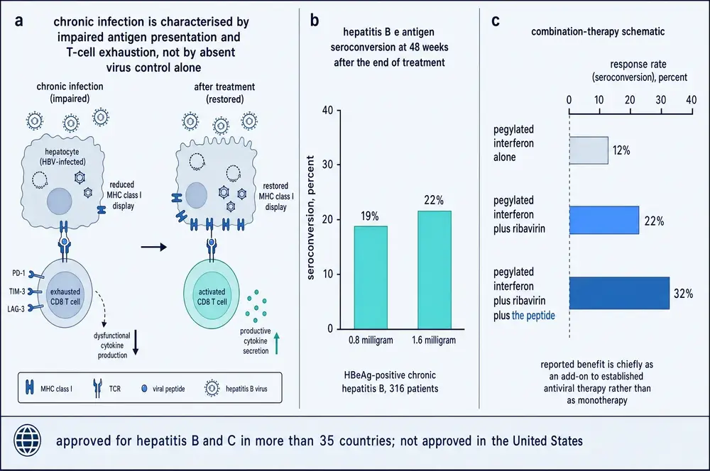
The hepatitis B story has a distinctive shape. Early trials were small and encouraging. A three-arm randomised trial in Taiwan reported a complete virological response of 40.6 per cent after twenty-six weeks of treatment against 9.4 per cent in untreated controls, assessed at eighteen months (Chien et al., 1998). Several open-label datasets described something unusual and appealing: benefit that appeared after treatment stopped, building through follow-up rather than fading. For an immunomodulator that is exactly what you would predict, and it became the signature claim of the compound.
Then the trial that should have settled it did not go the same way. A double-blind, placebo-controlled study of 97 patients found response rates of 14 per cent against 4 per cent, which did not reach statistical significance (P = 0.084); its authors wrote that their results did not confirm the earlier reports (Mutchnick et al., 1999). And when the delayed-response phenomenon itself was made a pre-specified endpoint under blinding, it occurred in 10 per cent of patients on drug and 8 per cent on placebo. The most interesting claim in the literature survived every open-label test and failed the one blinded one.
Hepatitis C went worse. As monotherapy in a placebo-controlled trial there were no viral clearances at all (Andreone et al., 1996). Added to interferon, it produced end-of-treatment gains that repeatedly failed to become sustained ones. In the pegylated-interferon era a 552-patient double-blind phase III found sustained virological response of 12.7 per cent against 10.5 per cent (P = 0.407), and the authors concluded that the peptide seems to play no role in the primary therapy of the disease (Ciancio et al., 2012).
Two further limits deserve stating plainly. First, adding the peptide on top of a modern nucleos(t)ide backbone such as entecavir improves surrogate markers at twenty-four weeks and no longer does so by forty-eight to fifty-two weeks. Second, and more important, no trial has shown a reduction in cirrhosis, decompensation, liver cancer or death. The two datasets actually powered to look — a 690-patient randomised trial and a 937-patient prospective cohort, both in compensated hepatitis B cirrhosis — were null (Wu et al., 2018; Wu et al., 2021). Meanwhile the therapeutic landscape moved: nucleos(t)ide analogues in hepatitis B and direct-acting antivirals in hepatitis C made the compound's original indication largely redundant, a point the review literature states in plain language.
One live signal remains and deserves replication. In hepatitis B-related acute-on-chronic liver failure — a setting where patients die of immune paralysis and secondary infection rather than of viral replication — a randomised study reported 90-day transplant-free survival of 75.0 per cent against 53.4 per cent (P = 0.030), with new infections falling from 58.6 to 32.1 per cent (P = 0.005) (Chen et al., 2022). That is mechanistically coherent and it is the most interesting positive result in the hepatitis literature.
12Sepsis: the best-designed failure
Sepsis is where the case was strongest on paper and has ended weakest in practice, and the arc is unusually well documented.
The rationale was never the problem. Severe sepsis is now understood to involve not only an inflammatory storm but a subsequent state of profound immune suppression — lymphocytes dying by apoptosis, monocytes losing their antigen-presenting machinery, patients succumbing to infections they should have cleared. That state is real, measurable and prognostically important. What it has never had is a therapy that reverses it and improves survival.
The mid-sized test came in 2013. ETASS randomised 361 patients with severe sepsis across six centres, single-blind against saline. Twenty-eight-day mortality was 26.0 per cent against 35.0 per cent — a relative risk of 0.74 whose confidence interval crossed one, at P = 0.062 (Wu et al., 2013). It did not meet its primary endpoint. In-hospital mortality and an unadjusted survival comparison did reach nominal significance, and monocyte HLA-DR rose more in the treated arm — though the treated arm also started with lower HLA-DR and was enrolled later, two baseline imbalances that both flatter the intervention on the endpoint each affects.
The definitive test came in 2025. TESTS randomised 1,106 patients across 22 centres, double-blind and placebo-controlled, under Sepsis-3 criteria, with 1.6 mg given subcutaneously every twelve hours for seven days.
The temptation is to file this as one trial against many, but that reading inverts the evidence. Read the sepsis literature in order of methodological quality and the effect shrinks at every step: small single-centre unblinded trials give odds ratios around 0.40; ETASS gives 0.74 at P = 0.062; the multicentre stratum of the 2025 meta-analysis gives 0.86; its high-quality stratum gives 0.82, no longer significant; a patient-level pool of both major trials gives a hazard ratio of 0.88; and a 2026 network meta-analysis of 76 randomised trials places the compound sixth of ten agents at GRADE low certainty (Gu et al., 2025; Luo et al., 2026). The subgroup-difference tests separating high- from low-quality trials are themselves statistically significant, which is the signature of bias rather than of biology, and Egger's test for publication bias comes out at P = 0.01. TESTS is not an outlier being outvoted. It is the endpoint of a gradient.
Three things keep this from being a closed file, and they should be said. The confidence intervals of ETASS and TESTS overlap, so a true relative risk near 0.85 remains consistent with both, and trial sequential analysis puts the information size needed at several times what has been accumulated. A subphenotype signal has surfaced from three methodologically independent directions, strongest in patients with cancer. And every trial ever conducted in this indication was conducted in one country. No sepsis guideline recommends the compound.
13COVID-19: the compound's largest moment
In early 2020 thymosin alpha-1 was prescribed widely, and the reason is easy to reconstruct: severe COVID-19 was accompanied by profound lymphopenia, lymphocyte count predicted death, and here was a licensed, cheap, apparently harmless drug that was supposed to restore lymphocytes.
The wave rested on a retrospective report of 76 severely ill patients in which mortality was 11.1 per cent against 30.0 per cent (Liu et al., 2020). Its accompanying immunological claim — that T-cell counts recovered — was a before-and-after comparison within treated patients, with no untreated comparator. When somebody finally made the between-group comparison in 178 patients, CD4 and CD8 counts recovered essentially identically with and without the drug (P = 0.851 and P = 0.842). The mechanism that justified a global prescribing wave did not survive its first proper control group.
What happened next is the clearest natural experiment available on how this compound's evidence gets made. One large cohort gave the drug to healthier patients and reported a nineteen-point crude mortality advantage that vanished completely on propensity matching. Another gave it to sicker patients and reported benefit. A 2,282-patient cohort found harm, which itself became non-significant once severity was properly modelled. Two studies from the same Shanghai hospital, over overlapping periods, reported that the drug both shortened and lengthened viral shedding, the difference tracing to analytic choices. The direction of confounding predicted the result better than any biological hypothesis.
Pooled properly, nothing was there. The larger and better-conducted meta-analysis — nine studies, 5,352 patients, registered in advance — found a relative risk for mortality of 1.03 (0.60–1.75), with null results for hospital stay, intensive-care stay and invasive ventilation, and concluded that the results do not support use of the compound in hospitalised adults with COVID-19 (Shang et al., 2023). The single blinded randomised trial rested on three deaths in 75 treated patients against five in 30 controls; an editorial in the same issue noted that one additional death would have rendered it non-significant, and it used four to six times the conventional amount.
14Cancer: preclinical promise, retrospective proof
Oncology is now the largest and fastest-growing part of this literature, and it has a two-layer structure that must be kept separate.
The randomised layer is old, small and mostly null. The only large randomised oncology trial — 488 patients with metastatic melanoma — met a response-rate criterion but missed statistical significance on overall survival (hazard ratio 0.80, P = .08) and on progression-free survival (Maio et al., 2010). A Cochrane systematic review of thymic peptides in cancer, covering 26 trials and 2,736 patients, found no significant survival benefit for thymosin alpha-1, with a pooled relative risk of 1.21 (0.94–1.56, P = 0.14) (Wolf et al., 2011).
The modern layer is large, positive and almost entirely retrospective: hazard ratios around 0.55 for survival after lung cancer resection, 0.31 after resection of hepatitis B-related liver cancer, 0.35 in small hepatocellular carcinoma. These come from single-institution, non-randomised, self-selected cohorts, several drawing on overlapping databases at the same hospital. More decisively, several define exposure by duration of treatment — more than 24 months, or continued to twelve months after chemoradiotherapy — which guarantees that longer-treated patients survived longer, because surviving is a prerequisite for continuing to be treated. That artefact, immortal time bias, is on its own directionally sufficient to produce the observed effects, and it is unaddressed in the studies that carry it.
There is a genuinely live line here, and it is recent. Radiotherapy destroys circulating lymphocytes, radiation-induced lymphopenia predicts worse cancer outcomes, and a series of studies from 2022 to 2026 report that the compound raises lymphocyte counts in irradiated patients and that this tracks with better outcomes. The lymphocyte effect is measurable and biologically plausible. The outcome link is not yet randomised evidence. This is where a properly designed trial would be most informative, and it does not yet exist.
One more finding cuts across the oncology rationale and deserves prominence rather than burial. In a mouse model of checkpoint-inhibitor colitis, the peptide's protective effect ran entirely through indoleamine 2,3-dioxygenase — the tolerance enzyme (Renga et al., 2020). That same pathway, driving regulatory T cells and myeloid-derived suppressor cells, is the canonical mechanism by which tumours escape immune attack. In one mouse tumour model the peptide given alone activated myeloid-derived suppressor cells and impaired antitumour activity. The compound is proposed simultaneously as a way to boost antitumour immunity and as a way to induce tolerance, and the reconciliation offered — that the effect is peritumoural rather than intratumoural — rests on a single murine line with no human tissue confirmation.
15The long tail
Beyond the four indications above the literature spreads thin, and one episode in it is more instructive than all the others combined.
In 2017 a paper in Nature Medicine reported that thymosin alpha-1 corrected the defective chloride channel that causes cystic fibrosis: a ten-fold increase in mature protein, a four-fold increase in channel opening, function restored to roughly half of normal, with a worked-out mechanism (Romani et al., 2017). It was a spectacular claim — a single small molecule fixing a disease that had defeated decades of work.
Within two years, six independent laboratories had failed to reproduce any of it, at the same concentration, in primary airway cells from multiple patients, with an established corrector working as a positive control every time (Tomati et al., 2018; Armirotti et al., 2019). The originating group offered two explanations — that the replicators' peptide lacked the N-terminal acetylation, and that their solvent had aggregated it. Both were tested directly and both were falsified: mass spectrometry confirmed the acetylation, and ion-mobility measurements found identical, non-aggregated species from either solvent. The original study had also disclosed that no blinding was applied, that the sample size was chosen empirically, and that only three of five patient cell donors responded.
The rest of the tail is thinner than its reputation. The influenza-vaccine adjuvant literature, frequently cited as established, rests clinically on a single pilot trial in haemodialysis patients. The multiple sclerosis work is cell-culture and serum-association only; one review states outright that there is no evidence on the compound's role in the standard animal model. The 2025 report of an antidepressant effect is an open-label, non-randomised study in five patients, two of whom relapsed during washout, with an effect size reported inconsistently within the paper itself (Aynekulu Mersha et al., 2025). The aging literature is mechanistic review rather than new data; the one solid human immunosenescence dataset measured endogenous peptide levels as a predictor of vaccine response durability in 98 men and administered no drug at all (Pozo-Balado et al., 2023).
16The quality gradient
Set the indications side by side and the same pattern appears in each of them, arrived at independently by different research communities working on different diseases.
In hepatitis B: small unblinded trials positive, the one adequately powered double-blind trial negative, hard endpoints null. In hepatitis C: monotherapy null, add-on gains that never became sustained, a 552-patient phase III negative. In sepsis: small single-centre trials strongly positive, a mid-sized single-blind trial at P = 0.062, a large double-blind trial flatly null, and pooled estimates that lose significance the moment quality is used as a filter. In COVID-19: uncontrolled before-and-after data positive, propensity matching erasing the effect, pooled relative risk 1.03. In oncology: randomised evidence null, retrospective evidence strongly positive with an unaddressed bias sufficient to produce it. In cystic fibrosis: one spectacular unblinded result, six independent replication failures.
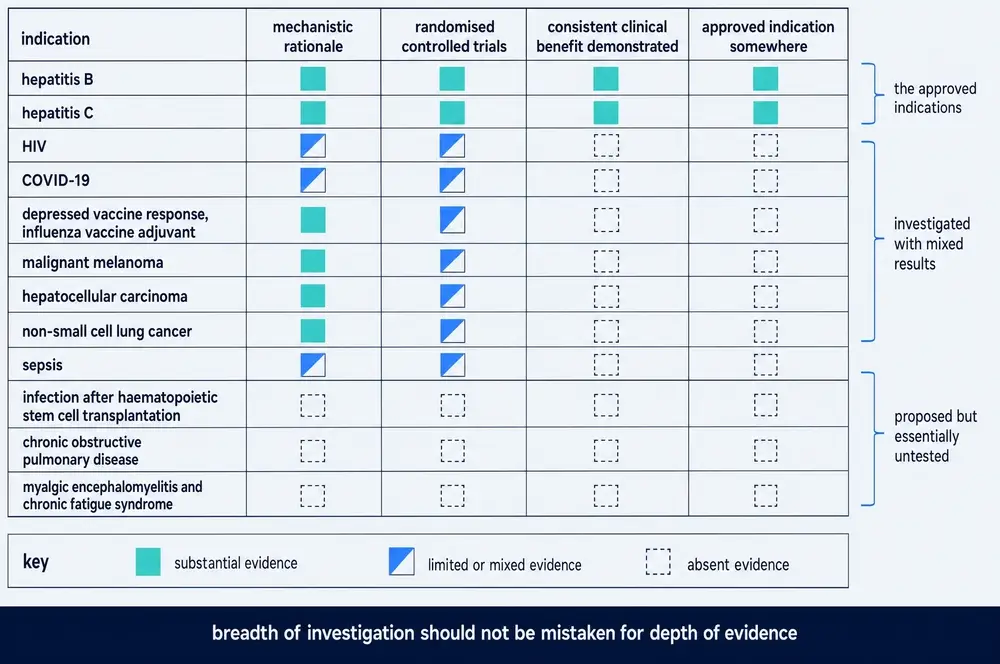
Six indications, six research communities, one shape. The apparent effect of thymosin alpha-1 is a function of study design. That is not a statement about the molecule; it is a statement about the literature, and it is the single most reliable finding this corpus contains.
17Where the trials were done, and who wrote them up
Four structural features of this evidence base do more to explain its shape than any biological hypothesis.
Geographic concentration. Three independent systematic reviews state that every trial they included was conducted in China; others say most were. Across the 215 full texts assembled for this monograph, 42.8 per cent mention China or Chinese authorship against 7.0 per cent for Italy. The field is structurally split in an unusual way: Italian academic groups produce nearly all the mechanistic immunology, Chinese groups produce nearly all the clinical trials, and the two literatures rarely share authors. The mechanism has therefore not been validated in the populations where the outcomes were measured, and the outcomes have not been replicated outside one health system. This is a methodological observation about replication, not a judgement about any country's research: single-region evidence is fragile evidence wherever the region is.
A review-to-trial ratio that manufactures the appearance of evidence. The metadata layer for this monograph contains 21 systematic reviews and 21 meta-analyses against 66 indexed randomised trials, of which only five records in 957 are phase III. The same small pool of trials is re-pooled repeatedly; narrative reviews then cite the meta-analyses, and later narrative reviews cite the earlier narrative reviews. Counting papers gives a badly inflated impression of how much has actually been tested.
Citation drift. The corpus contains documented instances of claims growing as they travel. A five-patient open-label pilot becomes an effect "demonstrated in a clinical study". A pilot trial with 94 patients becomes "large, randomized trials … without any safety alerts". An orphan-drug designation becomes FDA approval. A completers-only figure becomes a trial result. The number of countries where the compound is licensed is variously 35, 37, "over 35" and "approximately forty", with no regulatory citation attached to any of them.
Publication bias, measured. Egger's test on the pooled sepsis mortality data returns P = 0.01. That is not an inference from first principles; it is a positive test result.
18What can honestly be said about safety
Here the compound does well, and the finding should be stated as plainly as the negative ones.
Across roughly two and a half thousand randomised patients, short-course subcutaneous thymosin alpha-1 has shown no excess of adverse events, no drug-related serious adverse events, and essentially no discontinuations for toxicity. In the 1,089-patient sepsis trial, adverse events occurred in 66.4 per cent of treated patients against 67.6 per cent on placebo, and serious adverse events in 26.8 against 29.3 per cent — differences that are, if anything, numerically the wrong way for a harmful drug (Wu et al., 2025). In a 508-patient trial in severe pancreatitis, all three discontinuations driven by adverse events were in the placebo arm. The attributable adverse effect is injection-site reaction, and it is mild.
Four signals point the other way and an honest document names all four. In the large sepsis trial, a pre-specified subgroup analysis found higher mortality in patients under sixty (hazard ratio 1.67, 1.04–2.67) against no such effect in older patients, with a significant interaction test — in a trial whose overall result was null. A single case report describes severe multisystem immune-related toxicity with multiorgan failure beginning shortly after one dose given ten days after a checkpoint inhibitor, in a patient whose recent immunotherapy is a heavy confounder. In one COVID-19 cohort, treatment was associated with longer viral clearance. And in a mouse tumour model, the peptide alone activated immunosuppressive myeloid cells and impaired antitumour activity. Each is individually weak. Collectively they mark the edges of what the safety record actually covers.
19What would settle it
The interesting thing about this compound is that the question is not closed; it is under-informed, and the design that would inform it has been specified repeatedly and never executed.
Every recent synthesis converges on the same prescription: stop running uniform trials across whole disease populations, and enrich for the patients in whom the mechanism should apply. The candidate markers already exist. In sepsis, the pooled patient-level analysis of the two major trials found the cancer subgroup reaching a hazard ratio of 0.59 at moderate credibility on a formal subgroup-credibility instrument; a trial in severe pancreatitis found benefit confined to patients who were not lymphopenic, and the large sepsis trial enrolled a population whose median lymphocyte count sat below that threshold. In oncology, radiation-induced lymphopenia is a measurable, mechanistically appropriate enrichment criterion with an emerging signal attached to it. In hepatitis, acute-on-chronic liver failure is the one setting with a randomised result worth replicating.
None of these has been tested prospectively. The biomarker-enriched trial that the sepsis field called for in 2013, that ETASS's own authors called for in 2013, and that four separate syntheses have called for since, still does not exist. That is the actual state of play: not a compound that has been proven useless, but one that has been given, for fifty years, to populations chosen without reference to the mechanism it is supposed to work through — and that has failed, every time the trial was good enough to tell.
20Approval without proof
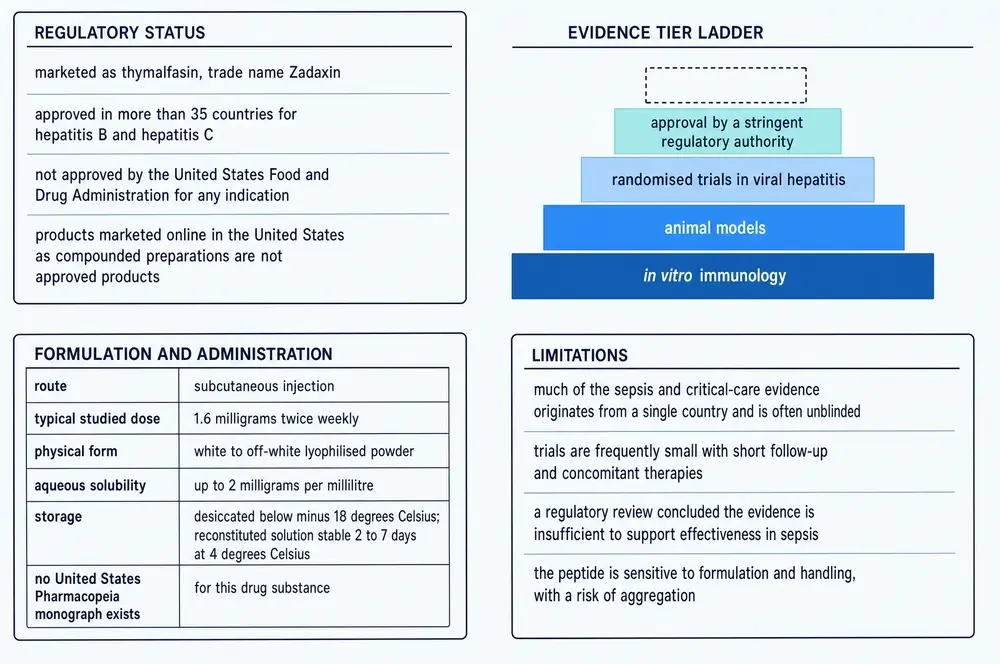
Thymosin alpha-1 occupies an unusual regulatory position. It is licensed in more than thirty-five countries, concentrated in Asia and Latin America, for chronic hepatitis B and in a subset of those for hepatitis C. It has never been approved by the United States Food and Drug Administration for any indication — a fact worth stating clearly, because the corpus assembled for this monograph contains at least one review that says otherwise, apparently by converting an orphan-drug designation into an approval.
The gap between that regulatory footprint and the trial record is the substantive point of this section. A licence in thirty-five countries reads, to most people, as an official finding that a drug works. What it records here is something narrower: that in the 1990s and 2000s, in jurisdictions where chronic hepatitis B was a major public-health problem and the alternatives were interferon and little else, a well-tolerated immunomodulator with modest surrogate-endpoint data cleared the bar that then applied. Those approvals have not been revisited in light of what came afterwards, because there is no mechanism that forces them to be.
One consequence sits outside the scientific literature entirely and should be named. The compound circulates in a research-chemical and compounding market in countries where it is not approved. The corpus is completely silent on this: not one of 215 full texts and 957 abstracts discusses grey-market supply, compounded preparations, counterfeiting or independent purity testing. Given that the molecule requires N-terminal acetylation for its membrane-anchoring behaviour, that solid-phase synthesis is the accepted route for clinical-grade production, and that the peptide is sensitive to formulation and handling, that silence is a substantive gap rather than a mere omission — and nothing in this evidence base can be used to fill it.
21Why it persists
A compound that has failed its best trials in four separate indications might reasonably have disappeared. This one has not; the publication rate is rising. The reasons are worth setting out, because they are structural rather than scientific, and because they generalise to other compounds.
It is genuinely safe, and safety lowers the bar for trying. The recurring argument in this literature runs: the compound is exceptionally well tolerated, therefore trying it costs little, therefore weak positive signals justify wider use. That reasoning is seductive and mostly sound, and it has one failure mode — it treats the cost of a treatment as its side-effect profile, when the real costs are the alternative not given, the trial not run, and the question left open for another decade.
The mechanism predicts benefit exactly where trials are hardest. Immunoparalysis in sepsis, exhaustion in chronic infection, lymphopenia after radiotherapy — these are real, measurable states in populations that are heterogeneous, severely ill and difficult to study. A mechanism aimed at them is attractive and hard to falsify.
Context dependence absorbs contradiction. A molecule whose direction of effect is said to be set by the host's inflammatory state can accommodate a positive trial, a negative trial and a harmful signal without revision. That is a serious epistemic problem, and it is the reason this document weights the blinded, adequately powered negatives as heavily as it does.
The evidence base is structured so that positive results are easier to produce than negative ones. Small single-centre trials, unblinded designs, surrogate endpoints, retrospective cohorts with exposure defined by treatment duration, before-and-after immunology without a control group, and a review-to-trial ratio above one to one. Every one of those features biases in the same direction.
22References
Generated from verified NCBI records rather than from recall. Author lists, journal names, volumes, pages and identifiers are taken from the PubMed record for each citation, and the build refuses to run if any identifier fails to resolve. This safeguard exists because it has been needed: in two earlier monographs in this series, reference lists drafted from memory contained identifiers that pointed at real but unrelated papers. It was needed again in drafting this one — four author attributions in the first draft named the wrong first author, and the generator caught all four.
- Andreone P, Cursaro C, Gramenzi A, Buzzi A, Covarelli MG, Di Giammarino L, et al.. A double-blind, placebo-controlled, pilot trial of thymosin alpha 1 for the treatment of chronic hepatitis C. Liver. 1996;16(3):207-10.
PMID 8873009 · doi:10.1111/j.1600-0676.1996.tb00729.x - Armirotti A, Tomati V, Matthes E, Veit G, Cholon DM, Phuan PW, et al.. Bioactive Thymosin Alpha-1 Does Not Influence F508del-CFTR Maturation and Activity. Scientific reports. 2019;9(1):10310.
PMID 31311979 · doi:10.1038/s41598-019-46639-1 · PMC6635361 - Aynekulu Mersha DG, Fromme SE, van Boven F, Arteaga-Henríquez G, Wijkhuijs A, van der Ent M, et al.. Indications for an antidepressive effect of thymosin alpha-1 in a small open-label proof of concept study in common variable immune deficiency patients with depression. Brain, behavior, & immunity - health. 2025;43:100934.
PMID 39867848 · doi:10.1016/j.bbih.2024.100934 · PMC11762651 - Bach JF, Dardenne M, Goldstein AL, Guha A, White A. Appearance of T-cell markers in bone marrow rosette-forming cells after incubation with thymosin, a thymic hormone. Proc Natl Acad Sci U S A. 1971;68(11):2734-8.
PMID 4399414 · doi:10.1073/pnas.68.11.2734 · PMC389512 - Bellet MM, Renga G, Pariano M, Stincardini C, D'Onofrio F, Goldstein AL, et al.. COVID-19 and beyond: Reassessing the role of thymosin alpha1 in lung infections. International immunopharmacology. 2023;117:109949.
PMID 36881979 · doi:10.1016/j.intimp.2023.109949 · PMC9977614 - Birr C, Stollenwerk U. Synthesis of thymosin alpha1, a polypeptide of the thymus. Angewandte Chemie (International ed. in English). 1979;18(5):394-5.
PMID 112886 · doi:10.1002/anie.197903941 - Chen JF, Chen SR, Lei ZY, Cao HJ, Zhang SQ, Weng WZ, et al.. Safety and efficacy of Thymosin α1 in the treatment of hepatitis B virus-related acute-on-chronic liver failure: a randomized controlled trial. Hepatology international. 2022;16(4):775-788.
PMID 35616850 · doi:10.1007/s12072-022-10335-6 - Chien RN, Liaw YF, Chen TC, Yeh CT, Sheen IS. Efficacy of thymosin alpha1 in patients with chronic hepatitis B: a randomized, controlled trial. Hepatology (Baltimore, Md.). 1998;27(5):1383-7.
PMID 9581695 · doi:10.1002/hep.510270527 - Ciancio A, Andreone P, Kaiser S, Mangia A, Milella M, Solà R, et al.. Thymosin alpha-1 with peginterferon alfa-2a/ribavirin for chronic hepatitis C not responsive to IFN/ribavirin: an adjuvant role?. Journal of viral hepatitis. 2012;19 Suppl 1:52-9.
PMID 22233415 · doi:10.1111/j.1365-2893.2011.01524.x - Costantini C, Bellet MM, Pariano M, Renga G, Stincardini C, Goldstein AL, et al.. A Reappraisal of Thymosin Alpha1 in Cancer Therapy. Frontiers in oncology. 2019;9:873.
PMID 31555601 · doi:10.3389/fonc.2019.00873 · PMC6742685 - Dalakas MC, Engel WK, McClure JE, Goldstein AL, Askanas V. Immunocytochemical localization of thymosin-alpha 1 in thymic epithelial cells of normal and myasthenia gravis patients and in thymic cultures. Journal of the neurological sciences. 1981;50(2):239-47.
PMID 7014787 · doi:10.1016/0022-510x(81)90170-2 - Dauphinee MJ, Talal N, Goldstein AL, White A. Thymosin corrects the abnormal DNA synthetic response of NZB mouse thymocytes. Proc Natl Acad Sci U S A. 1974;71(7):2637-41.
PMID 4546960 · doi:10.1073/pnas.71.7.2637 · PMC388521 - Elizondo-Riojas MA, Chamow SM, Tuthill CW, Gorenstein DG, Volk DE. NMR structure of human thymosin alpha-1. Biochemical and biophysical research communications. 2011;416(3-4):356-61.
PMID 22115779 · doi:10.1016/j.bbrc.2011.11.041 · PMC3419376 - Eschenfeldt WH, Manrow RE, Krug MS, Berger SL. Isolation and partial sequencing of the human prothymosin alpha gene family. Evidence against export of the gene products. The Journal of biological chemistry. 1989;264(13):7546-55.
PMID 2708378 - Freire M, Rey-Mendez M, Gomez-Marquez J, Arias P. Evidence for the synthesis of thymosin alpha 1 by calf thymocytes and the production of this peptide by natural processing. Archives of biochemistry and biophysics. 1985;239(2):480-5.
PMID 3873909 · doi:10.1016/0003-9861(85)90715-5 - Garaci E, Paci M, Matteucci C, Costantini C, Puccetti P, Romani L. Phenotypic drug discovery: a case for thymosin alpha-1. Frontiers in medicine. 2024;11:1388959.
PMID 38903817 · doi:10.3389/fmed.2024.1388959 · PMC11187271 - Goldstein AL, Slater FD, White A. Preparation, assay, and partial purification of a thymic lymphocytopoietic factor (thymosin). Proc Natl Acad Sci U S A. 1966;56(3):1010-7.
PMID 5230175 · doi:10.1073/pnas.56.3.1010 · PMC219962 - Goldstein AL, Low TL, McAdoo M, McClure J, Thurman GB, Rossio J, et al.. Thymosin alpha1: isolation and sequence analysis of an immunologically active thymic polypeptide. Proceedings of the National Academy of Sciences of the United States of America. 1977;74(2):725-9.
PMID 265536 · doi:10.1073/pnas.74.2.725 · PMC392366 - Gu B, Zhou Y, Nie Y, Wang L, Liang L, Liao Z, et al.. Efficacy of thymosin α1 for sepsis: a systematic review and meta-analysis of randomized controlled trials. Frontiers in cellular and infection microbiology. 2025;15:1673959.
PMID 40969554 · doi:10.3389/fcimb.2025.1673959 · PMC12440967 - Hannappel E, Huff T. The thymosins. Prothymosin alpha, parathymosin, and beta-thymosins: structure and function. Vitamins and hormones. 2003;66:257-96.
PMID 12852257 · doi:10.1016/s0083-6729(03)01007-0 - Hardy MA, Quint J, Goldstein AL, State D, White A. Effect of thymosin and an antithymosin serum on allograft survival in mice. Proc Natl Acad Sci U S A. 1968;61(3):875-82.
PMID 4879824 · doi:10.1073/pnas.61.3.875 · PMC305409 - Haritos AA, Tsolas O, Horecker BL. Distribution of prothymosin alpha in rat tissues. Proceedings of the National Academy of Sciences of the United States of America. 1984;81(5):1391-3.
PMID 6584887 · doi:10.1073/pnas.81.5.1391 · PMC344840 - Ke L, Zhou J, Mao W, Chen T, Zhu Y, Pan X, et al.. Immune enhancement in patients with predicted severe acute necrotising pancreatitis: a multicentre double-blind randomised controlled trial. Intensive care medicine. 2022;48(7):899-909.
PMID 35713670 · doi:10.1007/s00134-022-06745-7 · PMC9205279 - Li J, Liu CH, Wang FS. Thymosin alpha 1: biological activities, applications and genetic engineering production. Peptides. 2010;31(11):2151-8.
PMID 20699109 · doi:10.1016/j.peptides.2010.07.026 · PMC7115394 - Liu B, Gong X, Chang S, Sun P, Wu J. Generation of mature Nα-terminal acetylated thymosin α 1 by cleavage of recombinant prothymosin α. TheScientificWorldJournal. 2013;2013:387282.
PMID 24288480 · doi:10.1155/2013/387282 · PMC3830889 - Liu Y, Pan Y, Hu Z, Wu M, Wang C, Feng Z, et al.. Thymosin Alpha 1 Reduces the Mortality of Severe Coronavirus Disease 2019 by Restoration of Lymphocytopenia and Reversion of Exhausted T Cells. Clinical infectious diseases : an official publication of the Infectious Diseases Society of America. 2020;71(16):2150-2157.
PMID 32442287 · doi:10.1093/cid/ciaa630 · PMC7314217 - Low TL, Goldstein AL. The chemistry and biology of thymosin. II. Amino acid sequence analysis of thymosin alpha1 and polypeptide beta1. The Journal of biological chemistry. 1979;254(3):987-95.
PMID 762108 - Luo R, Xu S, Chen Y, Liu Z, Deng X, Li C, et al.. Comparative efficacy and safety of immunomodulatory therapies for sepsis: a systematic review and network meta-analysis. Frontiers in medicine. 2026;13:1808427.
PMID 42205841 · doi:10.3389/fmed.2026.1808427 · PMC13201415 - Maio M, Mackiewicz A, Testori A, Trefzer U, Ferraresi V, Jassem J, et al.. Large randomized study of thymosin alpha 1, interferon alfa, or both in combination with dacarbazine in patients with metastatic melanoma. Journal of clinical oncology : official journal of the American Society of Clinical Oncology. 2010;28(10):1780-7.
PMID 20194853 · doi:10.1200/JCO.2009.25.5208 - Mandaliti W, Nepravishta R, Pica F, Vallebona PS, Garaci E, Paci M. Thymosin α1 Interacts with Hyaluronic Acid Electrostatically by Its Terminal Sequence LKEKK. Molecules (Basel, Switzerland). 2017;22(11).
PMID 29077041 · doi:10.3390/molecules22111843 · PMC6150299 - Matteucci C, Minutolo A, Balestrieri E, Petrone V, Fanelli M, Malagnino V, et al.. Thymosin Alpha 1 Mitigates Cytokine Storm in Blood Cells From Coronavirus Disease 2019 Patients. Open forum infectious diseases. 2021;8(1):ofaa588.
PMID 33506065 · doi:10.1093/ofid/ofaa588 · PMC7798699 - Mutchnick MG, Lindsay KL, Schiff ER, Cummings GD, Appelman HD, Peleman RR, et al.. Thymosin alpha1 treatment of chronic hepatitis B: results of a phase III multicentre, randomized, double-blind and placebo-controlled study. Journal of viral hepatitis. 1999;6(5):397-403.
PMID 10607256 · doi:10.1046/j.1365-2893.1999.00181.x - Pozo-Balado MDM, Bulnes-Ramos Á, Olivas-Martínez I, Garrido-Rodríguez V, Lozano C, Álvarez-Ríos AI, et al.. Higher plasma levels of thymosin-α1 are associated with a lower waning of humoral response after COVID-19 vaccination: an eight months follow-up study in a nursing home. Immunity & ageing : I & A. 2023;20(1):9.
PMID 36879319 · doi:10.1186/s12979-023-00334-y · PMC9986663 - Renga G, Bellet MM, Pariano M, Gargaro M, Stincardini C, D'Onofrio F, et al.. Thymosin α1 protects from CTLA-4 intestinal immunopathology. Life science alliance. 2020;3(10).
PMID 32817121 · doi:10.26508/lsa.202000662 · PMC7441522 - Romani L, Oikonomou V, Moretti S, Iannitti RG, D'Adamo MC, Villella VR, et al.. Thymosin α1 represents a potential potent single-molecule-based therapy for cystic fibrosis. Nature medicine. 2017;23(5):590-600.
PMID 28394330 · doi:10.1038/nm.4305 · PMC5420451 - Savino W, Manganella G, Verley JM, Wolff A, Berrih S, Levasseur P, et al.. Thymoma epithelial cells secrete thymic hormone but do not express class II antigens of the major histocompatibility complex. The Journal of clinical investigation. 1985;76(3):1140-6.
PMID 2413072 · doi:10.1172/JCI112069 · PMC424007 - Severa M, Zhang J, Giacomini E, Rizzo F, Etna MP, Cruciani M, et al.. Thymosins in multiple sclerosis and its experimental models: moving from basic to clinical application. Multiple sclerosis and related disorders. 2019;27:52-60.
PMID 30317071 · doi:10.1016/j.msard.2018.09.035 · PMC7104151 - Shang W, Zhang B, Ren Y, Wang W, Zhou D, Li Y. Thymosin alpha1 use in adult COVID-19 patients: A systematic review and meta-analysis on clinical outcomes. International immunopharmacology. 2023;114:109584.
PMID 36527881 · doi:10.1016/j.intimp.2022.109584 · PMC9754924 - Simonova MA, Ivanov I, Shoshina NS, Komyakova AM, Makarov DA, Baranovskii DS, et al.. Aging and Thymosin Alpha-1. International journal of molecular sciences. 2025;26(23).
PMID 41373628 · doi:10.3390/ijms262311470 · PMC12692621 - Tao N, Xu X, Ying Y, Hu S, Sun Q, Lv G, et al.. Thymosin α1 and Its Role in Viral Infectious Diseases: The Mechanism and Clinical Application. Molecules (Basel, Switzerland). 2023;28(8).
PMID 37110771 · doi:10.3390/molecules28083539 · PMC10144173 - Tomati V, Caci E, Ferrera L, Pesce E, Sondo E, Cholon DM, et al.. Thymosin α-1 does not correct F508del-CFTR in cystic fibrosis airway epithelia. JCI insight. 2018;3(3).
PMID 29415893 · doi:10.1172/jci.insight.98699 · PMC5821210 - Wang SS, Makofske R, Bach A, Merrifield RB. Automated solid phase synthesis of thymosin alpha 1. International journal of peptide and protein research. 1980;15(1):1-4.
PMID 7358454 · doi:10.1111/j.1399-3011.1980.tb02543.x - Wolf E, Milazzo S, Boehm K, Zwahlen M, Horneber M. Thymic peptides for treatment of cancer patients. The Cochrane database of systematic reviews. 2011;2011(2):CD003993.
PMID 21328265 · doi:10.1002/14651858.CD003993.pub3 · PMC6481824 - Wu J, Zhou L, Liu J, Ma G, Kou Q, He Z, et al.. The efficacy of thymosin alpha 1 for severe sepsis (ETASS): a multicenter, single-blind, randomized and controlled trial. Critical care (London, England). 2013;17(1):R8.
PMID 23327199 · doi:10.1186/cc11932 · PMC4056079 - Wu J, Pei F, Zhou L, Li W, Sun R, Li Y, et al.. The efficacy and safety of thymosin α1 for sepsis (TESTS): multicentre, double blinded, randomised, placebo controlled, phase 3 trial. BMJ (Clinical research ed.). 2025;388:e082583.
PMID 39814420 · doi:10.1136/bmj-2024-082583 · PMC11780596 - Wu X, Shi Y, Zhou J, Sun Y, Piao H, Jiang W, et al.. Combination of entecavir with thymosin alpha-1 in HBV-related compensated cirrhosis: a prospective multicenter randomized open-label study. Expert opinion on biological therapy. 2018;18(sup1):61-69.
PMID 30063860 · doi:10.1080/14712598.2018.1451511 - Wu X, Zhou J, Sun Y, Ding H, Chen G, Xie W, et al.. Prediction of liver-related events in patients with compensated HBV-induced cirrhosis receiving antiviral therapy. Hepatology international. 2021;15(1):82-92.
PMID 33460002 · doi:10.1007/s12072-020-10114-1 - Yao Q, Doan LX, Zhang R, Bharadwaj U, Li M, Chen C. Thymosin-alpha1 modulates dendritic cell differentiation and functional maturation from human peripheral blood CD14+ monocytes. Immunology letters. 2007;110(2):110-20.
PMID 17532057 · doi:10.1016/j.imlet.2007.04.007 · PMC1986709 - Zatz MM, Oliver J, Samuels C, Skotnicki AB, Sztein MB, Goldstein AL. Thymosin increases production of T-cell growth factor by normal human peripheral blood lymphocytes. Proceedings of the National Academy of Sciences of the United States of America. 1984;81(9):2882-5.
PMID 6609371 · doi:10.1073/pnas.81.9.2882 · PMC345176 - Zhang P, Chan J, Dragoi AM, Gong X, Ivanov S, Li ZW, et al.. Activation of IKK by thymosin alpha1 requires the TRAF6 signalling pathway. EMBO reports. 2005;6(6):531-7.
PMID 15905851 · doi:10.1038/sj.embor.7400433 · PMC1369095 - Zisblatt M, Goldstein AL, Lilly F, White A. Acceleration by thymosin of the development of resistance to murine sarcoma virus-induced tumor in mice. Proc Natl Acad Sci U S A. 1970;66(4):1170-4.
PMID 5273447 · doi:10.1073/pnas.66.4.1170 · PMC335802 - [No authors listed]. The efficacy and safety of thymosin α1 for sepsis (TESTS): multicentre, double blinded, randomised, placebo controlled, phase 3 trial. BMJ (Clinical research ed.). 2025;389:r1098.
PMID 40447307 · doi:10.1136/bmj.r1098 · PMC12123687
23How this document was assembled
The corpus was built by a staged pipeline against project 05, the Therapeutic Peptide Research Library. Every file with a document extension in the project's document stores was opened and its extracted text searched for explicit mentions of the compound under any of its designations — thymosin alpha 1 in any hyphenation, thymosin α1, Tα1, thymalfasin or Zadaxin.
The alpha/beta distinction is load-bearing, and the pipeline was built around it. Thymosin beta-4 is a different peptide isolated from the same thymic fraction, sold in the research-chemical market as TB-500, and a large part of the available literature concerns it. A bare mention of thymosin, or of the thymic-peptide neighbourhood generally, was recorded as context but never counted as a match. That sweep opened 45,820 files and returned 494 raw matches, which collapsed to 404 after de-duplication.
Classifying those by kind of source is the step that matters. Only 51 were peer-reviewed scientific full texts. The other 353 were vendor catalogue material, consumer web content captured for style training, bulk acquisition files and internal working documents — none of which is evidence about the compound.
The external layer supplied the substance. A PubMed harvest returned 1026 records, of which 69 were rejected because neither title, abstract nor MeSH headings named this compound — overwhelmingly thymosin beta-4 papers caught by a deliberately broad query. That left 957 indexed records, of which 215 carried a PubMed Central identifier and were fetched as full text. Merging the local and fetched sets gives the reading corpus this monograph is written from: 236 unique scientific full texts, roughly 2,157 printed-page equivalents.
| Stage | What it does | Result |
|---|---|---|
| 01b | Targeted scan of the project's document stores | 45,820 files opened |
| 02 | PubMed harvest with a beta-4 relevance filter | 957 records kept |
| 03 | PubMed Central open-access full-text retrieval | 215 full texts |
| 04 | De-duplication, classification, inventory report | 236 unique |
| 05 | Reference list generation from verified records | 52 citations |
| 06 | Assembly of this document | 10 figures |
The reading corpus was then read in full by seven parallel passes, one per evidence domain — discovery and biochemistry, mechanism, hepatitis, sepsis, COVID-19 and respiratory, oncology, and safety with the long tail of minor indications. Each pass produced a structured evidence packet in which every extracted figure carries its own identifier and a study-type label, and each was required to keep a separate register of claims the corpus does not support. Those seven packets, not the reviews, are what this document was drafted from.
A note on one recurring trap. A PubMed article record contains reference and comment lists that are themselves full of identifier nodes belonging to other papers. Parsing those without scoping each lookup to the article's own subtree silently assigns a bibliography entry's identifiers to the article being read. Every lookup in this pipeline is scoped for that reason.
24Evidence handling
Findings in this document are labelled by the kind of study that produced them, in the sentence that reports them. Randomised trials, single-arm studies, retrospective cohorts, animal experiments, cell and tissue measurements, structural and computational work, and narrative reviews are different kinds of claim, and the differences are stated rather than left to the reader. Animal and in-vitro findings are never phrased so as to imply a human outcome.
Where evidence conflicts, both sides are given. The 2013 and 2025 sepsis trials are presented together with the reasons the newer null does not simply outvote the older positive, and with the quality gradient that connects them. The 2017 cystic fibrosis result and its six independent replication failures are presented as a live episode with its rebuttals tested. Where a pooled estimate loses significance once study quality is used as a filter, both estimates are given.
Supplied artwork is treated as evidence to check rather than decoration. Every value printed on the ten commissioned plates was audited against the reading corpus before publication. 118 printed items were checked: 54 were verified, 23 qualified, 17 could not be verified from this corpus, and 17 were contradicted by it. The outcome for each is recorded in figure-source-art/MAPPING.md, and every affected caption in this document carries a figure-audit rider naming what the artwork prints and what the evidence says instead. Where the two disagree, the running text is authoritative and the caption says so.
That result deserves to be stated rather than absorbed, because it is a finding in its own right. The plates were drawn from the same secondary literature this monograph is about, and they reproduce its characteristic errors: a negative trial rendered as a positive bar chart, a hypothesis printed as a mechanism, an orphan-drug designation promoted to an approval, an unresolved forty-year dispute captioned as settled. An audit that returns seventeen contradictions from a hundred and eighteen values is not a comment on the illustrator. It is the same measurement this document makes everywhere else, taken on a different instrument.
Continue exploring