
Incretins: GLP-1 and GIP
The gut hormones behind the incretin effect — why a glass of glucose triggers far more insulin than the same glucose dripped into a vein, and how that single piece of physiology became a class of medicines.
Abstract
An incretin is a gut hormone that amplifies insulin secretion in response to a meal. The phenomenon it produces — the incretin effect — is visible in a clean experiment: glucose taken by mouth triggers a substantially larger insulin response than the identical amount of glucose infused into a vein to reproduce the same blood-glucose curve, because hormones released from the gut, not the blood sugar itself, do the extra work. In healthy people this accounts for roughly half to two-thirds of the insulin secreted after oral glucose, and grows with the size of the load. Two hormones carry most of the effect: glucagon-like peptide-1 (GLP-1), from intestinal L-cells, and glucose-dependent insulinotropic polypeptide (GIP), from K-cells higher in the gut. Both stimulate insulin only when glucose is elevated — the reason incretin-based drugs carry little intrinsic risk of hypoglycemia — and both are inactivated within a minute or two by the enzyme DPP-4. GLP-1 does more than GIP: it suppresses glucagon, slows gastric emptying, and curbs appetite. In type 2 diabetes the incretin effect is blunted, chiefly because the pancreas stops responding to GIP while its response to GLP-1 is largely preserved — the biological reason the modern medicines lean on GLP-1.
Key findings
- The incretin effect is real and quantifiable: intravenous glucose produces only about 30–40% of the insulin that the same oral glucose does at a matched blood-sugar level, so gut hormones account for the remaining majority — commonly roughly 50–70% in healthy people, and larger with bigger glucose loads (Perley & Kipnis, 1967; Nauck et al., 1986).
- Two incretin hormones do the work: GLP-1, a 30-amino-acid peptide from intestinal L-cells, and GIP from K-cells higher in the gut; both are released within minutes of eating and act on the beta-cell (Kreymann et al., 1987; Baggio & Drucker, 2007; Holst, 2007).
- Their insulin action is glucose-dependent — it operates when blood glucose is high and fades as it normalizes — which is why incretin-based drugs carry little intrinsic risk of causing low blood sugar on their own (Baggio & Drucker, 2007; Nauck & Meier, 2016).
- The two hormones diverge: GLP-1 also suppresses glucagon, slows gastric emptying, and promotes satiety, while GIP acts mainly on fat and bone and does not suppress glucagon; GLP-1 is the more potent insulinotropic agent per molecule (Kreymann et al., 1987; Baggio & Drucker, 2007).
- Both are destroyed within a minute or two by the enzyme DPP-4 — only about 20% of infused GLP-1 remains intact — which is the degradation problem every incretin medicine was engineered around (Deacon et al., 1995; Holst, 2007).
- In type 2 diabetes the incretin effect is blunted (incretin-driven insulin ~73% in controls vs ~36% in patients), chiefly because the pancreas becomes unresponsive to GIP — its insulin effect falls by about half — while the response to GLP-1 is largely preserved; the defect is partly reversible with better glucose control (Nauck et al., 1986; Nauck et al., 1993; Nauck & Meier, 2016; Højberg et al., 2009).
Eat a meal and your gut does something a needle cannot. Within minutes of food reaching the small intestine, cells in its lining release hormones that travel ahead to the pancreas and prime it — so that when the sugar from the meal arrives in the blood, the insulin response is far larger than the same rise in blood sugar would produce on its own. Those gut hormones are called incretins, and the surplus insulin they summon is the incretin effect.
The effect is not subtle, and it is not a matter of interpretation. Give a person glucose to drink, then on another day drip glucose into a vein to reproduce the identical blood-sugar curve, and the drink triggers substantially more insulin than the infusion. The gap is the work of hormones released from the gut, not of the blood sugar itself. Two of those hormones — GLP-1 and GIP — carry most of that work, and they are the biological foundation beneath a class of medicines that has reshaped the treatment of diabetes and obesity.
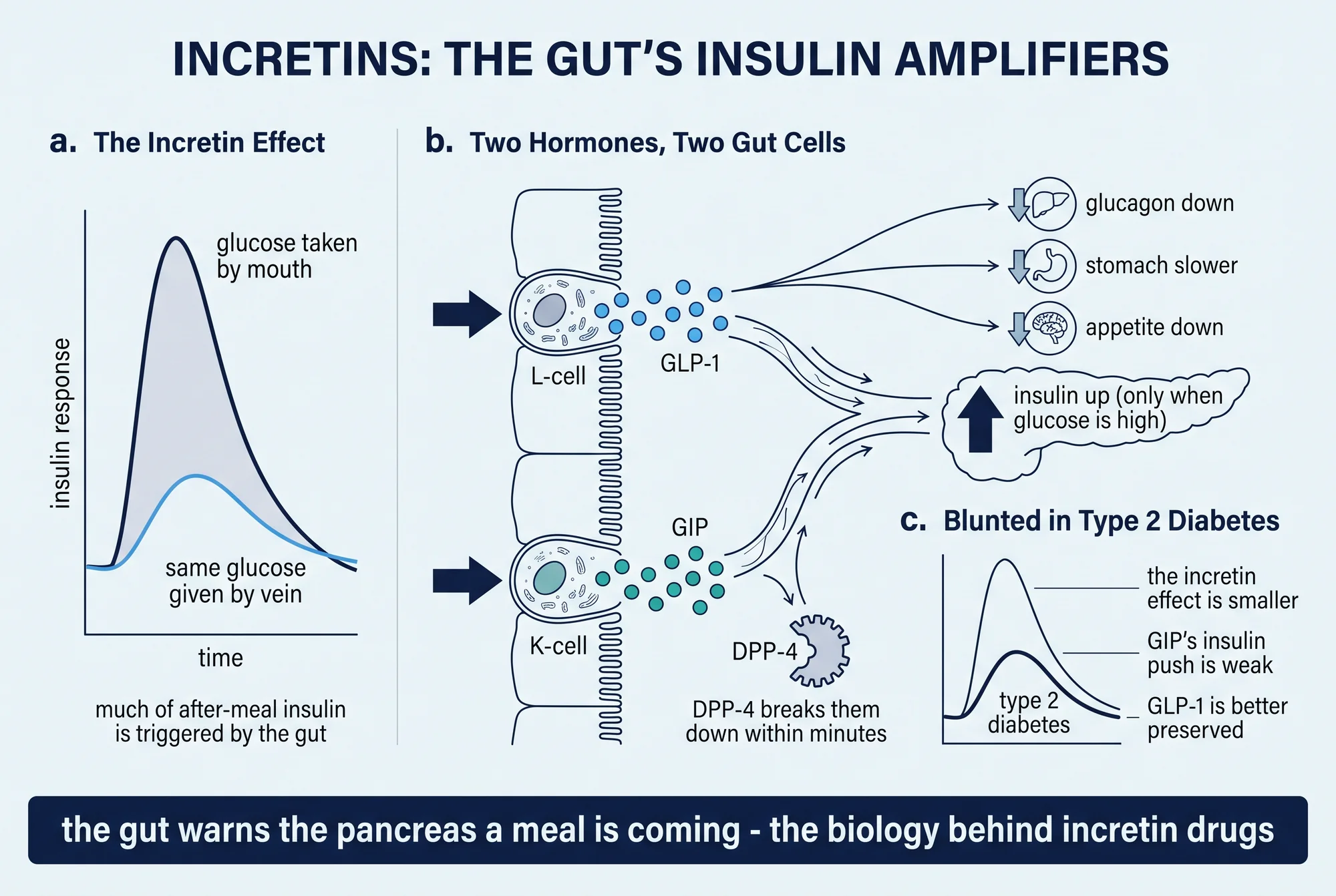
The incretin effect: why the mouth beats the vein
The whole subject rests on one experiment, and it is worth doing in the imagination before naming any hormone. Measure how much insulin a person secretes after drinking a measured dose of glucose. On a separate day, infuse glucose into a vein and adjust the drip so the blood-sugar curve traces the same path it took after the drink. The pancreas now faces an identical glucose stimulus by the only measure it is supposed to care about — the concentration of sugar in the blood. If blood sugar were the whole story, the two insulin responses would match. They do not. The oral route wins, and by a wide margin.
That result is old and durable. The first clear demonstration that oral glucose draws out more insulin than intravenous glucose came in the mid-1960s (Elrick et al., 1964). A landmark follow-up put a number on it: the plasma insulin response to intravenous glucose was only about 30 to 40% of the response to the same glucose given by mouth, which means a majority of the meal's insulin was being driven by something other than blood sugar — an "alimentary," gut-derived mechanism, and one the same study located in the intestinal tract (Perley & Kipnis, 1967). The difference between the oral and intravenous responses, attributed to factors other than glucose itself, is the definition of the incretin effect (Nauck & Meier, 2016).
How large is it? The honest answer is a range that depends on the size of the meal. Measuring insulin secretion directly across increasing oral glucose loads, the incretin contribution climbs as the load grows, reaching a majority of the secretory response at larger doses (Nauck et al., 1986, J Clin Endocrinol Metab). Pulling the human data together, the effect is commonly summarized as accounting for roughly 50 to 70% of the insulin secreted after oral glucose in healthy people, larger with a larger glucose load (Nauck & Meier, 2016). One measurement subtlety belongs beside that figure: estimates taken from insulin measured in a peripheral vein run higher than estimates based on C-peptide or on calculated secretion rates, because the liver clears a different fraction of insulin after oral versus intravenous glucose (Nauck et al., 1986, J Clin Endocrinol Metab). The precise percentage therefore shifts with the method, but the direction and the rough magnitude do not: in a healthy person, gut hormones account for a large share of the insulin a meal produces.
Two hormones, two cells
The "something other than blood sugar" turned out to be two hormones, released from two different specialized cells in the gut lining, both within minutes of nutrients arriving (Baggio & Drucker, 2007).
The first is glucagon-like peptide-1 (GLP-1), a 30-amino-acid peptide produced by intestinal endocrine cells called L-cells, concentrated in the lower small intestine and colon, by processing of a larger precursor protein called proglucagon (Holst, 2007). Its identity as a genuine human incretin was established by infusing it into volunteers at levels mimicking what a meal produces: insulin rose, glucose fell, and — a detail that matters later — it did so more powerfully than the other incretin (Kreymann et al., 1987).
The second is GIP, released from K-cells in the upper small intestine. Its acronym has survived a change of name. It was originally called gastric inhibitory polypeptide, for an inhibitory action on the stomach noted in early studies; as its insulin-releasing role became the important one, it was renamed glucose-dependent insulinotropic polypeptide — same letters, better description (Baggio & Drucker, 2007). In a healthy person, GIP is a major contributor to the incretin effect alongside GLP-1 (Baggio & Drucker, 2007; Nauck & Meier, 2016).
Two peptides, then, from two cell types, reading the arrival of food and converging on the same target: the insulin-producing beta-cell of the pancreas. What they do there is best understood as a single, elegant rule.
Insulin only when it is needed
Both hormones act on the beta-cell through their own receptors, and both share a defining property — their stimulation of insulin is glucose-dependent (Baggio & Drucker, 2007). The signal amplifies insulin release when blood glucose is elevated and fades as glucose returns toward normal. GLP-1's insulin action, in the founding human study, tracked the glucose level and tapered as glucose came down (Kreymann et al., 1987; Holst, 2007).
That single feature has an outsized consequence, and it is the reason incretin biology became attractive to drug developers. Because the insulin push switches off as glucose falls, incretin signaling does not keep forcing insulin into a normal or low bloodstream. It therefore carries little intrinsic risk of hypoglycemia — dangerously low blood sugar — a sharp contrast with therapies such as injected insulin or sulfonylureas, which raise insulin regardless of what glucose is doing (Baggio & Drucker, 2007; Nauck & Meier, 2016). This is a property of the mechanism, and it holds for the drugs built on it on their own; the risk can still rise when an incretin-based agent is combined with a drug that forces insulin independently of glucose. The point is not a clinical instruction but a piece of physiology: the incretin system is built to add insulin when a meal warrants it and to stand down when it does not.
Where the two hormones diverge
If GLP-1 and GIP only amplified insulin, one explainer would cover both. They do not. GLP-1 carries a wider portfolio, and the divergence is the reason it, rather than GIP, became the template for the blockbuster drugs.
Beyond insulin, GLP-1 does three additional things. It suppresses glucagon — the pancreatic hormone that tells the liver to release stored glucose — which further restrains the post-meal glucose rise. It slows gastric emptying, so a meal leaves the stomach more gradually and fullness lasts longer. And it acts on the brain as a satiety signal, reducing appetite and food intake (Baggio & Drucker, 2007; Holst, 2007). These four actions together are the subject of a companion explainer, what GLP-1 is and how it works; here the point is simply that GLP-1 is a multi-target hormone.
GIP's portfolio is different. It shares the glucose-dependent insulin action, but it does not suppress glucagon — under some conditions it raises it — and it does not meaningfully slow the stomach or curb appetite. Its extra actions lie elsewhere: GIP promotes energy storage through direct effects on fat tissue and supports bone formation (Baggio & Drucker, 2007). And on the shared job of stimulating insulin, GLP-1 is the more potent agent per molecule — the observation, present from the first human comparison, that GLP-1 is "more powerful than GIP" as an insulinotropic hormone (Kreymann et al., 1987; Nauck et al., 1993). Two hormones with a common origin story and a common target, then, but genuinely different jobs — a distinction that becomes decisive in disease.
Gone in a minute: the DPP-4 problem
There is a catch that shapes everything downstream. Both incretins are destroyed almost as fast as they are made. An enzyme called DPP-4 (dipeptidyl peptidase-4) clips two amino acids off the front of each hormone, inactivating it — and it does so extremely rapidly, beginning even before the hormone has left the gut (Holst, 2007). When GLP-1 is infused into people and its intact form measured directly, only about 20% of it remains undamaged, the rest already converted to an inactive fragment (Deacon et al., 1995). The functional half-life of native GLP-1 in the circulation is on the order of one to two minutes (Holst, 2007).
For a hormone the gut re-releases with every meal, that brevity is no problem. For a would-be medicine, it is fatal: a drug cannot be re-infused every ninety seconds. The rapid degradation of both incretins by DPP-4 is precisely what forced two engineering strategies, both of which became real drug classes — enzyme-resistant molecules that mimic GLP-1 and last for days, and inhibitors of DPP-4 itself that let the body's own incretins survive longer (Baggio & Drucker, 2007). The disposable design of the natural hormone is the reason the pharmacology exists.
The broken incretin effect in type 2 diabetes
The incretin effect is not a fixed constant of human biology. In type 2 diabetes it is blunted, and this is one of the more reliable findings in the field. Repeating the oral-versus-intravenous experiment in patients and matched controls, the share of the insulin response attributable to incretin factors fell from about 73% in healthy controls to about 36% in patients by one measure, and from about 58% to below 10% by a more direct measure of secretion (Nauck et al., 1986, Diabetologia). The gut's amplifier, in other words, is turned down in the disease.
The more revealing question is why, and the answer is not a uniform failure of both hormones. When GIP and GLP-1 are infused separately into people with type 2 diabetes under matched conditions, they behave very differently. GIP's ability to stimulate insulin is markedly impaired — its maximal effect roughly halved compared with healthy subjects (a reduction of about 54%). GLP-1's insulin action, by contrast, is largely preserved, reaching about 71% of the healthy response, a difference that did not reach statistical significance; and GLP-1 still suppressed glucagon in the patients, which GIP did not (Nauck et al., 1993). Modern reviews state the lesson plainly: in type 2 diabetes the pancreas remains responsive to GLP-1 but is no longer responsive to GIP, the most likely reason the overall incretin effect is reduced (Nauck & Meier, 2016).
Two honest refinements belong beside that headline. The first is that the defect lies more in how the pancreas responds to the hormones than in how much the gut secretes them. Secretion is only modestly affected: the meal-stimulated GLP-1 response is somewhat reduced in patients (Toft-Nielsen et al., 2001; Vilsbøll et al., 2001), while GIP secretion is largely intact (Vilsbøll et al., 2001). The amplifier is failing mostly because the beta-cell has stopped listening to GIP, not because the gut has gone quiet. The second refinement is that the unresponsiveness is not wholly fixed. Four weeks of near-normalizing blood glucose in patients restored beta-cell responsiveness to both GIP and GLP-1 by a factor of three to four (Højberg et al., 2009). That reversibility suggests high glucose itself contributes to the defect, and it leaves genuinely open the question of how much of the blunted incretin effect is a cause of the diabetic state versus a consequence of it.
From hormone to medicine
Everything above converges on a practical point. Because native incretins are potent but glucose-safe, and disposable within minutes, two strategies could turn the biology into treatment, and both are now established, FDA-approved drug classes for type 2 diabetes. DPP-4 inhibitors block the degrading enzyme and let the body's own incretins linger. GLP-1 receptor agonists are enzyme-resistant molecules that reproduce GLP-1's signal for days at a time; at higher doses they became the medicines now used for chronic weight management as well (Baggio & Drucker, 2007). These molecules — and the peptide chemistry behind them, covered in what a peptide is — are treated in depth in the explainers on what GLP-1 is and on semaglutide.
Why did the drugs lean on GLP-1 rather than GIP? For the reason the disease itself supplies: GLP-1's insulin action survives in type 2 diabetes while GIP's largely does not, so a GLP-1-based agent still works in the patients who need it (Nauck et al., 1993; Nauck & Meier, 2016). That made GIP look like a therapeutic dead end for years. The twist is that it was not. The most effective of the newer agents, tirzepatide, is a single molecule that activates both the GIP and the GLP-1 receptor, and it produces larger weight loss in trials than GLP-1 alone. Reconciling that with GIP's blunted action in diabetes is an active, unresolved area — part of the answer may be that lowering glucose restores GIP responsiveness (Højberg et al., 2009), and part may be that pharmacological GIP-receptor activity does things that a physiology experiment in untreated patients could not reveal (Campbell & Drucker, 2013).
One line has to stay bright through all of this. The incretin concept is physiology — a mechanism, measured in healthy volunteers and patients. It explains why these drugs were designed as they were and why they behave as a class. It does not, by itself, establish how much any individual benefits or is harmed by a given medicine; those are clinical outcomes, settled by trials of specific drugs, and they live in the drug-specific explainers, not in the mechanism. The link between the beta-cell's response to a gut hormone and a person's health is exactly where this article stops and the trial evidence begins — a boundary that also runs through the account of insulin resistance and the broader picture of metabolic health.
What remains uncertain
Several honest caveats travel with the confidence.
- The exact split between GIP and GLP-1 in the healthy incretin effect is not settled. Both contribute; their relative share is debated and hard to pin down, in part because the estimate depends on the measurement method — peripheral insulin overstates the effect relative to C-peptide or calculated secretion rates (Nauck et al., 1986, J Clin Endocrinol Metab; Nauck & Meier, 2016).
- Cause versus consequence in type 2 diabetes is unresolved. The incretin effect is reliably blunted in the disease, but the reversibility of the beta-cell's unresponsiveness with glucose control argues that at least part of the defect follows from hyperglycemia rather than causing it (Højberg et al., 2009). How much is upstream and how much downstream is not fully known.
- GIP's therapeutic role is genuinely open. Its insulin action is impaired in untreated diabetes, yet adding GIP-receptor activity to a GLP-1 drug improves results; both activating and blocking the GIP receptor have been pursued as strategies, a sign that the biology is not fully understood (Campbell & Drucker, 2013; Nauck & Meier, 2016).
- Mechanism is not outcome. The glucose-dependence that limits hypoglycemia risk, the actions on glucagon and appetite, and the differences between the two hormones are established physiology. What any of it means for a particular person on a particular drug is a separate, clinical question, and the same caution that applies to any therapy applies here.
This review covers the incretin effect and how it is measured, the two incretin hormones GLP-1 and GIP and the cells that make them, the glucose-dependence of their insulin action, how the two hormones differ, their rapid inactivation by DPP-4, and the blunted incretin effect in type 2 diabetes — drawn from primary human physiology studies and authoritative reviews retrieved from PubMed and PubMed Central. Evidence cutoff August 2026; the underlying physiology is mature and the anchoring studies span 1964 to 2016. It is educational and is not medical advice, a diagnosis, or a treatment recommendation, and it names no dose or protocol; the incretin-based drug classes referenced (DPP-4 inhibitors, GLP-1 receptor agonists, and the dual GIP/GLP-1 agonist tirzepatide) are FDA-approved for type 2 diabetes and, for some agents, chronic weight management, and their trial evidence lives in the drug-specific explainers. For related reading, see What Is GLP-1 and How Does It Work?, Semaglutide, Tirzepatide, and Insulin Resistance, or browse the Metabolic health and GLP-1s hubs.
References
- 1.Baggio LL, Drucker DJ. Biology of incretins: GLP-1 and GIP. Gastroenterology. 2007;132(6):2131-2157. doi:10.1053/j.gastro.2007.03.054
- 2.Campbell JE, Drucker DJ. Pharmacology, physiology, and mechanisms of incretin hormone action. Cell Metab. 2013;17(6):819-837. doi:10.1016/j.cmet.2013.04.008
- 3.Deacon CF, Nauck MA, Toft-Nielsen M, Pridal L, Willms B, Holst JJ. Both subcutaneously and intravenously administered glucagon-like peptide I are rapidly degraded from the NH2-terminus in type II diabetic patients and in healthy subjects. Diabetes. 1995;44(9):1126-1131. doi:10.2337/diab.44.9.1126
- 4.Elrick H, Stimmler L, Hlad CJ Jr, Arai Y. Plasma insulin response to oral and intravenous glucose administration. J Clin Endocrinol Metab. 1964;24:1076-1082. doi:10.1210/jcem-24-10-1076
- 5.Højberg PV, Vilsbøll T, Rabøl R, et al. Four weeks of near-normalisation of blood glucose improves the insulin response to glucagon-like peptide-1 and glucose-dependent insulinotropic polypeptide in patients with type 2 diabetes. Diabetologia. 2009;52(2):199-207. doi:10.1007/s00125-008-1195-5
- 6.Holst JJ. The physiology of glucagon-like peptide 1. Physiol Rev. 2007;87(4):1409-1439. doi:10.1152/physrev.00034.2006
- 7.Kreymann B, Williams G, Ghatei MA, Bloom SR. Glucagon-like peptide-1 7-36: a physiological incretin in man. Lancet. 1987;2(8571):1300-1304. doi:10.1016/s0140-6736(87)91194-9
- 8.Nauck MA, Homberger E, Siegel EG, et al. Incretin effects of increasing glucose loads in man calculated from venous insulin and C-peptide responses. J Clin Endocrinol Metab. 1986;63(2):492-498. doi:10.1210/jcem-63-2-492
- 9.Nauck M, Stöckmann F, Ebert R, Creutzfeldt W. Reduced incretin effect in type 2 (non-insulin-dependent) diabetes. Diabetologia. 1986;29(1):46-52. doi:10.1007/BF02427280
- 10.Nauck MA, Heimesaat MM, Ørskov C, Holst JJ, Ebert R, Creutzfeldt W. Preserved incretin activity of glucagon-like peptide 1 [7-36 amide] but not of synthetic human gastric inhibitory polypeptide in patients with type-2 diabetes mellitus. J Clin Invest. 1993;91(1):301-307. doi:10.1172/JCI116186
- 11.Nauck MA, Meier JJ. The incretin effect in healthy individuals and those with type 2 diabetes: physiology, pathophysiology, and response to therapeutic interventions. Lancet Diabetes Endocrinol. 2016;4(6):525-536. doi:10.1016/S2213-8587(15)00482-9
- 12.Perley MJ, Kipnis DM. Plasma insulin responses to oral and intravenous glucose: studies in normal and diabetic subjects. J Clin Invest. 1967;46(12):1954-1962. doi:10.1172/JCI105685
- 13.Toft-Nielsen MB, Damholt MB, Madsbad S, et al. Determinants of the impaired secretion of glucagon-like peptide-1 in type 2 diabetic patients. J Clin Endocrinol Metab. 2001;86(8):3717-3723. doi:10.1210/jcem.86.8.7750
- 14.Vilsbøll T, Krarup T, Deacon CF, Madsbad S, Holst JJ. Reduced postprandial concentrations of intact biologically active glucagon-like peptide 1 in type 2 diabetic patients. Diabetes. 2001;50(3):609-613. doi:10.2337/diabetes.50.3.609
Disclosures
Educational review of published evidence. Not medical advice, a diagnosis, or a treatment recommendation. Study parameters are reported with the population and design that produced them. Incretin-based drug classes named here (DPP-4 inhibitors, GLP-1 receptor agonists, and the dual GIP/GLP-1 agonist tirzepatide) are FDA-approved for type 2 diabetes and, for some agents, chronic weight management; this piece names no dose or protocol and does not compare products.